

Evolving evidence in the treatment of primary and recurrent posterior cruciate ligament injuries, part 1: anatomy, biomechanics and diagnostics
- PMID: 33201271
- PMCID: PMC7917041
- DOI: 10.1007/s00167-020-06357-y
Evolving evidence in the treatment of primary and recurrent posterior cruciate ligament injuries, part 1: anatomy, biomechanics and diagnostics
Abstract
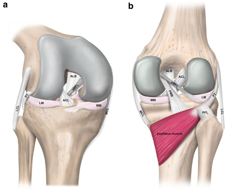
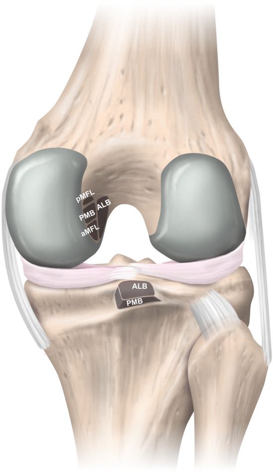
The posterior cruciate ligament (PCL) represents an intra-articular structure composed of two distinct bundles. Considering the anterior and posterior meniscofemoral ligaments, a total of four ligamentous fibre bundles of the posterior knee complex act synergistically to restrain posterior and rotatory tibial loads. Injury mechanisms associated with high-energy trauma and accompanying injury patterns may complicate the diagnostic evaluation and accuracy. Therefore, a thorough and systematic diagnostic workup is necessary to assess the severity of the PCL injury and to initiate an appropriate treatment approach. Since structural damage to the PCL occurs in more than one third of trauma patients experiencing acute knee injury with hemarthrosis, background knowledge for management of PCL injuries is important. In Part 1 of the evidence-based update on management of primary and recurrent PCL injuries, the anatomical, biomechanical, and diagnostic principles are presented. This paper aims to convey the anatomical and biomechanical knowledge needed for accurate diagnosis to facilitate subsequent decision-making in the treatment of PCL injuries.Level of evidence V.
Keywords: Anatomy; Biomechanics; Diagnostic workup; Knee; PCL; Posterior cruciate ligament; Posterior stress radiograph; Revision.
Conflict of interest statement
VM reports educational grants, consulting fees, and speaking fees from Smith & Nephew plc, educational grants from Arthrex, is a board member of the International Society of Arthroscopy, Knee Surgery and Orthopaedic Sports Medicine (ISAKOS), and deputy editor-in-chief of Knee Surgery, Sports Traumatology, Arthroscopy (KSSTA). In addition, VM has a patent Quantified injury diagnostics-U.S. Patent No. 9,949,684, ssued on April 24, 2018, issued to University of Pittsburgh.
Figures
References
-
- Ahmad CS, Cohen ZA, Levine WN, Gardner TR, Ateshian GA, Mow VC. Codominance of the individual posterior cruciate ligament bundles. An analysis of bundle lengths and orientation. Am J Sports Med. 2003;31:221–225. - PubMed
-
- Aman ZS, DePhillipo NN, Storaci HW, Moatshe G, Chahla J, Engebretsen L, et al. Quantitative and qualitative assessment of posterolateral meniscal anatomy: defining the popliteal hiatus, popliteomeniscal fascicles, and the lateral meniscotibial ligament. Am J Sports Med. 2019;47:1797–1803. - PubMed
-
- Amis AA, Gupte CM, Bull AM, Edwards A. Anatomy of the posterior cruciate ligament and the meniscofemoral ligaments. Knee Surg Sports Traumatol Arthrosc. 2006;14:257–263. - PubMed
-
- Anderson CJ, Ziegler CG, Wijdicks CA, Engebretsen L, LaPrade RF. Arthroscopically pertinent anatomy of the anterolateral and posteromedial bundles of the posterior cruciate ligament. J Bone Joint Surg Am. 2012;94:1936–1945. - PubMed
-
- Azar FM, Brandt JC, Miller RH, 3rd, Phillips BB. Ultra-low-velocity knee dislocations. Am J Sports Med. 2011;39:2170–2174. - PubMed
Publication types
MeSH terms
LinkOut - more resources
Full Text Sources
Medical
