. 2021 May;24(2):363-377.
doi: 10.1007/s10456-020-09757-3. Epub 2020 Nov 17.

Endothelium-specific deletion of Nox4 delays retinal vascular development and mitigates pathological angiogenesis

Affiliations

Endothelium-specific deletion of Nox4 delays retinal vascular development and mitigates pathological angiogenesis

Xixiang Tang et al. Angiogenesis. 2021 May.

Abstract

NADPH oxidase 4 (Nox4) is a major isoform of NADPH oxidases playing an important role in many biological processes. Previously we have shown that Nox4 is highly expressed in retinal blood vessels and is upregulated in oxygen-induced retinopathy (OIR). However, the exact role of endothelial Nox4 in retinal angiogenesis remains elusive. Herein, using endothelial cell (EC)-specific Nox4 knockout (Nox4EC-KO) mice, we investigated the impact of endothelial Nox4 deletion on retinal vascular development and pathological angiogenesis during OIR. Our results show that deletion of Nox4 in ECs led to retarded retinal vasculature development with fewer, blunted-end tip cells and sparser, dysmorphic filopodia at vascular front, and reduced density of vascular network in superficial, deep, and intermediate layers in postnatal day 7 (P7), P12, and P17 retinas, respectively. In OIR, loss of endothelial Nox4 had no effect on hyperoxia-induced retinal vaso-obliteration at P9 but significantly reduced aberrant retinal neovascularization at P17 and decreased the deep layer capillary density at P25. Ex vivo study confirmed that lack of Nox4 in ECs impaired vascular sprouting. Mechanistically, loss of Nox4 significantly reduced expression of VEGF, p-VEGFR2, integrin αV, angiopoietin-2, and p-ERK1/2, attenuating EC migration and proliferation. Taken together, our results indicate that endothelial Nox4 is important for retinal vascular development and contributes to pathological angiogenesis, likely through regulation of VEGF/VEGFR2 and angiopoietin-2/integrin αV/ERK pathways. In addition, our study suggests that endothelial Nox4 appears to be essential for intraretinal revascularization after hypoxia. These findings call for caution on targeting endothelial Nox4 in ischemic/hypoxic retinal diseases.

Keywords: Angiogenesis; Development; Endothelial cells; Nox4; Retina; Vascularization.


Conflict of interest statement

Conflict of Interests

The authors declare that they have no conflict of interests.

Figures
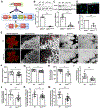
Figure 1.
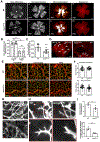
Figure 1.. Deletion of endothelial Nox4 impairs the development of retinal vasculature.
A. Schematic diagram of generation of EC-specific Nox4 knockout mice using Cre-Lox strategy. B-D. Brain microvascular endothelial cell (BMEC) were isolated from wild type (WT) and Nox4EC-KO mice (3-4 mice per group). B&C. Successful deletion of Nox4 in ECs was confirmed by significant reduction of Nox4 expression determined by qPCR (B) and western blot analysis (C). D. Representative images of BMECs stained with CellROX Deep Red Reagent (green) and DAPI (blue). Graph shows quantification of intracellular ROS. Scale bar: 20μm. Mean ± SD; **P<0.01. E. Representative images of retinal whole mounts stained with Isolectin B4 from WT and Nox4EC-KO mice (9-15 mice per group) at postnatal days 7 (P7). Scale bar: 100μm (iii and iv), 50μm (v and vi) and 10μm (vii-x). F&G. Quantification of radial expansion of superficial vascular plexus measured by the average distance from optic nerve head to the edge of retinal superficial vascular plexus (F) and the percentage of vascularized retina area measured by the ratio of vascular plexus area to the total retinal area (G). H. Quantification of branching points of the front sprouting vessels. I. Quantification of the number of filopodia bursts in retinal vasculature. Red arrows denote tip cells J. Quantification of numbers of filopodia per filopodia burst. Representative filopodia were denoted by red arrows in E (vii and viii). K. Average length of filopodia per filopodia burst. Representative average length of filopodia were denoted by yellow lines in E (vii and viii). M. Quantification of the distance from the base of filopodia to the closest nucleus, indicating cytoplasmic extension of tip cell at P7 (red line in E, ix & x). Mean ± SD; *P<0.05, **P<0.01.
Figure 2.
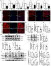
Figure 2.. Delayed retinal vasculature development and reduced vascular density in Nox4EC-KO mice.
A. Representative images of retinal whole mounts stained with Isolectin B4 from WT and Nox4EC-KO mice at P12, P17, and P25. Scale bar: 50μm. B. Quantification of the vessel density of superficial, intermediate and deep vascular network in P12, P17 and P25 retinas. Mean ± SD; n=3-10 mice per group; *P<0.05, **P<0.01.
Figure 3.
Figure 3.. Loss of endothelial Nox4 reduces pathological angiogenesis in OIR.
A. Representative images of retinal whole mounts stained with Isolectin B4 from WT and Nox4EC-KO mice with OIR or control, demonstrating vaso-obliteration at P9 and P17, peak of retinal NV at P17 and regression of NV at P25. In panels v/vi, white color signifies vaso-obliterated area and yellow color signifies retinal NV. Scale bar: 500μm. B&C. Quantification of areas of vaso-obliteration (B) and retinal NV (C); Mean ± SD; n=8-10 mice per group; *P<0.05, **P<0.01. D. Higher magnification images showing epi-retinal tufts (white arrows) that aggregate as large continuous areas of NV in WT retina and considerably smaller and fewer tufts in Nox4EC-KO retinas. Scale bar: 200μm. E. Representative images of retinal whole mounts stained with Isolectin B4 (red) and NG2 (marker for pericyte, green) from OIR mice at P25. Scale bar: 50μm. F. Graphs depict density of intermediate and deep layer vascular network in the area of central retina in P25 OIR mice. Mean ± SD; n=4 mice per group; **P<0.01 vs WT. G. Representative images showing tip cells (red arrow) and remodeling vessels (yellow arrow) in P25 OIR retinas. Scale bar: 20μm. H. Representative images of tip cells in P25 OIR retinas demonstrating disoriented tip cells with sparser, shorter, and thinner filopodia in Nox4EC-KO mice. Scale bar: 20μm. I. Quantification of numbers of filopodia per filopodia burst and the average length of filopodia per filopodia burst in P25 OIR retinas. Mean ± SD; n=4 mice per group; *P<0.05, **P<0.01 vs WT.
Figure 4.
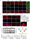
Figure 4.. Deletion of endothelial Nox4 reduces vascular sprouting, EC migration and proliferation.
A &B. Representative images of aorta rings from WT and Nox4EC-KO mice cultured in collagen I gel with 30ng/ml of VEGF for 8 days (A). Number of sprouts per aorta ring and average sprout length were quantified using Image J (B). Scale bar: 200μm; Mean ± SD; n=3 mice per group. C & D. EC migration assay. Representative images of wound areas at 0, 24, 48 hrs after scratching (C). Percentage of the scratched filled with cells was quantified over time (D). Scale bar: 200μm; Mean ± SD; n=4 mice per group; *P<0.05, **P<0.01 vs BMECWT without VEGF treatment; #P<0.05 vs BMECWT with VEGF treatment. E&F. Representative images of BMECs stained for Brdu (red) and DAPI (blue) after VEGF treatment for 12 hrs (E). Graph shows the percentage of Brdu positive cells (F), Scale bar: 100μm; Mean ± SD; n=5 mice per group; *P<0.05, **P<0.01 vs BMECWT without VEGF treatment; #P<0.05 vs BMECWT with VEGF treatment.
Figure 5.
Figure 5.. Nox4 deficiency reduces the expression of angiogenic genes and proteins in VEGF/VEGFR2 and angiopoietin 2/integrin αV pathways in ECs.
A. mRNA expression of Vegfr2, Vegfr1, Angpt2, Itgα5, Itgb1, Nos3, and Cxcr4 in BMECs isolated from Nox4EC-KO or WT mice. Values are presented as mean ± SD; n=3 mice per group; *P<0.05, **P<0.01; B-D. Immunofluorescence staining for VEGF (B), p-VEGFR2 (C), and integrin αV (D) in BMEC isolated from Nox4EC-KO or WT mice. Scale bar: 50μm (B and C) and 20μm (D). E-G: Quantification of the fluorescent intensity in VEGF (E), p-VEGFR2 (F), and integrin αV (G). Mean ± SD; n=3 mice per group; *P<0.05, **P<0.01. H&I. Representative western blots of VEGF, VEGFR2, Integrin αV, angiopoietin 2 and p-ERK1/2 protein levels in BMECs from WT or KO mice. Mean ± SD; n=5-8 mice per group; *P<0.05, **P<0.01. J. Representative western blots of p-VEGFR2, Integrin αV, angiopoietin 2 and p-ERK1/2 protein levels in retinas from WT or KO mice. Mean ± SD; n=3 mice per group; *P<0.05, **P<0.01.
Figure 6.
Figure 6.. Deletion of Nox4 inhibits VEGF-induced activation of VEGFR2 and ANGPT2 pathways.
BMECs from Nox4EC-KO or WT mice were treated with VEGF (10ng/ml) for 3 or 6 hrs. A. Representative images of BMECs stained for CD31 (red), p-VEGFR2 (green) and DAPI (blue). Insets show that p-VEGFR2 expression is both intracellular (white arrows) and at the cell surface (yellow arrows). Scale bar: 50μm. B&C. BMECs were stained for ANGPT2 (red) and DAPI (blue) (B). Image on the right shows enlarged area demonstrating ANGPT2 expression at the filopodia (white arrows) in WT BMECs after VEGF stimulation, which was not observed in KO cells. Total ANGPT2 expression was reduced in KO BMECs but not altered by VEGF treatment (C). Scale bar: 20μm. Mean ± SD; n=3 mice per group; *P<0.05 vs BMECWT without VEGF treatment; #P<0.05 vs BMECWT with VEGF treatment. D. Representative western blots of p-VEGFR2, integrin αV, angiopoietin 2 and p-ERK1/2 protein levels in retinas of OIR models at P9 and P17 from WT or KO mice. Mean ± SD; n=3 mice per group; *P<0.05, **P<0.01. E. Suggested mechanisms of endothelial Nox4-dependent regulation of angiogenesis. During retinal development and upon hypoxia, Nox4 is upregulated resulting in enhanced ROS production. ROS stimulates VEGF and ANGPT2 expression and the activation of VEGFR2 and integrin αV. This leads to the activation of downstream kinases including ERK1/2, promoting endothelial tip cell formation, proliferation, migration, and vascular sprouting.

Similar articles

Cited by

References

    1. Carmeliet P, Jain RK (2011) Molecular mechanisms and clinical applications of angiogenesis. Nature 473 (7347):298–307. doi:10.1038/nature10144 - DOI - PMC - PubMed
    1. Potente M, Carmeliet P (2017) The Link Between Angiogenesis and Endothelial Metabolism. Annual review of physiology 79:43–66. doi:10.1146/annurev-physiol-021115-105134 - DOI - PubMed
    1. Gerhardt H, Golding M, Fruttiger M, Ruhrberg C, Lundkvist A, Abramsson A, Jeltsch M, Mitchell C, Alitalo K, Shima D, Betsholtz C (2003) VEGF guides angiogenic sprouting utilizing endothelial tip cell filopodia. J Cell Biol 161 (6):1163–1177. doi:10.1083/jcb.200302047 - DOI - PMC - PubMed
    1. Geudens I, Gerhardt H (2011) Coordinating cell behaviour during blood vessel formation. Development 138 (21):4569–4583. doi:10.1242/dev.062323 - DOI - PubMed
    1. Jakobsson L, Franco CA, Bentley K, Collins RT, Ponsioen B, Aspalter IM, Rosewell I, Busse M, Thurston G, Medvinsky A, Schulte-Merker S, Gerhardt H (2010) Endothelial cells dynamically compete for the tip cell position during angiogenic sprouting. Nat Cell Biol 12 (10):943–953. doi:10.1038/ncb2103 - DOI - PubMed

Publication types

MeSH terms