Multicenter Study
. 2021 Feb;298(2):E98-E106.
doi: 10.1148/radiol.2020203465. Epub 2020 Nov 17.

Chest CT in the Emergency Department for Diagnosis of COVID-19 Pneumonia: Dutch Experience

Affiliations
Multicenter Study

Chest CT in the Emergency Department for Diagnosis of COVID-19 Pneumonia: Dutch Experience

Steven Schalekamp et al. Radiology. 2021 Feb.

Abstract

Background Clinicians need to rapidly and reliably diagnose coronavirus disease 2019 (COVID-19) for proper risk stratification, isolation strategies, and treatment decisions. Purpose To assess the real-life performance of radiologist emergency department chest CT interpretation for diagnosing COVID-19 during the acute phase of the pandemic, using the COVID-19 Reporting and Data System (CO-RADS). Materials and Methods This retrospective multicenter study included consecutive patients who presented to emergency departments in six medical centers between March and April 2020 with moderate to severe upper respiratory symptoms suspicious for COVID-19. As part of clinical practice, chest CT scans were obtained for primary work-up and scored using the five-point CO-RADS scheme for suspicion of COVID-19. CT was compared with severe acute respiratory syndrome coronavirus 2 reverse-transcription polymerase chain reaction (RT-PCR) assay and a clinical reference standard established by a multidisciplinary group of clinicians based on RT-PCR, COVID-19 contact history, oxygen therapy, timing of RT-PCR testing, and likely alternative diagnosis. Performance of CT was estimated using area under the receiver operating characteristic curve (AUC) analysis and diagnostic odds ratios against both reference standards. Subgroup analysis was performed on the basis of symptom duration grouped presentations of less than 48 hours, 48 hours through 7 days, and more than 7 days. Results A total of 1070 patients (median age, 66 years; interquartile range, 54-75 years; 626 men) were included, of whom 536 (50%) had a positive RT-PCR result and 137 (13%) of whom were considered to have a possible or probable COVID-19 diagnosis based on the clinical reference standard. Chest CT yielded an AUC of 0.87 (95% CI: 0.84, 0.89) compared with RT-PCR and 0.87 (95% CI: 0.85, 0.89) compared with the clinical reference standard. A CO-RADS score of 4 or greater yielded an odds ratio of 25.9 (95% CI: 18.7, 35.9) for a COVID-19 diagnosis with RT-PCR and an odds ratio of 30.6 (95% CI: 21.1, 44.4) with the clinical reference standard. For symptom duration of less than 48 hours, the AUC fell to 0.71 (95% CI: 0.62, 0.80; P < .001). Conclusion Chest CT analysis using the coronavirus disease 2019 (COVID-19) Reporting and Data System enables rapid and reliable diagnosis of COVID-19, particularly when symptom duration is greater than 48 hours. © RSNA, 2020 Online supplemental material is available for this article. See also the editorial by Elicker in this issue.

Keywords: AUC = area under the receiver operating characteristic curve; CO-RADS = COVID-19 Reporting and Data System; COVID-19 = coronavirus disease 2019; RT-PCR = reverse-transcription polymerase chain reaction; SARS-CoV-2 = severe acute respiratory coronavirus 2.


Figures

Flowchart clinical reference standard. Abbreviations: COVID-19 = coronavirus disease 2019; SARS-CoV-2 = severe acute respiratory syndrome coronavirus 2; PCR = polymerase chain reaction; ICU = intensive unit; BAL = bronchoalveolar lavage.
Figure 1:
Flowchart clinical reference standard. Abbreviations: COVID-19 = coronavirus disease 2019; SARS-CoV-2 = severe acute respiratory syndrome coronavirus 2; PCR = polymerase chain reaction; ICU = intensive unit; BAL = bronchoalveolar lavage.
Inclusion flowchart. Patient were excluded when they were not diagnosed in the emergency department, had no or only mild symptoms, did not have a RT-PCR, and if patient was RT-PCR proven at time of CT scan or no CO-RADS reporting system was used in the original report. * 10th of April for center F. Abbreviations: COVID-19 = coronavirus disease 2019; RT-PCR = real-time reversetranscription polymerase chain reaction; CO-RADS = COVID-19 reporting and data system.
Figure 2:
Inclusion flowchart. Patient were excluded when they were not diagnosed in the emergency department, had no or only mild symptoms, did not have a RT-PCR, and if patient was RT-PCR proven at time of CT scan or no CO-RADS reporting system was used in the original report. * 10th of April for center F. Abbreviations: COVID-19 = coronavirus disease 2019; RT-PCR = real-time reversetranscription polymerase chain reaction; CO-RADS = COVID-19 reporting and data system.
Performance per CO-RADS category. Bar chart of percentage ‘proven’ COVID-19, ‘probable’, ‘possible’, and ‘no’ COVID-19 per CO-RADS CT score. Abbreviations: COVID-19 = coronavirus disease 2019; CO-RADS = COVID-19 reporting and data system.
Figure 3:
Performance per CO-RADS category. Bar chart of percentage ‘proven’ COVID-19, ‘probable’, ‘possible’, and ‘no’ COVID-19 per CO-RADS CT score. Abbreviations: COVID-19 = coronavirus disease 2019; CO-RADS = COVID-19 reporting and data system.
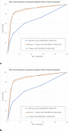
ROC analysis based on durations of symptoms. (a) ROC analysis based on durations of symptoms for CO-RADS against RT-PCR. (b) ROC analysis based on durations of symptoms for CO-RADS against clinical reference standard. Blue line: <48 hours complaints (n=52/220 RT-PCR positive); red line: 48 hours – 7 days complaints (n=239/430 RT-PCR positive); grey line: >7 days complaints (n=232/376 RT-PCR positive). Abbreviations: ROC = receiver operating characteristics; RT-PCR = realtime reverse-transcription polymerase chain reaction; AUC = area under the curve.
Figure 4:
ROC analysis based on durations of symptoms. (a) ROC analysis based on durations of symptoms for CO-RADS against RT-PCR. (b) ROC analysis based on durations of symptoms for CO-RADS against clinical reference standard. Blue line: <48 hours complaints (n=52/220 RT-PCR positive); red line: 48 hours – 7 days complaints (n=239/430 RT-PCR positive); grey line: >7 days complaints (n=232/376 RT-PCR positive). Abbreviations: ROC = receiver operating characteristics; RT-PCR = realtime reverse-transcription polymerase chain reaction; AUC = area under the curve.
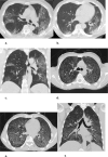
Example chest CT scans of patients with a true positive (case 1) and false positive (case 2) CO-RADS 4 score. (case 1). True positive CO-RADS 4 chest CT: (a,b) two axial slices and (c) a coronal slice of a 79-year-old female with 9 days of symptoms. CT shows diffuse ground-glass opacities close to visceral pleural surfaces but superimposed on emphysematous changes. Also note the widened esophagus. Coronavirus disease 2019 was RT-PCR confirmed. (case 2) False positive CO-RADS 4 chest CT: (d,e) two axial slices and (f) a coronal slice of a 51- year-old female with 2 days of symptoms. CT shows bilateral multifocal areas of consolidation with halo and subtle areas of ground glass without contact to visceral pleural surfaces. RT-PCR for SARS-CoV2 was repeatedly negative and an alternative diagnosis was established with a blood culture confirmed line sepsis.
Figure 5:
Example chest CT scans of patients with a true positive (case 1) and false positive (case 2) CO-RADS 4 score. (case 1). True positive CO-RADS 4 chest CT: (a,b) two axial slices and (c) a coronal slice of a 79-year-old female with 9 days of symptoms. CT shows diffuse ground-glass opacities close to visceral pleural surfaces but superimposed on emphysematous changes. Also note the widened esophagus. Coronavirus disease 2019 was RT-PCR confirmed. (case 2) False positive CO-RADS 4 chest CT: (d,e) two axial slices and (f) a coronal slice of a 51- year-old female with 2 days of symptoms. CT shows bilateral multifocal areas of consolidation with halo and subtle areas of ground glass without contact to visceral pleural surfaces. RT-PCR for SARS-CoV2 was repeatedly negative and an alternative diagnosis was established with a blood culture confirmed line sepsis.
Example chest CT scans of patients with a true positive (case 3) and false positive (case 4) CO-RADS 5 score. (case 3) True positive CO-RADS 5 chest CT: two axial slices (a,b) and a coronal slice (c) of a 69-year-old male with 7 days of symptoms. CT shows bilateral multifocal areas of groundglass and consolidation in vicinity of the visceral pleural surface. Also, few thickened vessels in areas of groundglass are observed. Coronavirus disease 2019 was RT-PCR confirmed. (case 4) False positive CO-RADS 5 chest CT: two axial slices (d,e) and a coronal slice (f) of a 42-year-old male with more than 7 days of symptoms. CT shows diffuse groundglass opacities in the close vicinity of visceral pleural surfaces. Also, a crazy paving pattern is observed. RT-PCR for SARS-CoV2 was negative, and a diagnosis of pneumocystis jirovecii pneumonia was made based on bronchoalveolar lavage fluid.
Figure 6:
Example chest CT scans of patients with a true positive (case 3) and false positive (case 4) CO-RADS 5 score. (case 3) True positive CO-RADS 5 chest CT: two axial slices (a,b) and a coronal slice (c) of a 69-year-old male with 7 days of symptoms. CT shows bilateral multifocal areas of groundglass and consolidation in vicinity of the visceral pleural surface. Also, few thickened vessels in areas of groundglass are observed. Coronavirus disease 2019 was RT-PCR confirmed. (case 4) False positive CO-RADS 5 chest CT: two axial slices (d,e) and a coronal slice (f) of a 42-year-old male with more than 7 days of symptoms. CT shows diffuse groundglass opacities in the close vicinity of visceral pleural surfaces. Also, a crazy paving pattern is observed. RT-PCR for SARS-CoV2 was negative, and a diagnosis of pneumocystis jirovecii pneumonia was made based on bronchoalveolar lavage fluid.

Comment in

References

    1. Zou L, Ruan F, Huang M, et al. . SARS-CoV-2 Viral Load in Upper Respiratory Specimens of Infected Patients. N Engl J Med 2020;382(12):1177-1179. - PMC - PubMed
    1. Corman VM, Landt O, Kaiser M, et al. . Detection of 2019 novel coronavirus (2019-nCoV) by realtime RT-PCR. Euro Surveill 2020;25(3):10.2807/1560-7917.ES.2020.25.3.2000045. - PMC - PubMed
    1. Ai T, Yang Z, Hou H, et al. . Correlation of Chest CT and RT-PCR Testing in Coronavirus Disease 2019 (COVID-19) in China: A Report of 1014 Cases. Radiology 2020:200642. - PMC - PubMed
    1. Wang W, Xu Y, Gao R, et al. . Detection of SARS-CoV-2 in Different Types of Clinical Specimens. JAMA 2020;323(18):1843-1844. - PMC - PubMed
    1. Li Y, Yao L, Li J, et al. . Stability issues of RT-PCR testing of SARS-CoV-2 for hospitalized patients clinically diagnosed with COVID-19. J Med Virol 2020 - PMC - PubMed

Publication types