. 2020 Nov 17;15(11):e0242380.
doi: 10.1371/journal.pone.0242380. eCollection 2020.

G ATA2 mediates the negative regulation of the prepro-thyrotropin-releasing hormone gene by liganded T3 receptor β2 in the rat hypothalamic paraventricular nucleus

Affiliations

G ATA2 mediates the negative regulation of the prepro-thyrotropin-releasing hormone gene by liganded T3 receptor β2 in the rat hypothalamic paraventricular nucleus

Go Kuroda et al. PLoS One. .

Abstract

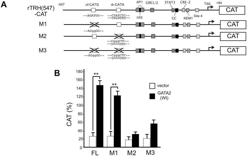
Thyroid hormone (T3) inhibits thyrotropin-releasing hormone (TRH) synthesis in the hypothalamic paraventricular nucleus (PVN). Although the T3 receptor (TR) β2 is known to mediate the negative regulation of the prepro-TRH gene, its molecular mechanism remains unknown. Our previous studies on the T3-dependent negative regulation of the thyrotropin β subunit (TSHβ) gene suggest that there is a tethering mechanism, whereby liganded TRβ2 interferes with the function of the transcription factor, GATA2, a critical activator of the TSHβ gene. Interestingly, the transcription factors Sim1 and Arnt2, the determinants of PVN differentiation in the hypothalamus, are reported to induce expression of TRβ2 and GATA2 in cultured neuronal cells. Here, we confirmed the expression of the GATA2 protein in the TRH neuron of the rat PVN using immunohistochemistry with an anti-GATA2 antibody. According to an experimental study from transgenic mice, a region of the rat prepro-TRH promoter from nt. -547 to nt. +84 was able to mediate its expression in the PVN. We constructed a chloramphenicol acetyltransferase (CAT) reporter gene containing this promoter sequence (rTRH(547)-CAT) and showed that GATA2 activated the promoter in monkey kidney-derived CV1 cells. Deletion and mutation analyses identified a functional GATA-responsive element (GATA-RE) between nt. -357 and nt. -352. When TRβ2 was co-expressed, T3 reduced GATA2-dependent promoter activity to approximately 30%. Unexpectedly, T3-dependent negative regulation was maintained after mutation of the reported negative T3-responsive element, site 4. T3 also inhibited the GATA2-dependent transcription enhanced by cAMP agonist, 8-bromo-cAMP. A rat thyroid medullary carcinoma cell line, CA77, is known to express the preproTRH mRNA. Using a chromatin immunoprecipitation assay with this cell line where GATA2 expression plasmid was transfected, we observed the recognition of the GATA-RE by GATA2. We also confirmed GATA2 binding using gel shift assay with the probe for the GATA-RE. In CA77 cells, the activity of rTRH(547)-CAT was potentiated by overexpression of GATA2, and it was inhibited in a T3-dependent manner. These results suggest that GATA2 transactivates the rat prepro-TRH gene and that liganded TRβ2 interferes with this activation via a tethering mechanism as in the case of the TSHβ gene.


Conflict of interest statement

The authors have declared that no competing interests exist.

Figures

Fig 1
Fig 1. GATA2 protein is expressed in the TRH neurons in the rat hypothalamic PVN.
(A) The specificity of anti-GATA2 antibody. CV1 cells were transfected with empty vector (a and b) or mouse GATA2-expression plasmid (c and d). These cells were stained with 0.1% anti-GATA2 antibody (B9922A, Perseus Proteomics, Japan) (a and c) or DAPI (b and d). Several GATA2-positive cells were detected in (c) but not in (a), while the numbers of nuclei stained with DAPI were comparable between (b) and (d). Transfection efficiency was approximately 5% (compare c and d). (B) (a, b, and c) Immunohistochemical staining of rat PVN with 0.2% anti-TRH antibody. Magnification: ×10 (a), ×40 (b), and ×100 (c). The cytoplasms were stained brown (open arrowhead). The boxed areas in (a) and (b) were magnified in (b) and (c), respectively. (d, e and f) Immunohistochemical staining of the rat PVN with anti-mouse 0.1% GATA2 antibody. Magnification: ×10 (d), ×40 (e) and ×100 (f). The nuclei were strongly stained blue (solid arrowhead). The boxed areas in (d) and (e) were magnified in (e) and (f), respectively. (C) Immunohistochemical double staining of the rat PVN with 0.2% anti-TRH antibody and 0.1% anti-mouse GATA2 antibody (a, b and c). Magnification: ×10 (a), ×40 (b), and ×100 (c). The cytoplasms of TRH neuron were stained brown with anti-TRH antibody (open arrowhead) and nuclei were stained blue with anti-mouse GATA2 antibody (solid arrowhead). The boxed areas in (a) and (b) were magnified in (b) and (c), respectively.
Fig 2
Fig 2. The rat prepro-TRH gene is activated by GATA2.
(A) The rat prepro-TRH promoter (nt. -547 to nt. +84) was fused with CAT-reporter gene to generate rTRH(547)-CAT, from which the pUC-derived AP-1 site was deleted (see main text). Conserved region among species (nt. -150 to TSS) is indicated [16]. The candidate GATA sequences, upstream forward GATA (uf-GATG) and downstream reverse GATA (dr-GATA), are indicated. In addition to these GATA sequences, there are multiple short DNA elements that can be recognized by various transcription factors (see main text). Site 4; the reported negative T3 responsive element (nTRE) in prepro-TRH promoter. We also generated several deletion mutants (Del1, Del2, Del3 and Del4) of rTRH(547)-CAT. (B) In CV1 cells, CAT activities of rTRH(547)-CAT were enhanced by the transfection of mouse GATA2 expression plasmid (pcDNA3-mGATA2) but not by its mutant (C349A, inset). (C) GATA2-dependent transcription was abolished in the deletion construct (Del3 and Del4) suggesting the presence of functional GATA-RE(s) between nt. -400 and nt. -342. Using the Lipofectamine method, 2.0 μg of rTRH(547)-CAT (B) or its deletion mutants (C) were transfected into CV1 cells that were plated at a density of 2×10⁵ cells per well in a six-well plate along with the expression plasmids for GATA2 or its mutant (C349A) (0.1 μg). **, P<0.01 for vector vs. GATA2 expression plasmids. CAT activity for pCMV-CAT (5.0 ng/well) was taken as 100%. Data are expressed as the mean ± S.E. of three to five independent experiments.
Fig 3
Fig 3. In the rat prepro-TRH promoter, downstream reverse GATA sequence (dr-GATA) is the functional GATA-RE.
(A) Schematic representation of rTRH(547)-CAT and its mutant, M1, M2 and M3. (B) GATA2-induced transcription was significantly reduced by the mutation of dr-GATA (M2 and M3) but not uf-GATG (M1). Wild-type and mutated sequences are indicated as upper and lower case letters, respectively. Using the Lipofectamine method, 2.0 μg of rTRH(547)-CAT or its mutants (M1, M2, and M3) were transfected into CV1 cells that were plated at a density of 2×10⁵ cells per well in a six-well plate along with GATA2 expression plasmid (0.1 μg). **, P<0.01 for vector vs. GATA2 expression plasmids. CAT activity for pCMV-CAT (5.0 ng/well) was taken as 100%. Data are expressed as the mean ± S.E. of three to five independent experiments.
Fig 4
Fig 4. The GATA2-dependent activation of the rat prepro-TRH promoter is inhibited by liganded TRβ2 and the reported nTRE, site 4, is dispensable for this inhibition.
(A) rTRH(547)-CAT was transfected into CV1 cells with GATA2 and TRβ2 expression plasmids and the cells were treated with 0 to 10 nM T3. The activity of this reporter gene was reduced by T3 treatment. *, P<0.05 vs. T3 (-) and **, P<0.01 vs. T3 (-) (B) Site 4 in rTRH(547)-CAT was mutated to generate site 4m. CV1 cells were transfected with this reporter gene and GATA2 and TRβ2 expression plasmids and the cells were treated 10 nM T3. In site 4m, the basal activity before T3 treatment is significantly reduced; however, T3-dependent negative regulation was maintained. **, P<0.01 vs. T3 (-) and #, P<0.05 vs. rTRH(547)-CAT.
Fig 5
Fig 5. PKA signaling pathway shows an additive effect on the T3-dependent negative regulation of the rat preproTRH gene by T3.
(A) Treatment with 1 mM 8-bromo-cAMP displayed an additive effect on the GATA2-dependent activity of rTRH(547)-CAT. rTRH(547)-CAT was transfected into CV1 cells with GATA2 expression plasmid in the presence or absence of 8-bromo-cAMP. **, P<0.01 vs. 8-bromo-cAMP (-). #, P<0.05 vs. vector. (B) GATA2-dependent activation in the presence of 8-bromo-cAMP (1 mM) was significantly inhibited by 10 nM T3 in the presence of TRβ2, indicating that the PKA signaling pathway may modulate the T3-dependent negative feedback regulation of the prepro-TRH gene. **, P<0.01 vs. T3 (-). #, P<0.05 vs. vector. The experimental procedures were the same as for Figs 2–4 but were conducted in the presence or absence of 8-bromo-cAMP and/or 10 nM T3.
Fig 6
Fig 6. GATA2 recognizes the dr-GATA sequence.
(A) Schematic representation of the rat prepro-TRH gene. The positions of primers for ChIP assay and the oligo-DNAs for gel shift assay (probe drG and its mutant probe M) are indicated. (B) A representative result of ChIP assays using an anti-GATA2 antibody and CA77 cells transfected with GATA2. Data are expressed as the mean ± S.E. of five independent experiments (p = 0.214 for control vs. dr-GATA transfected with GATA2 expression plasmid). (C) Gel shift assay with probe drG and its mutant probe M. A single band (arrowhead) was observed when 32P-radiolabeled probe drG was incubated with nuclear extract of CV1 cells transfected with mouse GATA2 expression plasmid. The specific binding signal (lane 2) was abolished by a 50-fold amount of cold probe drG (lane 3) but not by probe M (lane 4) or nonspecific double strand oligo-DNA (NS, lane 5). The signal was also reduced when GATA2 protein was mixed with the anti-GATA2 antibody before incubation with 32P-radiolabeled probe drG (supershift, lane 6). Arrow: free 32P-radiolabeled probe drG.
Fig 7
Fig 7. T3-dependent negative regulation of the rat prepro-TRH promoter can be observed in rat thyroid medullary carcinoma cell line, CA77, transfected with a GATA2 expression plasmid.
(A) Endogenous TRβ2 in CA77 cells were detected by western blot of whole cell extract of CA77 cells using an anti-TR antibody. As a control, whole cell extract of CV1 cells transfected with empty vector (lane 1), human TRβ1 (hTRβ1, lane 2), or human TRβ2 (hTRβ2, lane 3) are shown. Endogenous rat TRβ2 (arrowhead) was detected by anti-TR antibody in 299 or 598 μg/lane of whole cell extract of CA77 cells. The difference of mobilities is thought to be the different molecular weights between human and rat TRβ2s. (B) rTRH(547)-CAT was transfected with a mouse GATA2 expression plasmid into CA77 ells. The additive effect by overexpression of GATA2 was observed and this activity is inhibited by T3, presumably via endogenous TRβ2. *, P<0.05 vs. vector. **, P<0.01 vs. T3 (-).
Fig 8
Fig 8. A schematic representation of the transcriptional regulation of the rat prepro-TRH and TSHβ genes.
In the TRH neuron of the hypothalamic PVN (top) and thyrotroph of anterior pituitary lobe (bottom), TRβ2 associates with the Zn-finger region of GATA2 via protein-protein interactions, resulting in the T3-dependent interference of GATA2-dependent transactivation (tethering mechanism). The prepro-TRH gene is activated by multiple transcription factors stimulated by cognate cell membrane receptor signals (see main text), resulting in the fine-tuning of the set point of H-P-T axis. Broken lines indicate the signaling pathway via cell-membrane receptors/protein kinases. Although site 4 is dispensable for the T3-dependent negative regulation of the preproTRH gene, it may have physiological relevance at least in CV1 cells. In TRH neuron of the PVN, mature TRH (pyro-Glu-His-Pro-NH2) is synthesized via multiple processing steps. In thyrotrophs, liganded TRH receptor stimulates the TSHβ and CGA gene expression and TSH secretion via the PKC pathway. In the TSHβ gene, Pit1 protects the transactivation function of GATA2 from the inhibitory effect by suppressor region-binding protein (SRBP) [8]. PKC, protein kinase C. JAK2, Janus kinase 2. PKA, protein kinase A. CA, catecholamine. αMSH, α-melanocyte stimulating hormone. GRE, glucocorticoid-responsive element. AP-1, AP-1 binding site. CRE, c-AMP-responsive element. TRH-R, TRH receptor. α, CGA. β, TSHβ. nTRE, the reported negative T3 responsive element in the TSHβ gene.

Similar articles

Cited by

References

    1. Hollenberg AN. The role of the thyrotropin-releasing hormone (TRH) neuron as a metabolic sensor. Thyroid: official journal of the American Thyroid Association. 2008;18(2):131–9. Epub 2008/02/19. 10.1089/thy.2007.0251 . - DOI - PubMed
    1. Nillni EA. Regulation of the hypothalamic thyrotropin releasing hormone (TRH) neuron by neuronal and peripheral inputs. Frontiers in neuroendocrinology. 2010;31(2):134–56. Epub 2010/01/16. 10.1016/j.yfrne.2010.01.001 . - DOI - PMC - PubMed
    1. Fekete C, Lechan RM. Central regulation of hypothalamic-pituitary-thyroid axis under physiological and pathophysiological conditions. Endocrine reviews. 2014;35(2):159–94. Epub 2014/01/16. 10.1210/er.2013-1087 . - DOI - PMC - PubMed
    1. Joseph-Bravo P, Jaimes-Hoy L, Charli JL. Advances in TRH signaling. Reviews in endocrine & metabolic disorders. 2016;17(4):545–58. Epub 2016/08/16. 10.1007/s11154-016-9375-y . - DOI - PubMed
    1. Sugrue ML, Vella KR, Morales C, Lopez ME, Hollenberg AN. The thyrotropin-releasing hormone gene is regulated by thyroid hormone at the level of transcription in vivo. Endocrinology. 2010;151(2):793–801. Epub 2009/12/25. 10.1210/en.2009-0976 . - DOI - PMC - PubMed

Publication types

MeSH terms