. 2020 Nov;40 Suppl 1(Suppl 1):3-81.
doi: 10.1111/scd.12511.

Clinical practice guidelines: Oral health care for children and adults living with epidermolysis bullosa

Affiliations

Clinical practice guidelines: Oral health care for children and adults living with epidermolysis bullosa

Susanne Krämer et al. Spec Care Dentist. 2020 Nov.

Abstract

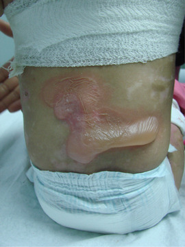
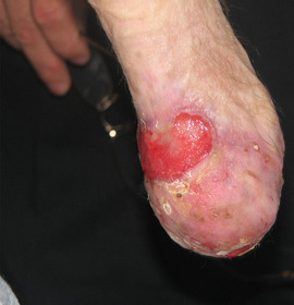
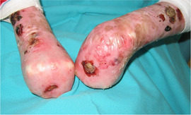
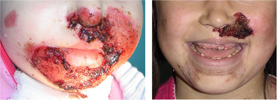
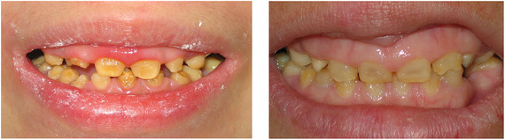
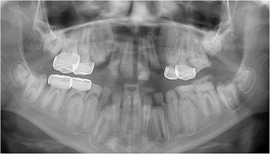
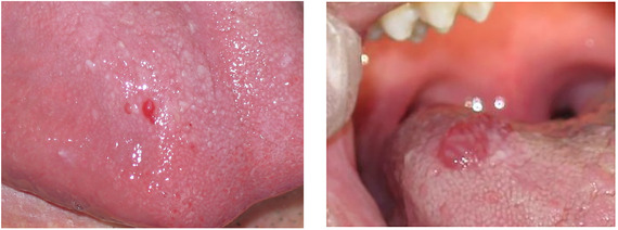
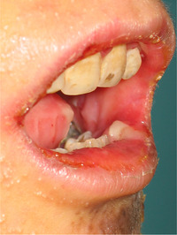
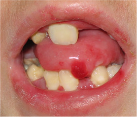
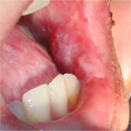
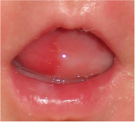
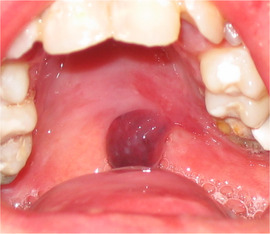
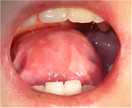
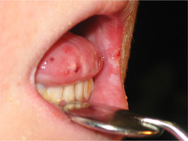
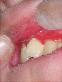
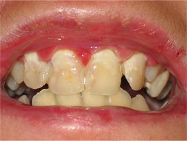
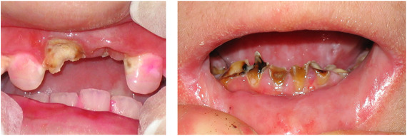
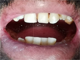
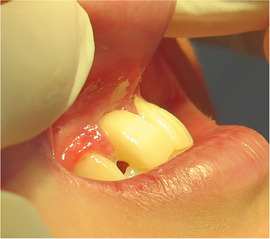
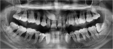
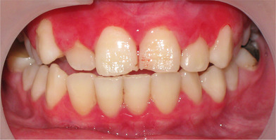
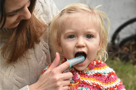
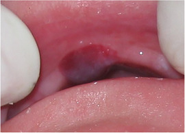
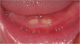
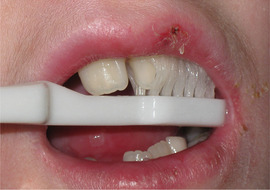
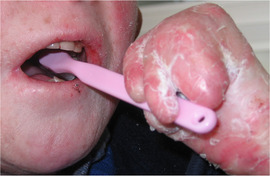
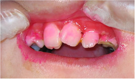
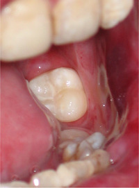
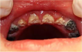
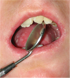
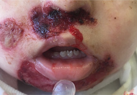
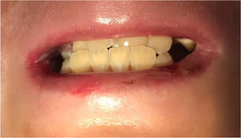
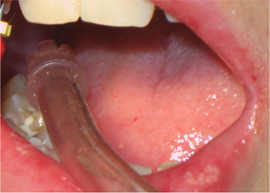
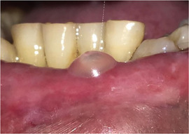
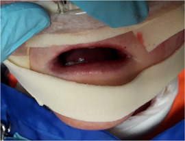
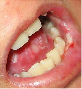
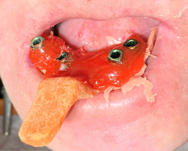
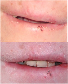
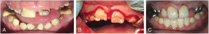
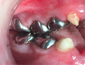
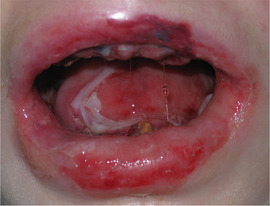
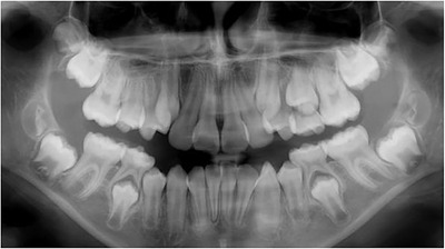
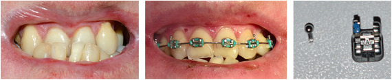
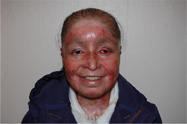
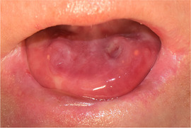
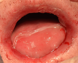
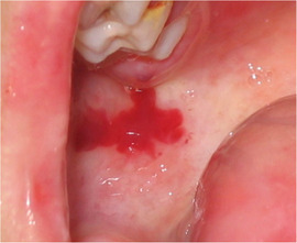
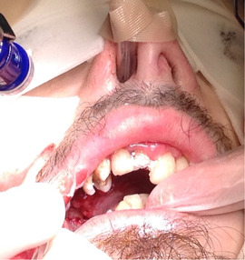
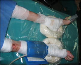
Background: Inherited epidermolysis bullosa (EB) is a genetic disorder characterized by skin fragility and unique oral features.

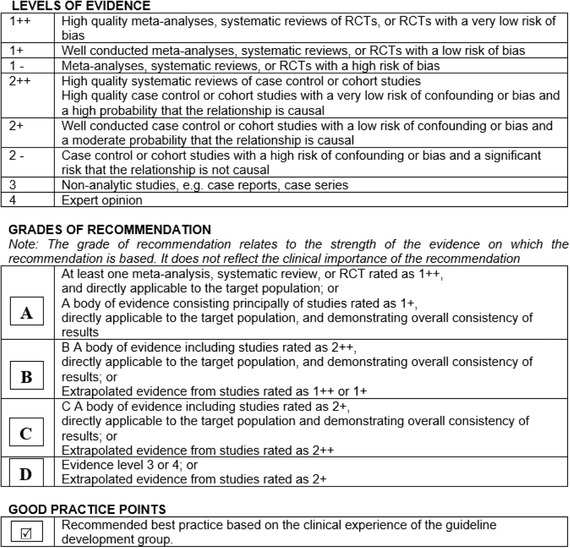
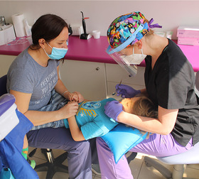
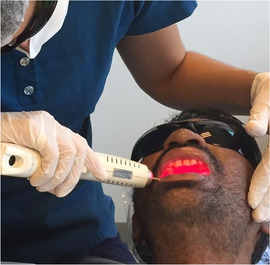
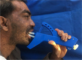
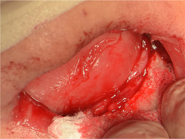
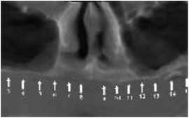
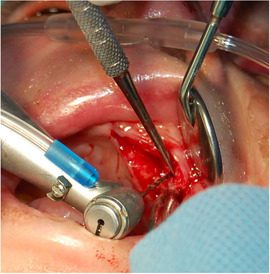
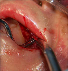
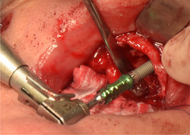
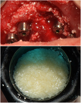
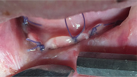
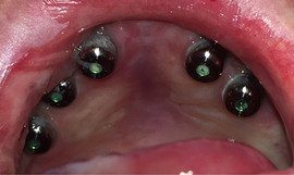
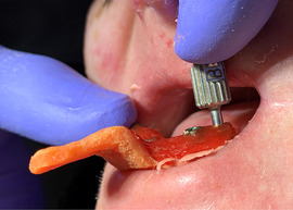
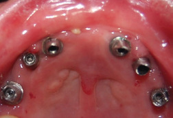
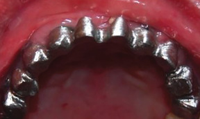
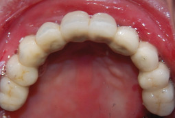
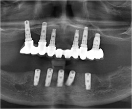
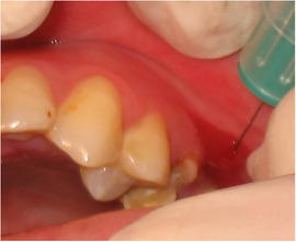
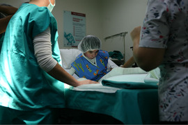
Aims: To provide (a) a complete review of the oral manifestations in those living with each type of inherited EB, (b) the current best practices for managing oral health care of people living with EB, (c) the current best practices on dental implant-based oral rehabilitation for patients with recessive dystrophic EB (RDEB), and (d) the current best practice for managing local anesthesia, principles of sedation, and general anesthesia for children and adults with EB undergoing dental treatment.

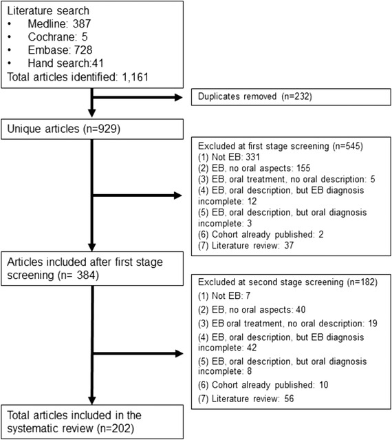
Methods: Systematic literature search, panel discussion including clinical experts and patient representatives from different centers around the world, external review, and guideline piloting.

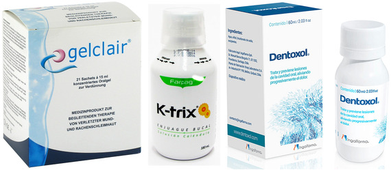
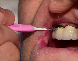
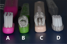
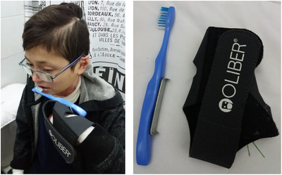
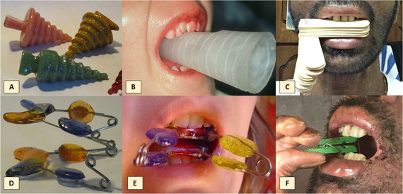
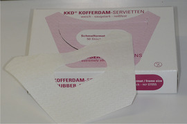
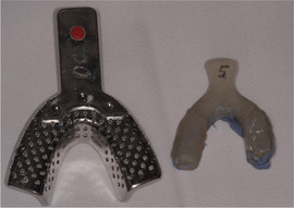
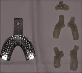
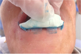
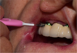
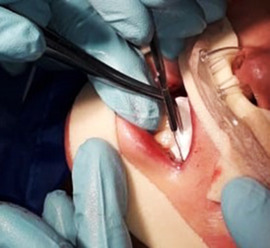
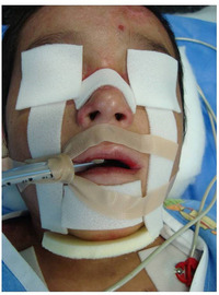
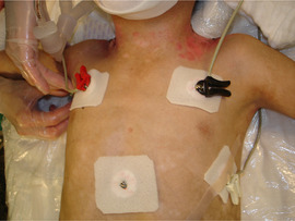
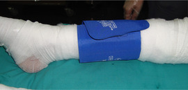
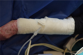
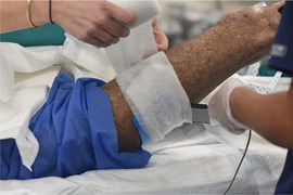
Results: This article has been divided into five chapters: (i) general information on EB for the oral health care professional, (ii) systematic literature review on the oral manifestations of EB, (iii) oral health care and dental treatment for children and adults living with EB-clinical practice guidelines, (iv) dental implants in patients with RDEB-clinical practice guidelines, and (v) sedation and anesthesia for adults and children with EB undergoing dental treatment-clinical practice guidelines. Each chapter provides recommendations on the management of the different clinical procedures within dental practice, highlighting the importance of patient-clinician partnership, impact on quality of life, and the importance of follow-up appointments. Guidance on the use on nonadhesive wound care products and emollients to reduce friction during patient care is provided.

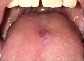
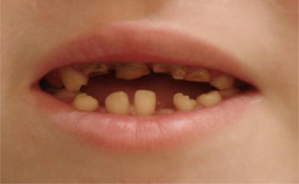
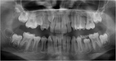
Conclusions: Oral soft and hard tissue manifestations of inherited EB have unique patterns of involvement associated with each subtype of the condition. Understanding each subtype individually will help the professionals plan long-term treatment approaches.

Keywords: clinical practice guideline; dental implants; dental treatment; dystrophic epidermolysis bullosa; epidermolysis bullosa; epidermolysis bullosa simplex; general anesthesia; junctional epidermolysis bullosa; kindler epidermolysis bullosa; oral care; oral rehabilitation; recessive dystrophic epidermolysis bullosa; sedation.


Conflict of interest statement

None of the authors declared conflict of interest. None of the authors has any connection to manufacturers.

Figures

IMAGE 1.1
IMAGE 1.1
Extensive bullae covering the back of a patient with RDEB
IMAGE 1.2
IMAGE 1.2
Squamous cell carcinoma in RDEB
IMAGE 1.3
IMAGE 1.3
Severe esophageal stenosis in a patient with RDEB
IMAGE 1.4
IMAGE 1.4
Mitten deformities in RDEB
FIGURE 2.1
FIGURE 2.1
Flow chart of selected articles
IMAGE 2.1 and 2.2
IMAGE 2.1 and 2.2
Areas of perioral granulation tissue in a 2‐ and 10‐year‐old patients with severe JEB
IMAGE 2.3 and 2.4
IMAGE 2.3 and 2.4
Generalized enamel hypoplasia in patients with JEB
IMAGE 2.5
IMAGE 2.5
Panoramic radiograph showing thin, abnormal, severely hypoplastic enamel on both dentitions of a 10‐year‐old patient with JEB and generalized enamel hypoplasia
IMAGE 2.6 and 2.7
IMAGE 2.6 and 2.7
Blood filled bullae on the tongue in patients with DDEB
IMAGE 2.8
IMAGE 2.8
Limited mouth opening in RDEB
IMAGE 2.9
IMAGE 2.9
Blood filled bullae on the tongue of a patient with RDEB
IMAGE 2.10
IMAGE 2.10
Bullae on the buccal mucosa of a patient with RDEB
IMAGE 2.11
IMAGE 2.11
Serous bullae covering 3/5 of the tongue of a newborn with RDEB
IMAGE 2.12
IMAGE 2.12
Blood filled bullae on the palate of a patient with RDEB
IMAGE 2.13
IMAGE 2.13
Absence of tongue papillae in RDEB
IMAGE 2.14
IMAGE 2.14
Ankyloglossia in RDEB
IMAGE 2.15
IMAGE 2.15
Obliteration of the labial vestibule in RDEB
IMAGE 2.16
IMAGE 2.16
Extensive plaque deposits and gingival inflammation in RDEB
IMAGE 2.17 and 2.18
IMAGE 2.17 and 2.18
Severe caries in a 12‐year‐old and a 20 years old patient with RDEB
IMAGE 2.19
IMAGE 2.19
Severe microstomia in a 33‐year‐old patient with Kindler EB
IMAGE 2.20
IMAGE 2.20
Obliteration of the labial vestibule in a 15‐year‐old patient with Kindler EB
IMAGE 2.21
IMAGE 2.21
Severe periodontitis in a 33‐year‐old patient with Kindler EB
IMAGE 2.22
IMAGE 2.22
Generalized gingival inflammation in a 13‐year‐old patient with Kindler EB
FIGURE 3.1
FIGURE 3.1
Method used for formulating the recommendations as described on the 50 Guideline Developer's Handbook, NHS Scottish Intercollegiate Guidelines Network SIGN. Revised Edition January 2008 2
IMAGE 3.1
IMAGE 3.1
Early oral hygiene instruction with a finger guard brush
IMAGE 3.2
IMAGE 3.2
Two‐week‐old newborn with severe RDEB. Early diagnosis and education to parents on bullae management
IMAGE 3.3
IMAGE 3.3
Early diagnosis of generalized enamel hypoplasia in an 8 months old child with JEB
IMAGE 3.4
IMAGE 3.4
Knee‐to‐knee position for examining a 3‐year‐old patient
IMAGE 3.5
IMAGE 3.5
Thirty‐three‐year‐old patient with EB having low‐level laser therapy (LLLT) after oral surgery to reduce pain
IMAGE 3.6
IMAGE 3.6
(A)‐(C) Mouth washes and oral gels aimed at wound healing
IMAGE 3.7
IMAGE 3.7
Forty‐one‐year‐old patient with intermediate RDEB cleaning her fixed denture with an interdental brush
IMAGE 3.8
IMAGE 3.8
Examples of toothbrushes available for patients with limited mouth opening: (A) standard toothbrush. (B) Collis Curve baby toothbrush, (C) Collis Curve Junior Toothbrush, and (D) Pro Super‐fine (Esro AG) Toothbrush
IMAGE 3.9
IMAGE 3.9
Collis Curve™ toothbrush (Collis‐Curve Toothbrush, TX, USA) cleans the palatal and buccal sides of the teeth simultaneously
IMAGE 3.10
IMAGE 3.10
Thirty‐year‐old patient with RDEB and pseudosyndactyly performing oral hygiene with a small handle
IMAGE 3.11
IMAGE 3.11
A 19‐year‐old patient with RDEB and complete pseudosyndactyly performing oral hygiene with the Oliber® orthotic
IMAGE 3.12
IMAGE 3.12
Use of disclosing solution in a patent with RDEB to educate on brushing technique
IMAGE 3.13
IMAGE 3.13
Fissure sealing in a lower second molar of a 16‐year‐old patient with RDEB
IMAGE 3.14
IMAGE 3.14
Caries arrest from SDF (note black stain of the teeth) in 6‐year‐old patients with severe RDEB and severe early childhood caries (ECCs)
IMAGE 3.15
IMAGE 3.15
Microstomia makes access to the oral cavity difficult
IMAGE 3.16
IMAGE 3.16
Adult with Kindler EB performing mouth opening exercises with a commercial device: TheraBite® (Atos Medical, Malmö, Sweden)
IMAGE 3.17
IMAGE 3.17
Custom and homemade appliances to perform mouth opening exercises. (A) and (B) Acrylic cones, (C) wooden spatulas, (D) and (E) mouth trainer, and (F) clothes peg to gently exercise opening
IMAGE 3.18
IMAGE 3.18
Blister on the tongue of a patient with EBS due to plectin mutation
IMAGE 3.19
IMAGE 3.19
Two‐year‐old patient with JEB and perioral granulation tissue
IMAGE 3.20
IMAGE 3.20
Five‐year‐old patient with JEB: generalized enamel hypoplasia. Perioral granulation tissue has healed without scarring
IMAGE 3.21
IMAGE 3.21
Lips well lubricated with Linovera® during dental treatment in a 9‐year‐old patient with RDEB
IMAGE 3.22
IMAGE 3.22
Suction tip leaned on tooth surface to avoid mucosal sloughing
IMAGE 3.23
IMAGE 3.23
Fluid‐filled bulla that arose on the lower lip of a 33‐year‐old patient during dental treatment. It should be drained immediately
IMAGE 3.24
IMAGE 3.24
Nonadhesive foam dressing protecting the contact areas
IMAGE 3.25
IMAGE 3.25
Lip wound caused by the removal of a cotton roll that was not lubricated or soaked with water
IMAGE 3.26
IMAGE 3.26
A rubber dam napkin
IMAGE 3.27
IMAGE 3.27
Orthopantomography of a 10‐year‐old patient with severe RDEB
IMAGE 3.28
IMAGE 3.28
Impression using silicone and a custom‐made tray in a 36‐year‐old patient with severe RDEB
IMAGE 3.29
IMAGE 3.29
Stock tray and custom‐made acrylic tray (size comparison)
IMAGE 3.30
IMAGE 3.30
Sectioned individual impression tray
IMAGE 3.31
IMAGE 3.31
Impression using the silicone putty as a tray
IMAGE 3.32
IMAGE 3.32
The smile of a 30‐year‐old patient with severe RDEB and severe microsomia before and after fixed crown oral rehabilitation
IMAGE 3.33
IMAGE 3.33
A 9‐year‐old boy with junctional EB and generalized hypoplastic enamel being treated with gingivectomy and complete oral rehabilitation of the hypoplastic teeth in one session under sedation. (A) Before treatment (upper right central incisor has a temporary restoration), (B) first stage of treatment: gingivectomy, and (C) 1 week after the clinical session: all incisors are crowned
IMAGE 3.34
IMAGE 3.34
Stainless steel crowns on a 6‐year‐old boy with junctional EB and generalized hypoplastic enamel
IMAGE 3.35
IMAGE 3.35
Bullae, ulcers, and mucosal sloughing after surgical extractions
IMAGE 3.36
IMAGE 3.36
Eleven‐year‐old patient with severe RDEB. Serial extractions of the first upper premolars were planned to allow eruption of the canines
IMAGE 3.37
IMAGE 3.37
Twenty‐three‐year‐old patient with severe RDEB: (A) Before and (B) during orthodontic treatment. (C) The hook of the canine bracket had to be removed as it caused trauma to the lip. Note changes in upper lip, as the patient also received vestibuloplasty during the same period
FIGURE 4.1
FIGURE 4.1
Method used for formulating the recommendations as described on the 50 Guideline Developer's Handbook, NHS Scottish Intercollegiate Guidelines Network SIGN. Revised edition January 2008 19
IMAGE 4.1
IMAGE 4.1
Thirty‐seven‐year‐old woman with severe recessive DEB who acknowledges improved aesthetics after complete dental implant supported rehabilitation
IMAGE 4.2
IMAGE 4.2
Severe microstomia limiting access to surgical field, mucosal sloughing can be observed on the tongue surface
IMAGE 4.3
IMAGE 4.3
Limited alveolar height on a maxillary bone of a patient with RDEB
IMAGE 4.4
IMAGE 4.4
Panoramic radiograph (A) and cone‐beam tomography (B and C) are the main preoperative images needed for surgery planning
IMAGE 4.5
IMAGE 4.5
Nonadherent dressings protecting the skin and eyes. Lips are well lubricated
IMAGE 4.6
IMAGE 4.6
Lips lubricated with petrolatum
IMAGE 4.7
IMAGE 4.7
Local infiltration should not be superficial, as it could separate the mucosal layers
IMAGE 4.8
IMAGE 4.8
Aspirator or suction tip should be leaned on hard tissue (bone)
IMAGE 4.9
IMAGE 4.9
Blood‐filled bulla drained with a sterile scissor
IMAGE 4.10
IMAGE 4.10
Supracrestal incision
IMAGE 4.11
IMAGE 4.11
Mucoperiostal detachment
IMAGE 4.12
IMAGE 4.12
Low‐velocity drilling
IMAGE 4.13
IMAGE 4.13
Expansion osteotomes
IMAGE 4.14
IMAGE 4.14
Implant insertion
IMAGE 4.15
IMAGE 4.15
Rotatory technique with little saline irrigation. Suction tip leaned on the bone
IMAGE 4.16
IMAGE 4.16
Tricalcium phosphate synthetic particulate
IMAGE 4.17
IMAGE 4.17
Implants left submerged after surgery. Resorbable sutures
IMAGE 4.18
IMAGE 4.18
Exposed healing caps after surgery and prior to rehabilitation
IMAGE 4.19
IMAGE 4.19
Implant caps indents on the tongue during osseointegration period
IMAGE 4.20
IMAGE 4.20
Oral hygiene cleaning the intaglio surface with interproximal brush
IMAGE 4.21
IMAGE 4.21
Customized acrylic tray used for an open tray technique. Copings were secured with acrylic resin. Patients’ limited mouth opening is a challenge for screw manipulation
IMAGE 4.22
IMAGE 4.22
Implant abutments
IMAGE 4.23
IMAGE 4.23
Metal framework
IMAGE 4.24
IMAGE 4.24
Fixed short arch metalceramic complete oral rehabilitation
IMAGE 4.25
IMAGE 4.25
Tongue mucosa sloughing during implant surgery
IMAGE 4.26
IMAGE 4.26
One‐year follow‐up panoramic radiograph
FIGURE 5.1
FIGURE 5.1
Method used for formulating the recommendations as described on the 50 Guideline Developer's Handbook, NHS Scottish Intercollegiate Guidelines Network SIGN. Revised Edition January 2008 2
IMAGE 5.1
IMAGE 5.1
Blood filled bulla at an injection site
IMAGE 5.2
IMAGE 5.2
Drainage of a blood‐filled bulla with a sterile needle
IMAGE 5.3
IMAGE 5.3
Limited oral access due to microstomia during a general anesthesia session using nasal intubation
IMAGE 5.4
IMAGE 5.4
Image taken during a sedation session: Skin areas touched by the surgeons and in contact with nasal cannula are well protected with soft silicone foam dressings. Lips are well lubricated with an emollient
IMAGE 5.5
IMAGE 5.5
Patient transferring to operating table
IMAGE 5.6
IMAGE 5.6
Pressure point padding during general anesthesia
IMAGE 5.7
IMAGE 5.7
Eye protection with nonadherent dressing. All the areas to be touched by the surgeon are protected with nonadherent dressing
IMAGE 5.8
IMAGE 5.8
Eye protection in a patient with incomplete eyelid closure: ophthalmic ointment is applied and covered with nonadherent pads. Procedure is repeated every hour or as needed to maintain eye moisture
IMAGE 5.9
IMAGE 5.9
Finger pulse oximeter probe secured with a nonadhesive tape
IMAGE 5.10
IMAGE 5.10
Pulse oximeter on a toe covered with Mepitel® (Mölnlycke, Gothenburg, Sweden) to protect skin
IMAGE 5.11
IMAGE 5.11
ECG leads secured with nonadhesive dressings
IMAGE 5.12
IMAGE 5.12
Noninvasive blood pressure cuff applied on a protected leg
IMAGE 5.13
IMAGE 5.13
Intravenous catheter secured with gauze
IMAGE 5.14
IMAGE 5.14
Fixing the electrosurgery pad with nonadhesive technique
IMAGE 5.15
IMAGE 5.15
Fiber optic bronchoscopy in a patient with challenging intubation due to severe microstomia
IMAGE 5.16
IMAGE 5.16
Suction tip leaned on tooth surface to prevent mucosal sloughing
IMAGE 5.17
IMAGE 5.17
Mucosal sloughing after extensive dental surgery

References

    1. Krämer SM, Serrano MC, Zillmann G, et al. Oral health care for patients with epidermolysis bullosa ‐ best clinical practice guidelines. Int J Paediatr Dent. 2012;22:1‐35. - PubMed
    1. Has C, Bauer JW, Bodemer C, et al. Consensus reclassification of inherited epidermolysis bullosa and other disorders with skin fragility. Br J Dermatol. 2020;bjd.18921 10.1111/bjd.18921. - DOI - PubMed
    1. Uitto J. Toward treatment and cure of epidermolysis bullosa. Proc Natl Acad Sci U S A.2019;116:26147‐26149. 10.1073/pnas.1919347117. - DOI - PMC - PubMed
    1. Pearson RW. Studies on the pathogenesis of epidermolysis bullosa. J Invest Dermatol. 1962;39(6):551‐575. - PubMed
    1. Fine JD, Eady RAJ, Bauer EA, et al. The classification of inherited epidermolysis bullosa (EB): Report of the Third International Consensus Meeting on Diagnosis and Classification of EB. J Am Acad Dermatol. 2008;58(6):931‐950. - PubMed

Publication types