. 2020 Dec 2;142(48):20340-20350.
doi: 10.1021/jacs.0c06767. Epub 2020 Nov 17.

Unique Hydrogen Bonding of Adenine with the Oxidatively Damaged Base 8-Oxoguanine Enables Specific Recognition and Repair by DNA Glycosylase MutY

Affiliations

Unique Hydrogen Bonding of Adenine with the Oxidatively Damaged Base 8-Oxoguanine Enables Specific Recognition and Repair by DNA Glycosylase MutY

Chandrima Majumdar et al. J Am Chem Soc. .

Abstract

The DNA glycosylase MutY prevents deleterious mutations resulting from guanine oxidation by recognition and removal of adenine (A) misincorporated opposite 8-oxo-7,8-dihydroguanine (OG). Correct identification of OG:A is crucial to prevent improper and detrimental MutY-mediatedadenine excision from G:A or T:A base pairs. Here we present a structure-activity relationship (SAR) study using analogues of A to probe the basis for OG:A specificity of MutY. We correlate observed in vitro MutY activity on A analogue substrates with their experimental and calculated acidities to provide mechanistic insight into the factors influencing MutY base excision efficiency. These data show that H-bonding and electrostatic interactions of the base within the MutY active site modulate the lability of the N-glycosidic bond. A analogues that were not excised from duplex DNA as efficiently as predicted by calculations provided insight into other required structural features, such as steric fit and H-bonding within the active site for proper alignment with MutY catalytic residues. We also determined MutY-mediated repair of A analogues paired with OG within the context of a DNA plasmid in bacteria. Remarkably, the magnitudes of decreased in vitro MutY excision rates with different A analogue duplexes do not correlate with the impact on overall MutY-mediated repair. The feature that most strongly correlated with facile cellular repair was the ability of the A analogues to H-bond with the Hoogsteen face of OG. Notably, base pairing of A with OG uniquely positions the 2-amino group of OG in the major groove and provides a means to indirectly select only these inappropriately placed adenines for excision. This highlights the importance of OG lesion detection for efficient MutY-mediated cellular repair. The A analogue SARs also highlight the types of modifications tolerated by MutY and will guide the development of specific probes and inhibitors of MutY.


Figures
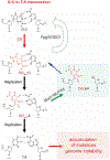
Figure 1.
Figure 1.. G:C to T:A transversion mutation and ‘GO’ repair pathway.
The BER glycosylases Fpg and MutY act upon their respective base substrates, OG:C and OG:A, and downstream BER enzymes (e.g. AP endonuclease, polymerase, ligase) restore the correct G:C bp.
Figure 2.
Figure 2.. Probing the structural features required for proper A recognition and excision.
(a) Abbreviated mechanism of MutY mediated adenine excision. Protonation of the N7 of A promotes N-glycosidic bond scission and departure of A as a neutral leaving group. The resultant oxacarbenium ion is stabilized as a covalent intermediate by Asp144, enabling stereospecific attack by a water molecule to form an abasic site product. (b) Crystal structure of Gs MutY (gray) bound to the non-cleavable substrate OG:FA showing that within the active site, A (purple) is oriented such that the N7 is aligned with the catalytic Glu43 and the C1' of the sugar is aligned with the catalytic Asp144 (dark blue sticks) (PDB ID: 3G0Q) (c) H-bonding network formed by A with the active site residues; atom numbering on A is shown in red; inset, electrostatic potential map of A. (d) Chemical structures and electrostatic potential maps of the adenine analogs used in this study (isovalue 0.020, density 0.0004).
Figure 3.
Figure 3.. Tautomerization of 2OA to 2OA-cyto can stably base pair with OGanti through a C-like hydrogen bonding face.
The Watson-Crick base pairing face of C and 2OA-cyto are indicated in blue.
Figure 4.
Figure 4.. MutY-catalyzed excision of A nucleobase analogs opposite OG in duplex DNA.
(a) 30 bp DNA duplex used for in vitro assays (b) Minimal kinetic scheme used to described MutY processing the OG:Y substrate (DNAOG:Y) to the abasic site product (DNAOG:AP), where there are three basic steps, substrate binding (KD), base excision (k2) and DNA-product release (k3). (c) Representative single exponential fits showing time dependent formation of 14 nt product resulting from the removal of A analogs by MutY followed by quenching with 0.2 M NaOH. The experiments were performed under single turnover (STO) conditions at 37°C using 20 nM DNA substrate and 40 nM active WT E. coli MutY. The analogs ADA and BA are not plotted as minimal cleavage was observed in the glycosylase assay in 60 minutes.
Figure 5.
Figure 5.. Relationship between overall repair and rate of base excision.
The overall repair plotted on the y-axis represents the normalized percent G:C conversion of each analog in muty+ cells above background levels in muty cells, as measured by extent of restriction digest by BmtI. The gradient bar indicates the extent of repair ranging from poor (red; <40%), moderate (yellow; ca. 40-60%) to well (green; >60%) repaired.
Figure 6.
Figure 6.. High quality control of MutY engendered at multiple checkpoints.
(a) Summary of SAR highlighting the importance of the different structural features of A in OG:A mismatch recognition and base excision. (b) Positioning of the 2-amino group in the major groove of the helix enables initial recognition of OG:A in a cellular context; left inset, crystal structure of an OG:A mispair (PDB ID 178D) shows that canonical adenine H-bonds with OG and stabilizes the syn conformation, which facilitates recognition; right inset, analogs that improperly base pair with OG and displace the 2-amino group from its major groove position leading to inefficient recognition by MutY.

References

    1. Friedberg EC DNA Damage and Repair. Nature 2003, 421 (6921), 436–440. 10.1038/nature01408. - DOI - PubMed
    1. Neeley WL; Essigmann JM Mechanisms of Formation, Genotoxicity, and Mutation of Guanine Oxidation Products. Chem. Res. Toxicol 2006, 19 (4), 491–505. 10.1021/tx0600043. - DOI - PubMed
    1. McGoldrick JP; Yeh YC; Solomon M; Essigmann JM; Lu AL Characterization of a Mammalian Homolog of the Escherichia Coli MutY Mismatch Repair Protein. Mol. Cell. Biol 1995, 15 (2), 989–996. - PMC - PubMed
    1. Banda DM; Nuñez NN; Burnside MA; Bradshaw KM; David SS Repair of 8-OxoG:A Mismatches by the MUTYH Glycosylase: Mechanism, Metals and Medicine. Free Radic. Biol. Med 2017, 107, 202–215. https://doi.org/10.1016/j.freeradbiomed.2017.01.008. - DOI - PMC - PubMed
    1. Drabløs F; Feyzi E; Aas PA; Vaagbø CB; Kavli B; Bratlie MS; Peña-Diaz J; Otterlei M; Slupphaug G; Krokan HE Alkylation Damage in DNA and RNA—Repair Mechanisms and Medical Significance. DNA Repair (Amst). 2004, 3 (11), 1389–1407. https://doi.org/10.1016/j.dnarep.2004.05.004. - DOI - PubMed

Publication types