. 2021 Feb;64(2):196-207.
doi: 10.1165/rcmb.2020-0112OC.

MicroRNA-574-5p Attenuates Acute Respiratory Distress Syndrome by Targeting HMGB1

Affiliations

MicroRNA-574-5p Attenuates Acute Respiratory Distress Syndrome by Targeting HMGB1

Binchan He et al. Am J Respir Cell Mol Biol. 2021 Feb.

Abstract

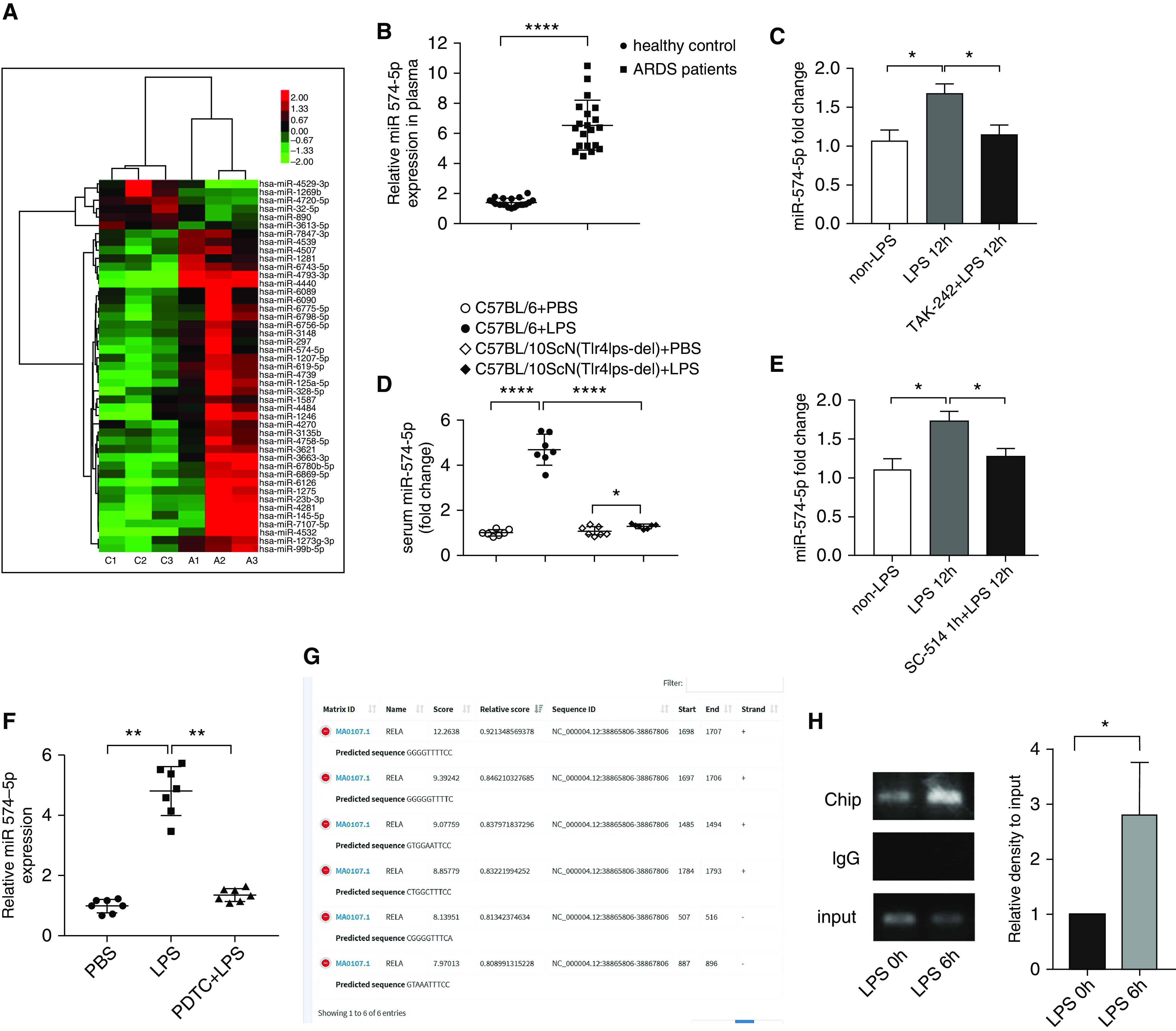
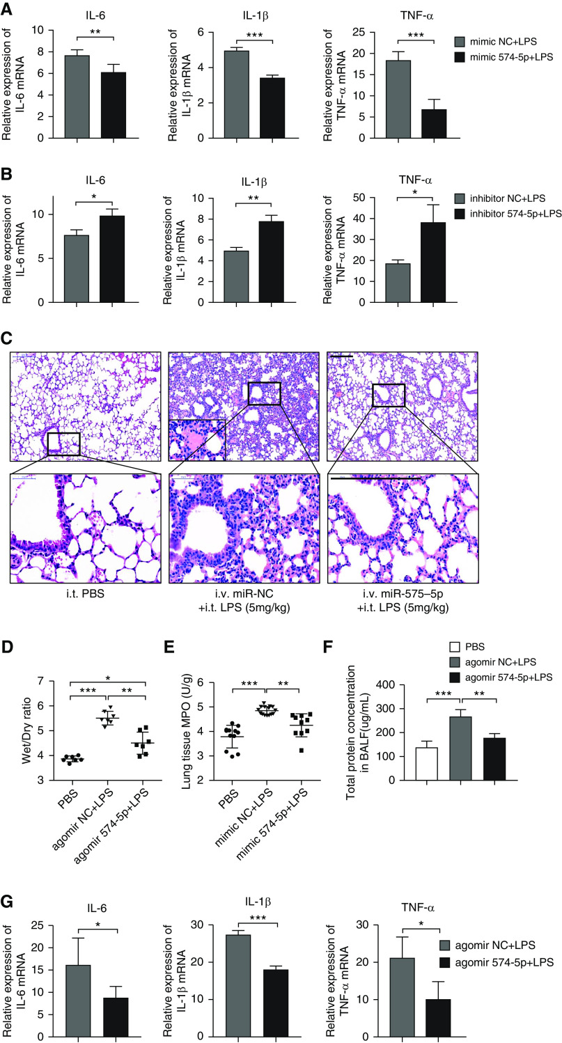
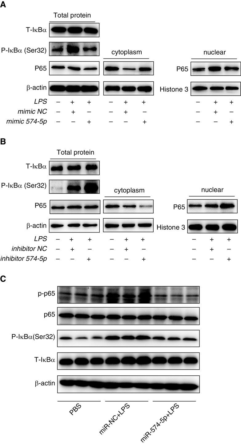
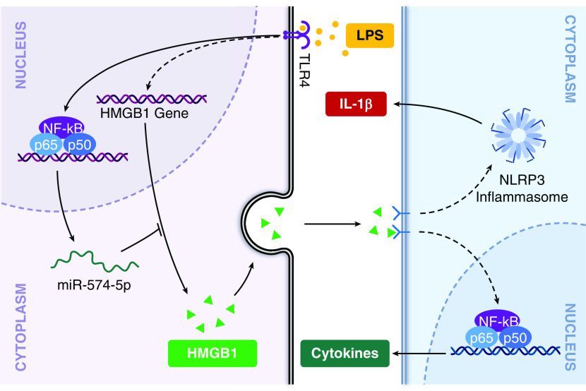
Acute respiratory distress syndrome (ARDS) is a critical condition with high mortality. HMGB1 (high-mobility group protein B1) is one of the key proinflammatory factors in the ARDS "inflammatory storm." According to previous studies, some microRNAs (miRNAs) play important roles in this process. We aimed to determine the contributing miRNAs targeting the expression and release of HMGB1. miRNA expression in the peripheral blood of patients with ARDS was measured by miRNA microarray. miRNAs targeting HMGB1 were screened and explored for further study. In LPS-induced cell and mouse ARDS models, we explored the effect of this miRNA on the expression and secretion of HMGB1 by Western blot, real-time qPCR, and ELISA. The effects of this miRNA on the NF-κB signaling pathway, proinflammatory cytokines, and NLRP3 (nod-like receptor protein 3) inflammasome were detected by Western blot and real-time qPCR. In ARDS models, microRNA-574-5p (miR-574-5p) expression could be induced by the TLR4/NF-κB pathway upon LPS stimulation. It could suppress the inflammatory response by targeting HMGB1. Enforcing the expression of miR-574-5p or HMGB1 siRNA silencing inhibits the activation of NF-κB signaling pathway and the NLRP3 inflammasome. Moreover, overexpression of HMGB1 reversed the antiinflammatory effect of miR-574-5p. In ARDS mice, overexpression of miR-574-5p suppresses alveolar leukocytes infiltration, interstitial edema, protein effusion, and inflammation. This study demonstrated that miR-574-5p provided negative feedback to LPS-induced inflammation and relieved ARDS. It may provide new therapeutic strategies for ARDS.

Keywords: HMGB1; NF-κB; NLRP3 inflammasome; miR-574-5p.


Figures

Figure 1.
Figure 1.
miR-574-5p expression was upregulated in acute respiratory distress syndrome (ARDS) and LPS-stimulated human pulmonary microvascular endothelial cells (HPMECs) and was induced by TLR4 via the NF-κB signaling pathway. (A) Microarray analysis of miRNA expression in healthy subjects (C1, C2, and C3) versus patients with ARDS (A1, A2, and A3) using a red, black, and green color scale (red indicates high expression, green indicates low expression, and black indicates unchanged expression). (B) Real-time qPCR analysis of miR-574-5p expression in the plasma of 20 healthy people and 20 patients with ARDS. (C) HPMECs were cultured for 45 minutes with medium only or LPS in the presence or absence of TAK-242. The expression of miR-574-5p was analyzed by real-time qPCR analysis (n = 7). (D) Wild-type and TLR4−/− mice were administered PBS or LPS and killed at 24 hours after infection. Serum was obtained, and the concentrations of miR-574-5p were measured (n = 7). (E) HPMECs were cultured for 1 hour with medium only or LPS in the presence or absence of SC-514. The expression of miR-574-5p was analyzed by real-time qPCR analysis (n = 7). (F) Wild-type and ammonium pyrrolidine dithiocarbamate (PDTC)-pretreated mice were administered LPS and killed at 24 hours after infection. Serum was obtained, and the concentrations of miR-574-5p were measured (n = 7). (G) The predicted potential binding sites between NF-κB p65 and the promoter of the miR-574-5p gene. (H) Binding of the NF-κB p65 subunit to the promoter of the miR-574-5p gene. IgG was used as a negative control in the chromatin IP assay (n = 3). The values presented are the mean ± SD. Comparisons were made by t test in B, D, and H. Comparisons were made by one-way ANOVA followed by Dunnett Multiple Comparison in C, E, and F. *P < 0.05, **P < 0.01, and ****P < 0.0001. Chip = chromatin IP; miR-574-5p = microRNA-574-5p; WT = wild-type.
Figure 2.
Figure 2.
miR-574-5p inhibited LPS-induced ARDS in vitro and in vivo. (A and B) HPMECs were transfected with miR-574-5p mimics (A) or inhibitor (B) for 24 hours followed by stimulation with LPS for 6 hours. IL-6, IL-1β, and TNF-α mRNA concentrations were measured by real-time qPCR (n = 7). (C) Mice were intravenously injected with miR negative control agomir or miR-574-5p agomir; 24 hours later, the mice were intratracheally instilled with PBS or 5 mg/ml LPS. Twenty-four hours later, lung inflammation was assessed by morphologic analysis. The lungs were embedded in formalin, and sections were analyzed by hematoxylin and eosin staining. Scale bars, 200 mm (n = 7). (D and E) Lungs were harvested to measure wet/dry (D) ratio and myeloperoxidase (E) activity (n = 7). (F) BAL fluid was collected, and the protein concentration was measured (n = 7). (G) Real-time qPCR analysis of the IL-6, IL-1β, and TNF-α mRNA concentrations in lung tissues (n = 7). The values presented are the mean ± SD. Comparisons were made by t test in A, B, and G. Comparisons were made by one-way ANOVA followed by Dunnett Multiple Comparison in DF. *P < 0.05, **P < 0.01, and ***P < 0.001. MPO = myeloperoxidase; NC = negative control.
Figure 3.
Figure 3.
miR-574-5p suppressed the activation of the NF-κB signaling pathway in vitro and in vivo. (A and B) HPMECs were transfected with miR-574-5p mimics (A) or inhibitor (B) for 48 hours followed by stimulation with LPS for 6 hours and Western blot analysis of the total and phosphorylated IκBα in HPMECs and the translocation of NF-κB p65 in HPMECs. (C) Mice were intravenously injected with miR-NC agomir or miR-574-5p agomir, and 24 hours later, the mice were intratracheally instilled with PBS or 5 mg/ml LPS. Twenty-four hours later, the lungs were harvested for total and phosphorylated IκBα and NF-κB p65 analysis. All images are representatives of five independent experiments.
Figure 4.
Figure 4.
miR-574-5p suppressed the activation of the NLRP3 (nod-like receptor protein 3) inflammasome in vitro and in vivo. (A) Mice were intravenously injected with miR-NC agomir or miR-574-5p agomir, and 24 hours later, the mice were intratracheally instilled with PBS or 5 mg/ml LPS. Twenty-four hours later, the lungs were harvested, and NLRP3, pro–capsase-1, cleaved caspase-1 were analyzed by Western blot. (B) IL-1β was analyzed by ELISA. All images are representatives of five independent experiments. The values presented are the mean ± SD. Comparisons were made by one-way ANOVA followed by Dunnett Multiple Comparison. *P < 0.05 and **P < 0.01.
Figure 5.
Figure 5.
HMGB1 is the functional target of miR-574-5p. (A) The HMGB1 3ʹ untranslated region (UTR) contains four predicted miR-574-5p binding sites. The figure shows the predicted binding interactions between the HMGB1 3ʹ UTR (middle) and miR-574-5p (bottom). The sites of targeted mutagenesis (top) are also indicated. (B) Normalized luciferase activity of a reporter containing the WT or point-mutated (MUT1, MUT2, MUT3, and MUT4) 3ʹ UTR reporter constructs of HMGB1 in 293T cells cotransfected with miR-NC mimics or miR-574-5p mimics (n = 5). (C) Twenty-four hours later, the cells were stimulated with LPS for 6 hours. Real-time qPCR analysis of HMGB1 mRNA concentrations in HPMECs transfected with miR-NC mimics or miR-574-5p mimics (n = 5). (D) Twenty-four hours after transfection of HPMECs with miR-NC mimics or miR-574-5p mimics, 5 μg/ml actinomycin D was added, the cell samples were collected at different time points, and HMGB1 mRNA abundance in HPMECs was detected by real-time qPCR (n = 5). (E and F) Western blot analysis of HMGB1 protein concentrations in HPMECs. HPMECs were transfected with miR-574-5p mimics or miR-574-5p inhibitors (n = 5). (G) HPMECs were transfected with miR-574-5p mimics for 48 hours followed by stimulation with LPS for 6 hours and ELISA analysis of HMGB1 protein concentrations in cell culture supernatants (n = 5). (H) Western blot analysis of HMGB1 protein in lung tissues (n = 5). (I and J) ELISA analysis of HMGB1 protein expression in serum (I) and BAL fluid (J) of ARDS mice (n = 5). The values presented are the mean ± SD. Comparisons were made by t test in C and D. Comparisons were made by one-way ANOVA followed by Dunnett Multiple Comparison in GJ. *P < 0.05 and **P < 0.01. NS = not significant.
Figure 6.
Figure 6.
miR-574-5p regulates the inflammatory response through HMGB1. (A) Real-time qPCR analysis of HMGB1, IL-6, IL-1β, and TNF-α mRNA concentrations in HPMECs transfected with NC-siRNA or HMGB1-siRNA followed by LPS stimulation (n = 5). (B) Western blot analysis of HMGB1, total and phosphorylated NF-κB p65 and IκBα, NLRP3, and cleaved caspase-1 in HPMECs transfected with NC-siRNA or HMGB1-siRNA followed by LPS stimulation (n = 3). (C) HPMECs were cotransfected with miR-574-5p mimics/miR-NC mimics and pEGFP-N1-HMGB1/pEGFP-N1. After 48 hours, the cells were treated with LPS for 6 hours. The HMGB1, NLRP3, and cleaved caspase-1 protein concentrations were evaluated by Western blotting (n = 3). (D) HPMECs were cotransfected with miR-574-5p inhibitor/miR-NC inhibitor and si-HMGB1/si-NC. After 48 hours, the cells were treated with LPS for 6 hours. The HMGB1, NLRP3, and cleaved caspase-1 protein concentrations were evaluated by Western blotting (n = 3). (E) mRNA concentrations of IL-6, IL-1β, and TNF-α were measured by real-time qPCR in HPMECs treated as described in C (n = 5). (F) mRNA concentrations of IL-6, IL-1β, and TNF-α were measured by real-time qPCR in HPMECs treated as described in D. The values presented are the mean ± SD. Comparisons were made by t test in A. Comparisons were made by one-way ANOVA followed by Dunnett Multiple Comparison in E and F. *P < 0.05, **P < 0.01, ***P < 0.001, and ****P < 0.0001.
Figure 7.
Figure 7.
Schematic model of the critical role of miR-574-5p in ARDS pathogenesis. miR-574-5p is induced by TLR4 through the NF-κB signaling pathway upon LPS stimulation. It functions as a negative feedback regulator in NF-κB signaling and NLRP3 inflammation activation by targeting HMGB1.

Comment in

Similar articles

Cited by

References

    1. Villar J, Blanco J, Kacmarek RM. Current incidence and outcome of the acute respiratory distress syndrome. Curr Opin Crit Care. 2016;22:1–6. - PubMed
    1. Confalonieri M, Salton F, Fabiano F. Acute respiratory distress syndrome. Eur Respir Rev. 2017;26:160116. - PMC - PubMed
    1. Huang C, Wang Y, Li X, Ren L, Zhao J, Hu Y, et al. Clinical features of patients infected with 2019 novel coronavirus in Wuhan, China. Lancet. 2020;395:497–506. - PMC - PubMed
    1. Matthay MA, Zemans RL. The acute respiratory distress syndrome: pathogenesis and treatment. Annu Rev Pathol. 2011;6:147–163. - PMC - PubMed
    1. Millar FR, Summers C, Griffiths MJ, Toshner MR, Proudfoot AG. The pulmonary endothelium in acute respiratory distress syndrome: insights and therapeutic opportunities. Thorax. 2016;71:462–473. - PubMed

Publication types

LinkOut - more resources