. 2020 Dec 17;80(6):980-995.e13.
doi: 10.1016/j.molcel.2020.10.023. Epub 2020 Nov 16.

Gene- and Species-Specific Hox mRNA Translation by Ribosome Expansion Segments

Affiliations

Gene- and Species-Specific Hox mRNA Translation by Ribosome Expansion Segments

Kathrin Leppek et al. Mol Cell. .

Abstract

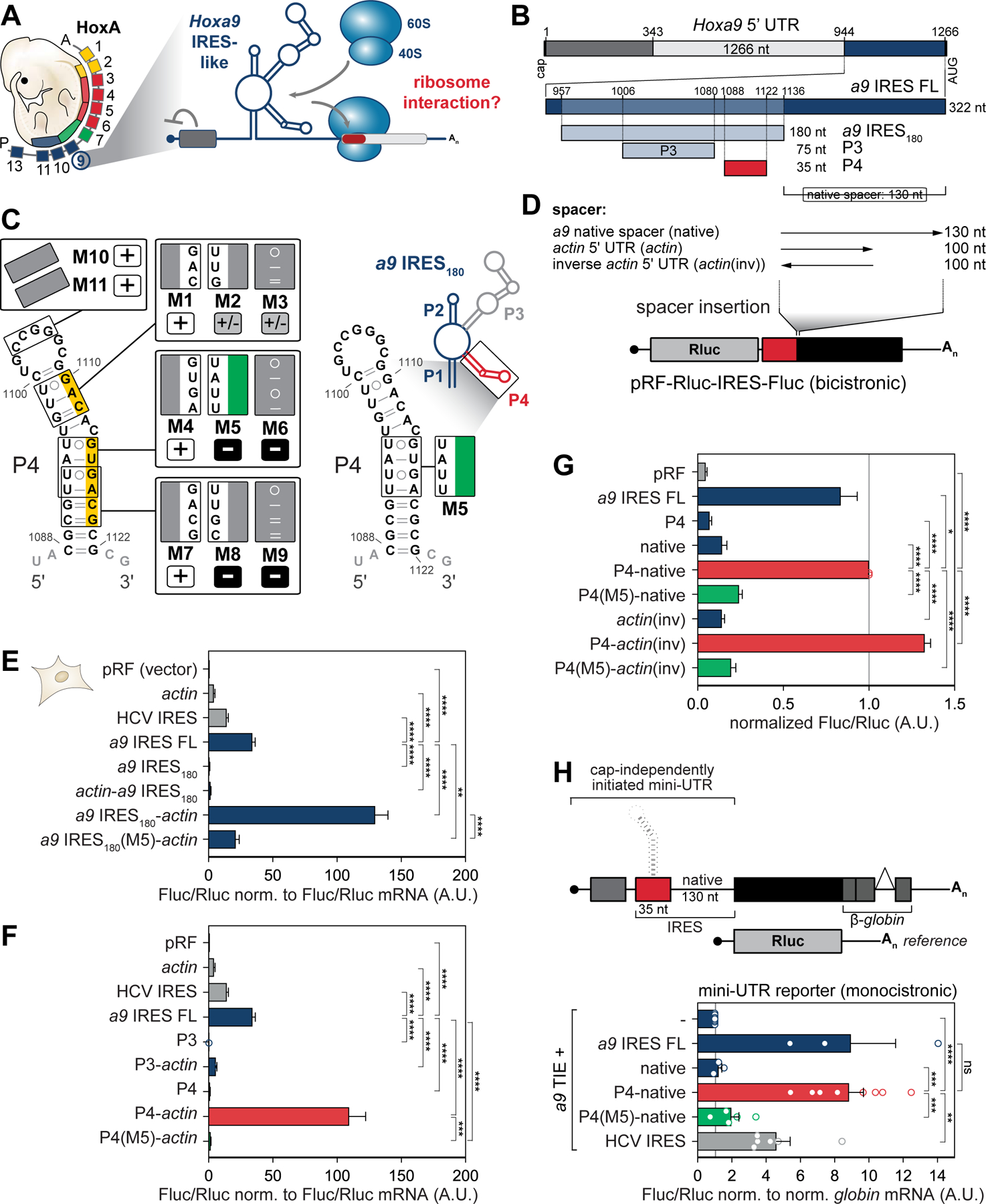
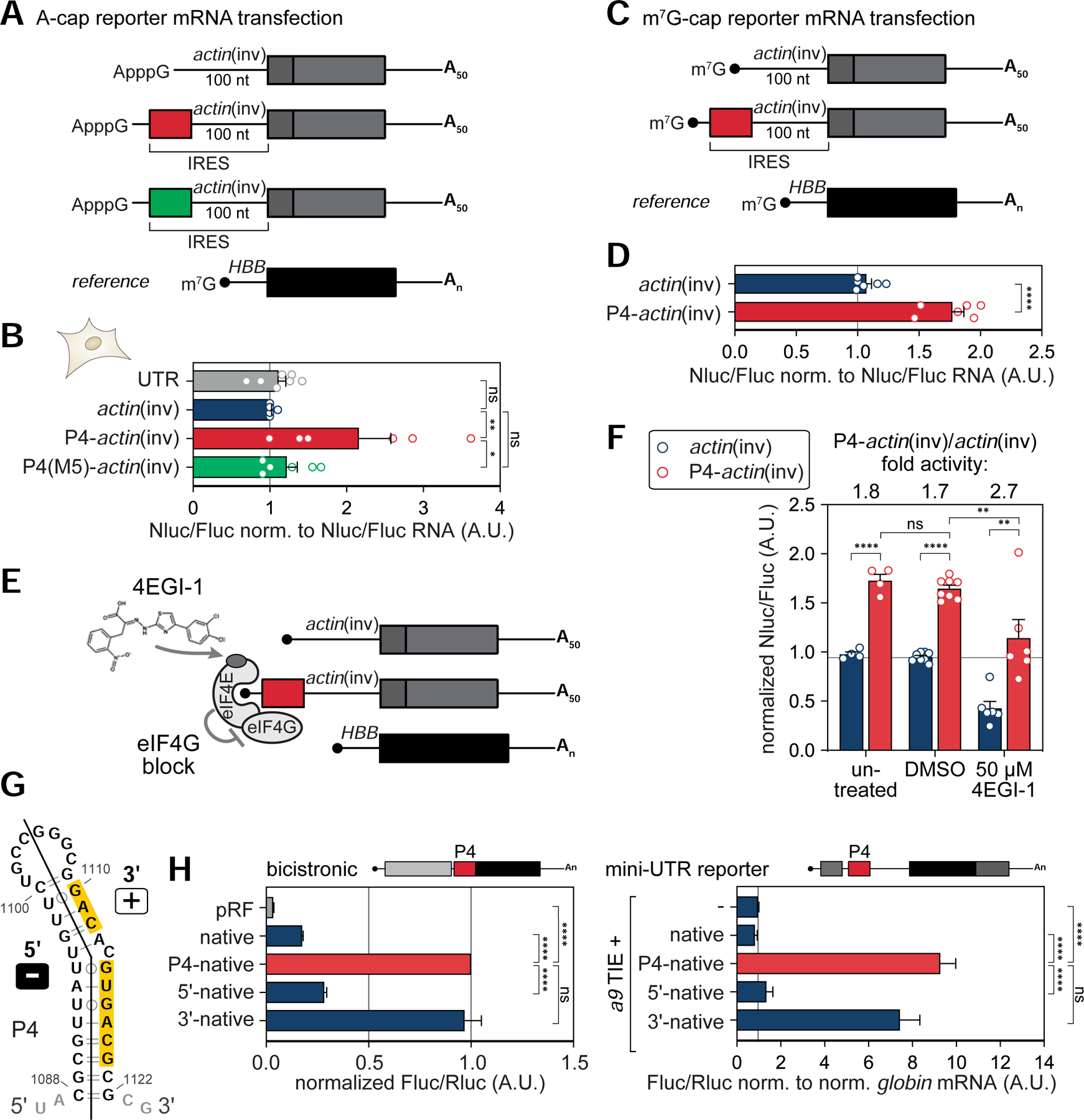
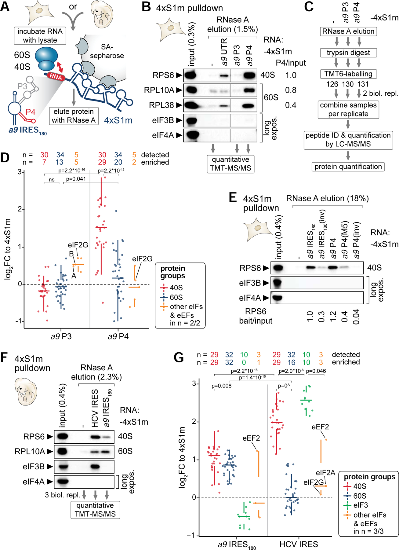
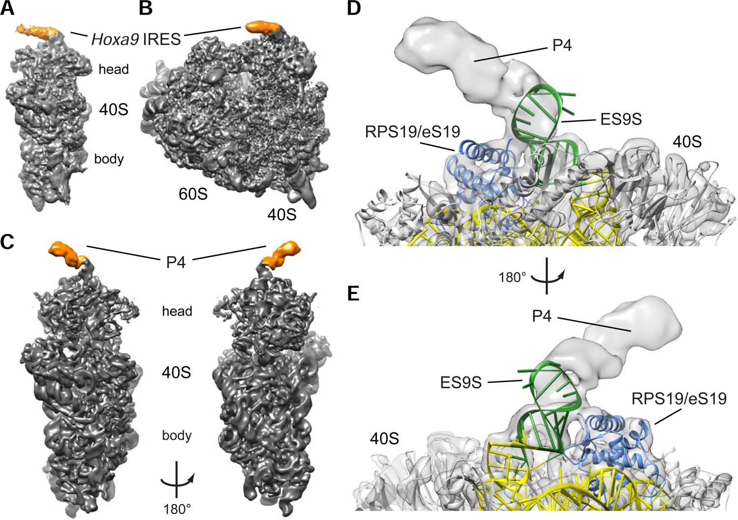
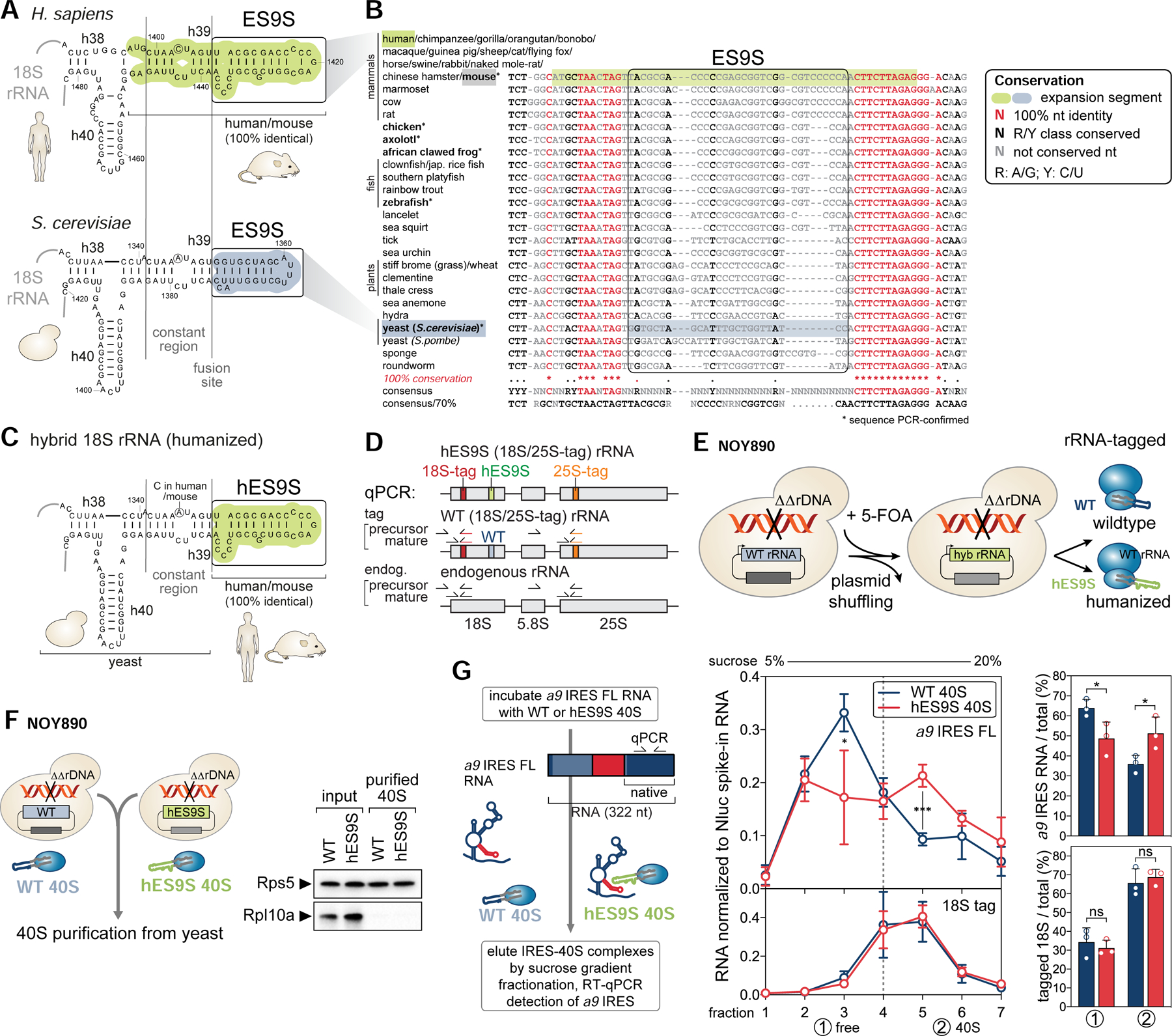
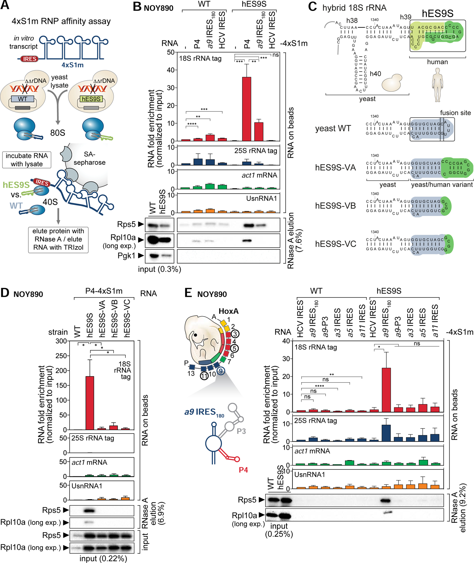
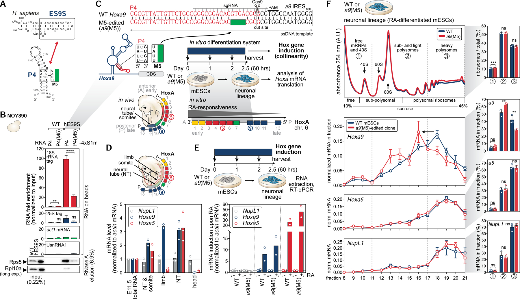
Ribosomes have been suggested to directly control gene regulation, but regulatory roles for ribosomal RNA (rRNA) remain largely unexplored. Expansion segments (ESs) consist of multitudes of tentacle-like rRNA structures extending from the core ribosome in eukaryotes. ESs are remarkably variable in sequence and size across eukaryotic evolution with largely unknown functions. In characterizing ribosome binding to a regulatory element within a Homeobox (Hox) 5' UTR, we identify a modular stem-loop within this element that binds to a single ES, ES9S. Engineering chimeric, "humanized" yeast ribosomes for ES9S reveals that an evolutionary change in the sequence of ES9S endows species-specific binding of Hoxa9 mRNA to the ribosome. Genome editing to site-specifically disrupt the Hoxa9-ES9S interaction demonstrates the functional importance for such selective mRNA-rRNA binding in translation control. Together, these studies unravel unexpected gene regulation directly mediated by rRNA and how ribosome evolution drives translation of critical developmental regulators.

Keywords: ES9S; Hox cluster; RNA structure; RNA-protein interaction; expansion segment; internal initiation; internal ribosome entry site; mRNA translation; ribosomal RNA; ribosome; ribosome engineering; stem-loop; yeast.


Conflict of interest statement

Declaration of Interest K.L. and M.B. are inventors on patents and submitted provisional patent applications related to the Hoxa9 P4 stem-loop and RNA therapeutics and their various uses.

Figures

Figure 1.
Figure 1.. A short stem-loop in the Hoxa9 5’ UTR is sufficient for recruitment of the ribosome.
(A) Model of functional RNA elements in the Hoxa9 5’ UTR that regulate the translation of subsets of Hoxa mRNAs in the embryo (Xue et al., 2015). TIE, translation inhibitory element; IRES, internal ribosome entry site. (B) Schematic of the topology of regulatory elements in the mouse Hoxa9 5’ UTR. The 180 nucleotides (nt)-long Hoxa9 IRES-like RNA element (a9 IRES180) harbors the P3 and P4 stem-loops and resides 130 nt upstream of the AUG (native spacer). (C) Secondary structure model of a9 IRES180, a zoomed-in view of the P4 stem-loop (red), and substitution mutations mapped onto the P4 structure. Nts mutated in P4(M5) (green). Numbers refer to nt positions in the Hoxa9 5’ UTR. Active P4 mutants (normalized Fluc/Rluc < 0.5 A.U.) are labeled “+”, moderately active mutants (Fluc/Rluc < 0.5, > 1.0 A.U.) are labeled “+/–”, and inactive mutants (Fluc/Rluc > 0.5 A.U.) are labeled “–”. Yellow: Sequence critical for IRES-like activity. See also Figure S1. (D) Spacer sequence requirement for a9 IRES-like element activity is tested by inserting spacers of different lengths downstream of an IRES-like element in a bicistronic reporter mRNA plasmid (pRF). Rluc, renilla luciferase; Fluc, firefly luciferase. (E) Bicistronic reporter genes were transiently transfected into mouse C3H/10T1/2 cells and expressed from plasmids. Cells from the same transfection were split in half for protein and mRNA analysis. Relative luciferase activity is expressed as a Fluc(IRES)/Rluc(cap-initiation) ratio normalized to respective Fluc/Rluc mRNA levels. Average IRES-like activity ± standard error of the mean (SEM), n = 4–15. pRF and actin 5’ UTR serve as negative controls, HCV IRES as an IRES control. a9 IRES FL: FL, full-length; pRF (vector), no insert in the intergenic region; A.U., arbitrary units. (F) Bicistronic reporter mRNAs were transiently expressed as described in (E). Average IRES-like activity normalized to respective Fluc/Rluc mRNA levels ± SEM, n = 4–15. (G) Bicistronic reporter mRNAs were transiently expressed as described in (E). actin(inv) serves as a spacer sequence control. Average Fluc/Rluc IRES-like activity ± SEM, n = 3–8. (H) Schematic of monocistronic “mini UTR” Fluc and control Rluc reporter mRNAs. IRES-like elements and spacer-derivatives were introduced into the Fluc 5’ UTR, and Fluc/Rluc luciferase activity was measured in transiently plasmid-transfected C3H/10T1/2 cells. A co-expressed Rluc reporter served as reference. Average Fluc/Rluc activity is normalized to respective globin/NupL1 mRNA levels ± SEM, n = 3–7; -, TIE alone; ns, not significant.
Figure 2.
Figure 2.. The P4 stem-loop is the minimal TE element in the Hoxa9 5’ UTR.
(A) In vitro transcribed, A-capped, and polyadenylated (A50-tail) HA-Nanoluc (Nluc) reporter mRNAs containing 5’ UTR elements. m7G-capped and polyadenylated HBB-Fluc mRNA served as reference. (B) A-capped reporter mRNAs shown in (A) were directly transfected into mouse C3H/10T1/2 cells. IRES-like activity of P4-actin(inv) derivatives were compared to a 46 nt-long scrambled control (UTR). m7G-capped HBB-Fluc mRNA was co-transfected as reference. Cells were transiently transfected with RNA for 6 hours, cells from the same transfection were split in half for protein and mRNA analysis, and average Nluc/Fluc luciferase activity was normalized to respective Nluc/Fluc mRNA levels ± SEM, n = 6–9; actin(inv) alone was set to 1. (C) In vitro transcribed, m7G-capped, and polyadenylated (A50-tail) HA-Nanoluc (Nluc) reporter mRNAs as in (A) that contain a conventional m7G cap. (D) m7G-capped reporter mRNAs shown in (C) were directly transfected into human HEK293T cells as in (B). Average Nluc/Fluc luciferase activity was normalized to respective Nluc/Fluc mRNA levels ± SEM, n = 6. (E) Schematic of the small molecule inhibitor 4EGI-1 that binds to eIF4E and blocks eIF4G association, thus uncouples cap-dependent initiation from P4 translation enhancer function. See also Figure S1. (F) Luciferase activity analysis in mouse C3H/10T1/2 cells was carried out as in (D). Cells were treated for 3 h, transiently transfected with RNA for 6 hours in presence of drug or carrier, and harvested for luciferase analysis. Average Nluc/Fluc luciferase activity ± SEM, n = 4–8; actin(inv), untreated was set to 1. (G) Secondary structure model of the 5’ and 3’ arms of the P4 stem-loop mapped onto the structure. Yellow: Sequence critical for P4 activity. (H) P4 derivatives fused to the native spacer were tested for IRES-like activity in bicistronic (left, average Fluc/Rluc IRES-like activity ± SEM, n = 4–6) or monocistronic mini-UTR reporter mRNAs (right, average Fluc/Rluc activity normalized to respective globin/NupL1 mRNA levels ± SEM, n = 3–8). Reporter mRNAs were transiently expressed from plasmids as described in Figure 1E, H. The inactive 5’ arm (Fluc/Rluc > 0.5 A.U.) is labeled “–”, and the active 3’ arm (normalized Fluc/Rluc < 0.5 A.U.) is labeled “+” in (G).
Figure 3.
Figure 3.. The Hoxa9 IRES-like element binds to the 40S ribosomal subunit via P4.
(A) Schematic of the 4xS1m pulldown to probe interactions of in vitro transcribed 4xS1m-aptamer fusion RNA with lysate components from C3H/10T1/2 cells or mouse embryos to form ribonucleoproteins (RNPs) in vitro. SA, streptavidin. (B) 4xS1m pulldown is performed by combining mouse Hoxa9 mRNA elements with C3H/10T1/2 cell lysates. The aptamer alone (−) served as negative control. RPs of the 40S and 60S subunit and eIFs were monitored by western blot (WB) analysis. The fraction loaded of input and elution samples is expressed as percentage of the original lysate volume. Representative of n = 2 is shown. RNase A elutions of the aptamer control, P3 and P4 were subjected to mass spectrometry (MS) analysis. Differential enrichment of RPs compared to input with P4 was normalized to RPS6 set to 1. UTR, full-length Hoxa9 5’ UTR. (C) Workflow for identifying and quantifying proteins in RNase A elutions by quantitative MS using tandem mass tag (TMT) peptide labelling. Proteins were trypsin-digested into peptides, labeled with a distinct TMT, mixed equally per replicate, and subjected to liquid chromatography (LC)-tandem MS (MS/MS) analysis for multiplex quantification. (D) Analysis of TMT-MS/MS data displayed as log2 fold change (FC) relative to the aptamer control (4xS1m) for a9 P3 and P4 shows the relative abundance of proteins detected and enriched in respective protein groups compared to their levels in the control. Samples correspond to the pulldown in (B) from C3H/10T1/2 cells. Only proteins detected in 2/2 biological replicates are shown. See also Table S4. (E) 4xS1m-pulldown as described in (B) comparing the a9 IRES180 and a9 P4 to control constructs. Lysates of C3H/10T1/2 cells were used as input. Representative of n = 3 is shown. Differential enrichment of RPS6 compared to input with RNA baits was normalized to a9 IRES180 set to 1. (F) 4xS1m-pulldown as described in (B) comparing the a9 IRES180 to an unrelated viral IRES, HCV. Lysates of FVB stage E11.5 mouse embryos were used as input. Representative of n = 3 is shown. RNase A elutions were subjected to TMT-MS/MS analysis. (G) Analysis of TMT-MS/MS data displayed as log2 fold change (FC) relative to 4xS1m for a9 IRES180 and HCV IRES as in (D). Samples correspond to the pulldown in (F) from FVB stage E11.5 mouse embryos. Only proteins detected in 3/3 biological replicates are shown. See also Table S5.
Figure 4.
Figure 4.. Cryo-EM reveals that the Hoxa9 IRES-like and P4 RNA bind to the ribosome via ES9S.
(A) Reconstruction of the human 40S ribosomal subunit with the mouse Hoxa9 IRES-like element (a9 IRES FL) at 3.9 Å resolution. Additional density for a9 IRES FL is indicated in orange. (B) Reconstruction of the human 80S ribosome at 4.40 Å resolution with the additional density for the mouse a9 IRES FL indicated in orange. (C) The a9 P4 stem-loop binds to the head of the small 40S ribosomal subunit. Reconstruction of the P4 stem-loop (orange) bound to human 40S ribosomal subunit (grey) at 4.1 Å for the 40S head and 3.1 Å resolution for the 40S body. The density is low-pass filtered to 7 Å to show RNA helical features of P4 (orange). (D) Cryo-EM analysis of the mouse a9 P4 stem-loop in complex with the human ribosome. Reconstruction of the 40S ribosomal subunit head in complex with P4 at 4.1 Å resolution. P4 binds to ES9S (green) of the 18S rRNA (yellow) near ribosomal protein RPS19/eS19 (blue). The tip of ES9S was modelled onto pdb 5a2q to better visualize the bound P4 element. (E) 180° rotation of the reconstruction in (D).
Figure 5.
Figure 5.. Engineering of chimeric hES9S-humanized ribosomes in yeast.
(A) Secondary structure models of the human (green) and yeast (blue) 18S rRNA region containing ES9S. The constant region (h39) and ES9S-fusion site selected for chimeric 18S rRNA engineering are indicated. (B) A 40-way multiple sequence alignment (MSA) and conservation analysis of the highly conserved 18S rRNA region in which the more variable ES9S is embedded. Nts are color coded according to conservation. For six species (bold, asterisk) annotated 18S rRNA sequences were confirmed by RT-PCR spanning the ES9S region. R, purine; Y, pyrimidine. (C) Structure model of the engineered yeast 18S rRNA after exchange of the yeast to human ES9S (hES9S, green). (D) The rRNA cassette that encodes 18S, 5.8S and 25S rRNA indicating the position of unique sequence tags in 18S (red) and 25S (orange) rRNA used for RT-qPCR to detect precursor and mature forms of endogenous and tagged yeast rRNA. (E) The plasmid shuffling approach to generate yeast strains that contain a homozygous knock-out of the rDNA locus (NOY890) and exclusively express plasmid-encoded tagged chimeric 18S rRNA as in (C). See also Figure S5. (F) 40S subunits of WT and hES9S yeast strains (NOY890) were purified by sequential sucrose gradient sedimentation (see also Figure S6). The purity of the isolated 40S was confirmed by WB analysis of RPs compared to the input lysate. RPL10A/uL1 is yeast Rpl1 (referred to as Rpl10a). (G) In vitro binding assays using purified WT and hES9S 40S subunits to test direct binding to the a9 IRES FL RNA. IRES-40S complexes were eluted by 5–20% sucrose gradient fractionation. Co-sedimentation of 40S (18S rRNA tag) and bound RNA (a9 IRES FL) was detected by RT-qPCR, normalized to a Nluc spike-in RNA (average ± SD, n = 3). IRES-40S co-sedimentation was assessed by integrating the gradient distribution and expressed for the free (1) and 40S (2) fractions as the percentage relative to the total (average ± SD, n = 3).
Figure 6.
Figure 6.. Chimeric hES9S-humanized yeast ribosomes reconstitute Hoxa9- and P4-ES9S binding.
(A) Schematic of the 4xS1m pulldown to probe interactions of IRES-4xS1m RNA with WT and hES9S ribosomes from yeast (NOY890) lysates. Ribosome enrichment is monitored by RT-qPCR for tagged rRNA and other RNA classes normalized to the input and WB analysis for RPs. (B) rRNA bound to 4xS1m-fused RNA is quantified with primers specific for 18S and 25S rRNA tags (RNA on beads). 4xS1m aptamer alone (−) and the HCV IRES serve as a negative and IRES control, respectively. The 4xS1m aptamer/WT sample was used to normalize for fold enrichment of detected RNA (set to 1). Yeast actin (act1) and yeast UsnRNA1 serve as negative controls for an mRNA and a non-coding RNA, respectively. Ribosome enrichment was assessed by WB analysis of same volumes of protein released from beads by RNase A. The fraction loaded of input and elution samples is expressed as percentage of the original lysate volume. Cytoplasmic enzyme PGK1 serves as a negative control. Average RNA fold enrichment, standard deviation (SD), n = 3; long exp., long exposure. See also Figure S6. (C) Structure model of the engineered (hES9S, green) yeast 18S rRNA with annotations of the tested sequences (dark green; white circle) for hES9S-variants VA-C which contain partial hES9S sequences. Structure models of hES9S variants were predicted using Vienna RNAfold which predicted the assumed correct RNA folds for the yeast and human WT ES9S. (D) Same analysis was performed as in (B), comparing hES9S variants VA-C for their ability to bind to P4. The full hES9S serves as a positive control. The P4–4xS1m/WT sample was used to normalize for fold enrichment (set to 1). Average RNA fold enrichment, SEM, n = 3. (E) Same analysis was performed as in (B), comparing IRES-like elements of the Hoxa cluster, a9 IRES180, a3, a5, a11, and a9 P3. The HCV IRES serves as a negative control. The structure model of a9 IRES180 with P3 and P4, and the HoxA gene cluster chromosomal arrangement is given. The HCV-4xS1m/WT sample was used to normalize for fold enrichment (set to 1). Average RNA fold enrichment, SD, n = 3.
Figure 7.
Figure 7.. The P4-ES9S interaction is important for endogenous Hoxa9 mRNA translation.
(A) Schematic of the secondary structures of human ES9S, which is identical to mouse, and the P4 stem-loop indicating the inactive M5 mutation used to test the functional relevance of their interaction for Hoxa9 mRNA translation. (B) 4xS1m pulldown analysis was performed as in Figure 6B, comparing a9 P4 with P4(M5). The P4–4xS1m/WT sample was used to normalize for fold enrichment (set to 1). Average RNA fold enrichment, SD, n = 4. (C) Schematic of targeted CRISPR/Cas9-editing of the 4 nt-mutation (TATT) of P4(M5) into the genomic Hoxa9 locus of mESCs. In vitro differentiation of mESCs by retinoic acid (RA)-treatment into the neuronal lineage (neural stem cells) induces colinear Hox gene expression. Genome-edited clones and WT cells were subjected to endogenous Hoxa mRNA translation analysis. See also Figure S7. (D) The expression levels of NupL1, Hoxa9, and Hoxa5 mRNAs relative to actin mRNA in whole stage E11.5 FVB mouse embryos (E11.5 total RNA) compared to embryonic tissues of the same stage. DNase-treated total RNA from a whole embryo (E11.5 total RNA) was set to 1, n = 2–3; NT, neural tube. (E) Schematic of Hox gene induction in WT and a9(M5)-edited mESCs (clone D6) upon 60 hours (2.5 days) of 33 nM RA-treatment or DMSO (control). mRNA induction of Hoxa9, Hoxa5, and NupL1 (control), normalized to actin mRNA in RA (+) and DMSO (−)-treated mESCs. Respective DMSO/WT or DMSO/a9(M5) samples were set to 1 to indicate mRNA induction, n = 2. (F) Sucrose gradient fractionation analysis of lysates derived from RA-treated WT and a9(M5)-edited cells on 10–45% sucrose gradients. RNA extraction of individual fractions, RT-qPCR specific for Hoxa9, as well as Hoxa5 and NupL1 mRNAs as controls, and normalization to a Fluc-Rluc spike-in RNA, reflects the normalized distribution of the endogenous mRNAs in fractions to determine their translation efficiencies (average ± SEM, n = 3). Gradient distribution was quantified by integrating free and 40S (1), sub- and light (2), heavy polysomes (3) which was expressed as the percentage relative to the total (average ± SD, n = 3).

References

    1. Acker MG, Kolitz SE, Mitchell SF, Nanda JS, and Lorsch JR (2007). Reconstitution of Yeast Translation Initiation. In Methods in Enzymology, pp. 111–145. - PubMed
    1. Anger AM, Armache JP, Berninghausen O, Habeck M, Subklewe M, Wilson DN, and Beckmann R (2013). Structures of the human and Drosophila 80S ribosome. Nature 497, 80–85. - PubMed
    1. Armache J-P, Jarasch A, Anger AM, Villa E, Becker T, Bhushan S, Jossinet F, Habeck M, Dindar G, Franckenberg S, et al. (2010). Cryo-EM structure and rRNA model of a translating eukaryotic 80S ribosome at 5.5-A resolution. Proc. Natl. Acad. Sci. U. S. A 107, 19748–19753. - PMC - PubMed
    1. Beltrame M, Hendry Y, and Tollervey D (1994). Mutational analysis of an essential binding site for the U3 snoRNA in the 5′ external transcribed spacer of yeast pre-rRNA. Nucleic Acids Res 22, 5138–5147. - PMC - PubMed
    1. Chen E, Sharma MR, Shi X, Agrawal RK, and Joseph S (2014). Fragile X mental retardation protein regulates translation by binding directly to the ribosome. Mol. Cell 54, 407–417. - PMC - PubMed

Publication types

MeSH terms