. 2020 Nov 14;10(11):950.
doi: 10.3390/diagnostics10110950.

Spontaneous Remission and Concomitant Progression in a Patient with DLBCL

Affiliations

Spontaneous Remission and Concomitant Progression in a Patient with DLBCL

Eun Ji Han et al. Diagnostics (Basel). .

Abstract

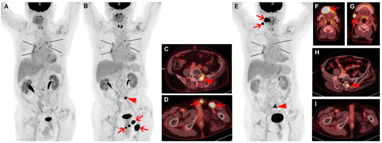
Diffuse large B-cell lymphoma (DLBCL) is the most common type of lymphoma. Although DLBCL can be cured in more than half of all patients, up to 50% of patients become refractory to initial treatment or relapse after complete remission. We present a case of complete spontaneous remission of some tumors and concomitant newly developed tumors observed in a patient with relapsed DLBCL. Spontaneous remission of lymphoma without treatment is a rare phenomenon and can occur at baseline as well as in relapsed DLBCL. However, most patients who initially experience spontaneous remission later develop relapse. Thus, careful follow-up is required, and fluorine-18-fluorodeoxyglucose (18F-FDG) positron emission tomography (PET)/computed tomography (CT) allows monitoring of multiple lesions.

Keywords: FDG; PET/CT; diffuse large B-cell lymphoma; spontaneous remission.


Conflict of interest statement

The authors declare no conflict of interest.

Figures

Figure 1
Figure 1
A 67-year-old male patient was diagnosed with diffuse large B-cell lymphoma (DLBCL) and had right orchiectomy followed by 8 cycles of rituximab, cyclophosphamide, hydroxydaunorubicin, vincristine, prednisone (R-CHOP) chemotherapy, and then radiotherapy, and complete response was achieved. Three years after the first line of therapy, the patient experienced relapse confirmed by left orchiectomy. Six cycles of dexamethasone, L-asparaginase, ifosfamide, carboplatin, etoposide (DL-ICE) chemotherapy, and radiotherapy followed, and the end-of-therapy fluorine-18-fluorodeoxyglucose (18F-FDG) positron emission tomography (PET)/ computed tomography (CT) findings indicated complete metabolic response again (A). FDG uptakes in the mediastinum and the peribronchial nodes showed similar patterns as in the previous 18F-FDG PET/CT studies without any size change and were assumed to be reactive nodal hyperplasia (A; black lines). Four months after the second line of therapy, the patient developed a palpable mass in left orchiectomy bed. Restaging 18F-FDG PET/CT demonstrated newly developed intense focal FDG uptakes in left orchiectomy bed (SUVmax 22.7), left inguinal area, and penis (B,D; arrows). In addition, localized FDG uptake was newly noted in sacrum and adjacent left L5/S1 intervertebral foramen (SUVmax 10.8; B,C; arrow heads). Clinically, these new lesions were considered to be second relapse of lymphoma. The patient refused further treatment and was lost to follow-up. Seven months later, the patient returned to the hospital complaining of discomfort in the oral cavity. Biopsy from right gingival lesion was performed. Immunohistochemistry confirmed tumor cells to be positive for CD79 and CD20 and negative for CD3, and the histology confirmed relapsed DLBCL. 18F-FDG PET/CT was performed for evaluation of overall disease status (E). Previously noted intense FDG uptakes in left orchiectomy bed, left inguinal area, penis, and sacrum had disappeared (E,I), without any form of treatment according to the patient and available medical records. Through multiple interviews, the patient and his family corroborated that the patient did not receive care at outside hospital. However, intense FDG uptakes were newly noted in right upper gingiva and adjacent maxilla (SUVmax 29.8; E,F; arrows), right cervical lymph node (E,G; arrows), and along the sacral canal at S2 level (SUVmax 12.3; E,H; arrow heads). FDG uptakes in the mediastinum and the peribronchial nodes showed little interval change (A,B,E; black lines).

References

    1. Li S., Young K.H., Medeiros L.J. Diffuse large B-cell lymphoma. Pathology. 2018;50:74–87. doi: 10.1016/j.pathol.2017.09.006. - DOI - PubMed
    1. Iacoboni G., Zucca E., Ghielmini M., Stathis A. Methodology of clinical trials evaluating the incorporation of new drugs in the first-line treatment of patients with diffuse large B-cell lymphoma (DLBCL): A critical review. Ann. Oncol. 2018;29:1120–1129. doi: 10.1093/annonc/mdy113. - DOI - PubMed
    1. Crump M., Neelapu S.S., Farooq U., Van Den Neste E., Kuruvilla J., Westin J., Link B.K., Hay A., Cerhan J.R., Zhu L., et al. Outcomes in refractory diffuse large B-cell lymphoma: results from the international SCHOLAR-1 study. Blood. 2017;130:1800–1808. doi: 10.1182/blood-2017-03-769620. - DOI - PMC - PubMed
    1. Gattiker H.H., Wiltshaw E., Galton D.A. Spontaneous regression in non-Hodgkin’s lymphoma. Cancer. 1980;45:2627–2632. doi: 10.1002/1097-0142(19800515)45:10<2627::AID-CNCR2820451023>3.0.CO;2-0. - DOI - PubMed
    1. Kumar R., Bhargava P., Zhuang H., Yu J.Q., Schuster S.J., Alavi A. Spontaneous regression of follicular, mantle cell, and diffuse large B-cell non-Hodgkin’s lymphomas detected by FDG-PET imaging. Clin. Nucl. Med. 2004;29:685–688. doi: 10.1097/00003072-200411000-00002. - DOI - PubMed

LinkOut - more resources