. 2020 Nov 13;9(11):1122.
doi: 10.3390/antiox9111122.

Effects of Rutin on Wound Healing in Hyperglycemic Rats

Affiliations

Effects of Rutin on Wound Healing in Hyperglycemic Rats

Li-You Chen et al. Antioxidants (Basel). .

Abstract

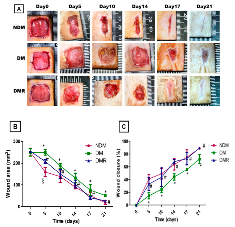
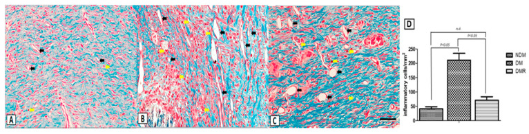
Long-term poor glycemic control negatively affects macrovascular and microvascular diseases, as well as wound restoration. Buckwheat is a good source of rutin (quercetin-3-O-rutoside) and has benefits in regulating blood sugar. This study was to evaluate the antioxidant and anti-inflammatory effects of rutin on wound healing in streptozotocin-induced hyperglycemic rats. Eighteen male Wistar rats were randomly divided into three groups: normal (NDM), hyperglycemic (DM), and hyperglycemic with rutin (DMR). After induction of hyperglycemia for 2 days, a 15 × 15 mm wound was induced on the back of each rat. Intraperitoneal injection of rutin significantly ameliorated diabetes-induced body weight loss and improved metabolic dysfunctions of hyperglycemic rats. Based on appearance and histopathological staining, rutin promotes wound healing and inhibits production of inflammatory cells. The immunoblotting data indicated that rutin promotes production of antioxidant enzymes induced by nuclear factor erythroid 2-related factor 2 (NRF2), inhibits the expression of matrix metalloproteinases (MMPs) regulated by NF-κB, and decreases the expression of vascular endothelial growth factor (VEGF). It also promotes the expression of neurogenic-related protein (UCH-L1). The aforementioned results indicated that rutin reduces oxidative stress and inflammatory response in hyperglycemic rats, promoting wound healing and subsequently reducing the risk of wound ulcers.

Keywords: anti-inflammatory; antioxidant; hyperglycemia; rutin; wound healing.


Conflict of interest statement

The authors declare no conflict of interest.

Figures

Figure 1
Figure 1
Histogram of serum biochemical markers and metabolic functions (AD), including AST and ALT (A), ALP (B), CHO and TG (C), HDL and LDL (D). Line chart of 21 days of continuous observation of weight change (E). * p < 0.05 hyperglycemic (DM) compared with the normal (NDM) group, § p < 0.05 hyperglycemic with rutin (DMR) compared with the NDM group, # p < 0.05 DMR compared with the DM group, n.d. means no difference (p > 0.05).
Figure 2
Figure 2
During the study period, blood glucose level (A) and serum insulin level (B) were monitored. * p < 0.05 DM compared with the NDM group, § p < 0.05 DMR compared with the NDM group, # p < 0.05 DMR compared with the DM group.
Figure 3
Figure 3
Continuous monitoring of 15 mm wound closure in each group over 21 days (A). The wound area (B) and wound closure (C) were calculated with NIH Image J analyzer by tracing the wound margin and calculating the pixel area. * p < 0.05 DM compared with the NDM group, § p < 0.05 DMR compared with the NDM group, # p < 0.05 DMR compared with the DM group.
Figure 4
Figure 4
Representative images of wound tissue (AC) on day 21 (H&E staining). The arrow indicates the width of re-epithelialization. The enlarged image shows the presence of inflammatory cells. Scale bar: 300 μm.
Figure 5
Figure 5
Masson’s trichrome staining of NDM (A), DM (B), and DMR (C) wound sections on day 21. There were significant strain-specific differences in collagen fibril deposition. The yellow arrow denotes inflammatory cells, while the black arrow denotes blood vessel. Quantitative analysis of inflammatory cells was performed using Image J analysis software (D). Scale bar: 50 μm.
Figure 6
Figure 6
The expression of NRF2-related antioxidant enzymes was analyzed by Western blotting (A,B). * p < 0.05 DM compared with the NDM group, § p < 0.05 DMR compared with the NDM group, # p < 0.05 DMR compared with the DM group.
Figure 7
Figure 7
The expression of NF-κB, MMP-2, MMP-9 and TGFβ-1 was analyzed by Western blotting (A,B). Distributions of MMP-9, NF-κB, TNF-α, IL-6 and IL-1β (arrow) expressions based on immunohistochemistry results (C). Brown represents positive staining for MMP-9 in NDM, DM and DMR cytoplasms (magnification, ×100). * p < 0.05 DM compared with the NDM group, § p < 0.05 DMR compared with the NDM group, # p < 0.05 DMR compared with the DM group.
Figure 8
Figure 8
The expression of growth factors was analyzed by Western blotting (A,B). Distributions of VEGF (C) and UCH-L1 (D) expressions based on immunohistochemistry and immunofluorescence staining results. Brown represents positive staining for VEGF in NDM, DM and DMR cytoplasms (magnification, ×200). UCH-L1 immunofluorescence staining of wounds in NDM, DM, and DMR groups on day 21. Nerve fibers stained with UCH-L1 exhibited small single varicosities in DM group in the area of re-epithelialization. UCH-L1-immunoreactive nerve fibers in the NDM and DMR groups were expressed in cross and longitudinal sections of large nerve bundles. Arrows indicate UCHL-1 positive nerve fibers (green, UCH-L1-nerve fiber; blue, DAPI) (magnification, ×400). * p < 0.05 DM compared with the NDM group, § p < 0.05 DMR compared with the NDM group, # p < 0.05 DMR compared with the DM group.

References

    1. Federation, Identiy. IDF Diabetes Atlas. [(accessed on 23 June 2020)];2019 Available online: https://diabetesatlas.org/en/sections/worldwide-toll-of-diabetes.html.
    1. Ekmektzoglou K.A., Zografos G.C. A concomitant review of the effects of diabetes mellitus and hypothyroidism in wound healing. World J. Gastroenterol. 2006;12:2721–2729. doi: 10.3748/wjg.v12.i17.2721. - DOI - PMC - PubMed
    1. Elbatreek M.H., Pachado M.P., Cuadrado A., Jandeleit-Dahm K., Schmidt H. Reactive Oxygen Comes of Age: Mechanism-Based Therapy of Diabetic End-Organ Damage. Trends Endocrinol. Metab. 2019;30:312–327. doi: 10.1016/j.tem.2019.02.006. - DOI - PubMed
    1. Giacco F., Brownlee M. Oxidative stress and diabetic complications. Circ. Res. 2010;107:1058–1070. doi: 10.1161/CIRCRESAHA.110.223545. - DOI - PMC - PubMed
    1. Chang M. Restructuring of the extracellular matrix in diabetic wounds and healing: A perspective. Pharm. Res. 2016;107:243–248. doi: 10.1016/j.phrs.2016.03.008. - DOI - PubMed

LinkOut - more resources