Review
. 2020 Nov 13;9(11):2475.
doi: 10.3390/cells9112475.

An Immediate and Long-Term Complication of COVID-19 May Be Type 2 Diabetes Mellitus: The Central Role of β-Cell Dysfunction, Apoptosis and Exploration of Possible Mechanisms

Affiliations
Review

An Immediate and Long-Term Complication of COVID-19 May Be Type 2 Diabetes Mellitus: The Central Role of β-Cell Dysfunction, Apoptosis and Exploration of Possible Mechanisms

Melvin R Hayden. Cells. .

Abstract

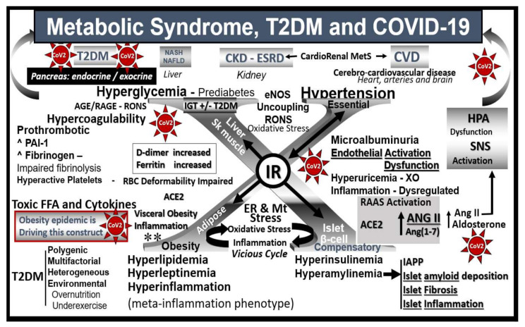
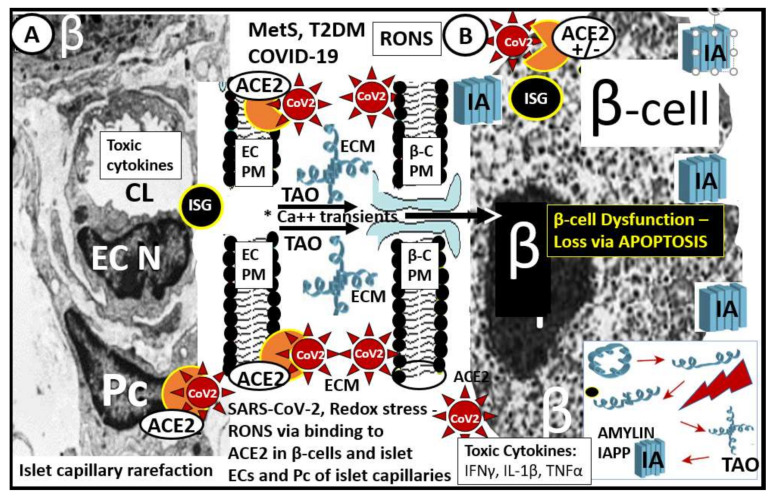
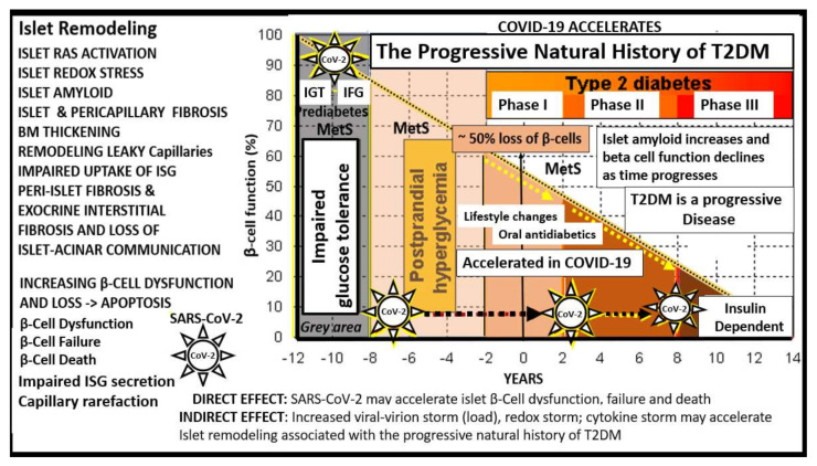
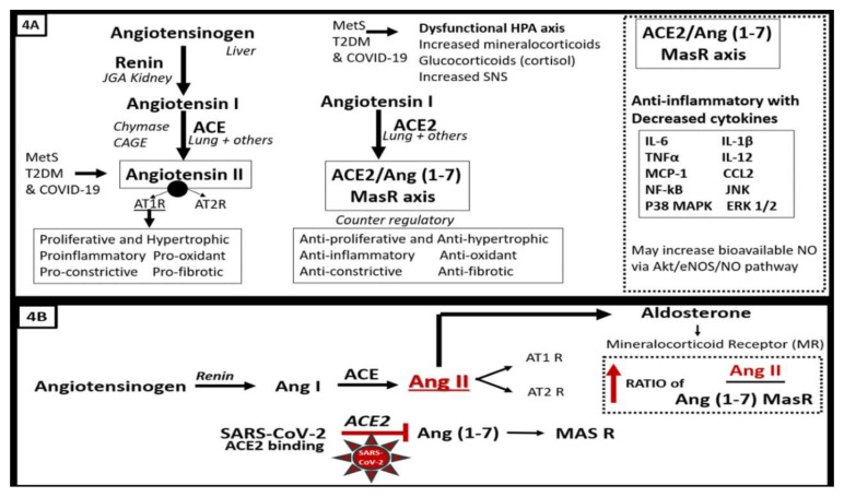
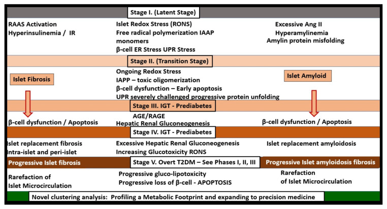
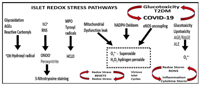
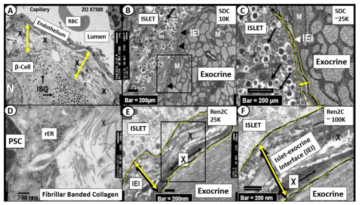
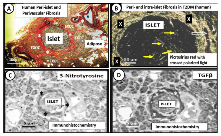
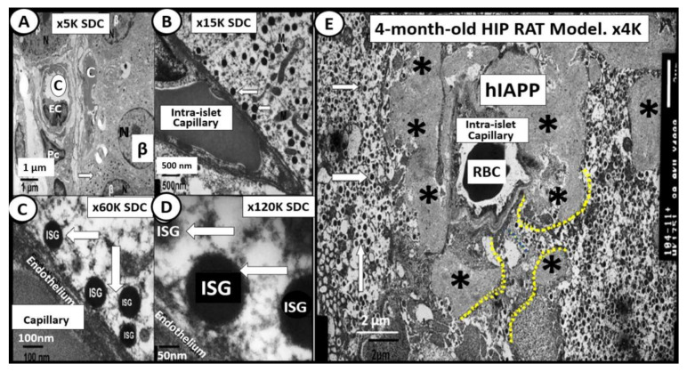
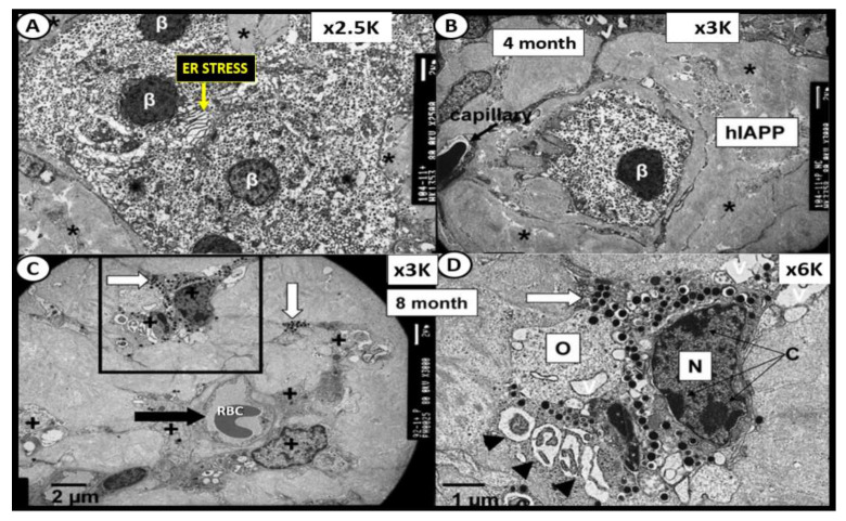
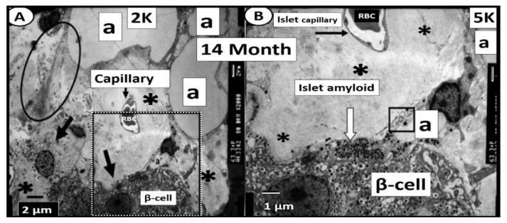
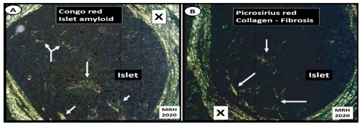
The novel coronavirus disease 2019 (COVID-19) caused by the severe acute respiratory syndrome coronavirus-2 (SARS-CoV-2) was declared a pandemic by the WHO on 19 March 2020. This pandemic is associated with markedly elevated blood glucose levels and a remarkable degree of insulin resistance, which suggests pancreatic islet β-cell dysfunction or apoptosis and insulin's inability to dispose of glucose into cellular tissues. Diabetes is known to be one of the top pre-existing co-morbidities associated with the severity of COVID-19 along with hypertension, cardiocerebrovascular disease, advanced age, male gender, and recently obesity. This review focuses on how COVID-19 may be responsible for the accelerated development of type 2 diabetes mellitus (T2DM) as one of its acute and suspected long-term complications. These observations implicate an active role of metabolic syndrome, systemic and tissue islet renin-angiotensin-aldosterone system, redox stress, inflammation, islet fibrosis, amyloid deposition along with β-cell dysfunction and apoptosis in those who develop T2DM. Utilizing light and electron microscopy in preclinical rodent models and human islets may help to better understand how COVID-19 accelerates islet and β-cell injury and remodeling to result in the long-term complications of T2DM.

Keywords: ACE2; SARS-CoV-2; amylin; fibrosis; islet; islet amyloid; metabolic syndrome; oxidative stress; renin–angiotensin–aldosterone-system; β-cell apoptosis.


Conflict of interest statement

The author declares that there is no conflict of interest.

Figures

Figure 1
Figure 1
Metabolic syndrome, T2DM and COVID-19 are multisystem diseases. This image illustrates how metabolic syndrome (MetS)/type 2 diabetes mellitus (T2DM) and coronavirus disease 2019 (COVID-19) are two multisystem diseases that can have a tremendous interaction, with multiple crosstalk when they intersect. The central “X” in this figure honors Jerry Reaven who initially coined the term Syndrome X and championed the concept that resistance to insulin-mediated glucose disposal was a characteristic of patients with T2DM and cardiovascular disease (CVD), which was later termed MetS. There are four arms to this letter X and each arm has a designated condition to further illustrate the “H” phenomenon, representing a “hyper” state, i.e., hyperlipidemia, lower left; islet β-cell hyperinsulinemia/hyperamylinemia, lower right; hypertension, upper right; hyperglycemia, upper left. Note how insulin resistance (IR) is central to each of the four arms. While each arm is important, one can note that hyperinsulinemia and hyperamylinemia are of great importance to this review, in that this arm represents the hormonal secretion by the pancreatic β-cells that have the ACE2 on their outer surface that is necessary for SARS-CoV-2 (red spiked icon with CoV-2 labeling) to enter the cells. Further, ACE2 is present on the intra-islet microcirculation capillary endothelial cells/pericytes and the peri-islet capillaries. In addition to intra-islet amyloid deposition and fibrosis, there is also peri-islet amyloid and fibrosis, redox stress oxidative/nitrosative stress (RONS) and inflammation that are in a vicious cycle with one another. MetS and T2DM are known to be associated with the renin–angiotensin–aldosterone system (RAAS) within the islet and there exists the possibility that further activation of islet RAAS may be due to the diminished ACE2/Ang(1–7)/MasR as a result of viral virion binding and contribute to ongoing remodeling over time following the recovery from COVID-19. Additionally, due the overriding effect of Ang II excess due to ACE2 binding, there will be increased vasospasm and hypoxia to the islets that may compound the COVID-19 islet injury. Endothelial activation/dysfunction due MetS, T2DM and COVID-19 may be responsible for further islet damage. Importantly, there is the known cytokine storm that could initially play a damaging role to the islet and its contents with loss of β-cells. Further, cerebrocardiovascular disease (CVD) and chronic kidney disease (CKD) together comprise the brain–heart–kidney axis that is involved when there is vascular stiffness associated with MetS and T2DM. It is very important to note that only body mass index (BMI)/obesity (not morbid obesity) turned out to be independently associated with the primary outcome of need for ventilation and/or death at 7 days post-admission in the French CORONADO study and thus implicates obesity as a major predicting phenotype associated with the need for supportive ventilation and or death as obesity is also noted to be driving MetS (lower left-hand side of figure **) see reference 21. ACE = angiotensin-converting enzyme; ACE2 = angiotensin-converting enzyme 2; AGE = advanced glycation end products; ANGII/Ang II = angiotensin II; Ang(1–7) = angiotensin 1–7; CKD = chronic kidney disease; CVD = cerebrocardiovascular disease; D-dimer = a fibrin degradation product and is named after two D fragments of the fibrin protein joined by a crosslink upon fibrinolysis; eNOS = endothelial nitric oxide synthase; ER = endoplasmic reticulum; ESRD = end-stage renal disease; FFA = free fatty acids; HPA = hypothalamic–pituitary–adrenal axis; IAPP = islet amyloid polypeptide; MasR = MAS-related G protein-coupled receptor; Mt = mitochondria; NAFLD = non-alcoholic fatty liver disease; NASH = non-alcoholic steatohepatitis; PAI-1 = plasminogen activator-1; RAGE = receptor for AGE; RBC = red blood cell; RONS = reactive oxygen and nitrogen (nitrosative stress) species; SNS = sympathetic nervous system.
Figure 2
Figure 2
Pancreatic islet β-cells undergo a multiple hit injury with MetS/T2DM and COVID-19. This illustration depicts the close relationship of the islet capillaries and the β-cells with transmission electronic microscopic (TEM) images of the pancreatic islet capillary Panel (A) and the β-cell Panel (B) in those infected by SARS-CoV-2. Panel (A) depicts an islet capillary in close proximity to an islet β-cell (preclinical HIP rodent model) and notes that CoV-2 may bind not only to the endothelial cell ACE2 protein of capillary endothelial cells but may also bind to pericytes, since ACE2 has recently been found to bind to pericytes in the brain and myocardial capillary specimens especially if the EC glycocalyx barrier function has been damaged allowing SARS-CoV-2 to enter the subendothelial space. Panel (B) illustrates the small electron dense dots that are the insulin secretory granules (ISG) within the β-cell cytoplasm (preclinical HIP rodent model). Additionally, amylin undergoes unfolding and misfolding to form islet amyloid polypeptide (IAPP) (panel (B)—lower right hand) when exposed to toxic oxidative stress—reactive oxygen and nitrogen (nitrosative stress) species (RONS). The early more intermediate-sized toxic amyloid oligomers (TAO) of amylin have a propensity to form membrane permeant channels in the islet β-cell plasma membrane, which allow for the entrance of calcium transients to enter the β-cell and result in not only β-cell dysfunction but also β-cell loss via apoptosis see reference 63 and 64. There is approximately a 50% decrease in β-cell function of those with T2DM and a 40–50% loss of β-cells in individuals with impaired glucose tolerance or prediabetes. Therefore, one can deduce that if there is already this much β-cell loss why SARS-CoV-2 could significantly add to this loss via SARS-CoV-2 binding to the ACE2 on β-cells (+/−) with further β-cell dysfunction, apoptosis and possibly accelerate the natural history of T2DM. Note the (+/−) regarding the presence of the ACE2 enzyme receptor since there is currently some controversy regarding its presence in pancreatic islet β-cells; see Section 6 reference [70,71,72,73]. Notably, systemic toxic cytokines liberated from pulmonary tissues and systemic immune cells can also be related to pancreatic islet injury mechanisms. Additionally, in preclinical T2DM rodent models, there is intra-islet capillary rarefaction that may contribute to β-cell dysfunction and death see reference 66. ACE2 = angiotensin-converting enzyme 2 (orange color); β = β-cell; β-C = β-cell; Ca++ = calcium; CoV-2 = SARS-CoV-2 (spiked red icon); CL = capillary lumen; EC = endothelial cell; ECM = extracellular matrix; IA = islet amyloid; IAPP = islet amyloid polypeptide deposition (blue fibril icon); IFN-γ = interferon gamma; Il-1β = interleukin 1 beta; ISG = insulin secretory granules; MetS = metabolic syndrome; N = nucleus; Pc = pericyte; PM = plasma membrane; RONS = reactive oxygen and nitrogen (nitrosative stress) species; TAO = toxic intermediate-sized amyloid oligomers; TNFα = tumor necrosis alpha; T2DM = type 2 diabetes mellitus.
Figure 3
Figure 3
T2DM is a progressive disease with multiple stages and phases in humans. At the time of diagnosis of T2DM, patients may already have a 50% reduction in β-cell function. As one moves through the various stages/phases, there is a progressive loss of β-cell function, while concurrently there is increased deposition of amylin-derived islet amyloid and islet fibrosis (islet remodeling—isletopathy). When SARS-CoV-2 (spiked icon) binds to the ACE2 receptor of β-cells and vascular EC/pericyte, cells there will be a detrimental increase in the ratio of the ANG II–ACE2–MasR axis. COVID-19 is known to be associated with a systemic and even local cytokine storm in addition to a redox storm (excessive oxidative stress—reactive oxygen and nitrogen (nitrosative stress) species (RONS)) and could be possibly based on the viral virion (viral load) storm associated with viremia. Therefore, COVID-19 could accelerate the natural history of T2DM. This image is based on a classic graph by Lebovitz HE. Diabetes Rev. 1999, 7, 139–153 and Hayden MR et al. JOP. J. Pancreas (online) 2002, 3(5), 126–138. BM = basement membrane; CoV-2 = abbreviation of SARS-CoV-2; EC = endothelial cell; IFG = impaired fasting glucose; IGT = impaired glucose tolerance; ISG = insulin secretory granule; MetS = metabolic syndrome; T2DM = type 2 diabetes mellitus.
Figure 4
Figure 4
(A,B). The protective role of the ACE2–Ang (1–7)–MasR axis is attenuated and/or Lost in COVID-19 due to SARS-CoV-2 binding of ACE2 in pancreatic islets. Since SARS-CoV-2 in COVID-19 infections will bind to the ACE2 enzyme in order to gain entry into the β-cells once viremia has occurred, the anti-inflammatory effects will be less available to help prevent the proinflammatory effects of ACE/Ang II and inflammation and fibrosis will prevail in the peri-islet and intra-islet regions along with the deposition of islet amyloid polypeptide (IAAP) and collagen types I and III (fibrosis). The binding of SARS-CoV-2 to ACE2 in the islet β-cells may be partially responsible for the elevated blood glucose during the acute/immediate phase of COVID-19 and also the increased insulin resistance that is known to occur in individuals with COVID-19. Further, in the convalescent period and into the late-complication phase of COVID-19, there may be an increased risk for the development of T2DM and progression from orally treated T2DM to an insulin-dependent type of T2DM. Therefore, the loss of the ACE2–Ang (1–7)–MasR axis is of critical importance in COVID-19 [19]. Dashed boxes indicate the loss of the ACE2–Ang (1–7)–MasR axis and their protective pathways. ACE = angiotensin-converting enzyme; AGE = advanced glycation end products; Ang II = angiotensin II; CAGE = chymostatin-sensitive angiotensin II-generating enzyme; ERK CCL2 = C-C motif chemokine 2; ERK 1/2 = extracellular signal-regulated kinase 1/2; HPA = hypothalamic–pituitary–adrenal axis; IL-1β = interleukin-1beta; IL-6 = interleukin-6; IL-12 = interleukin-12; JNK = c-Jun N-terminal kinase; MasR = MAS-related G protein-coupled receptor; MCP-1 = monocyte chemoattractant protein 1; NF-KB = nuclear factor-kappa B; p38 MAPK = p38 mitogen-activated protein kinase; RAGE = receptor for AGE; RONS = reactive oxygen and nitrogen (nitrosative stress) species; TNFα = tumor necrosis factor alpha.
Figure 5
Figure 5
T2DM is a progressive disease consisting of five stages in the natural history of T2DM. T2DM implicates metabolic syndrome, redox stress, islet amyloid, islet fibrosis and RAAS. T2DM is the end stage of a process that involves a definite loss of pancreatic β-cell function and β-cell apoptosis that has multiple stages (I–V) during its development of this end-stage isletopathy. This figure represents a putative model of these five stages and how they are associated with islet amyloid, islet fibrosis and involves not only a systemic cRAAS but also a local tRAAS with excess islet Ang II production. Most often, insulin resistance is associated early on in this process and relates to a central role or state of hyperinsulinemia and hyperamylinemia as noted in metabolic syndrome in Figure 1. Loss of β-cell function and/or loss via apoptosis eventually develops and blood glucose levels continue to rise. Stages I–V demonstrate that T2DM is a progressive disease as in Figure 3, which involves islet amyloid and islet fibrosis remodeling. Thus, the late complication of T2DM development in previous non-diabetic patients may depend on the stage of development of the individual when they develop COVID-19 in relation to the type of T2DM during the development of late complications since SARS-CoV-2 may either involve the islet and its β-cells directly or indirectly. Further, an individual that is controlled on oral medication and lifestyle modifications may indeed develop an insulin-dependent type of T2DM due to the acceleration of the underlying stage at the time of infection due to loss of β-cells and possibly due to novel hybrid forms of diabetes such as latent autoimmune diabetes in adults following COVID-19. T2DM is a heterogeneous, multifactorial spectrum disease. Therefore, not all individuals who develop T2DM will strictly follow this 5-stage roadmap in a lock-step manner. Importantly, recent findings utilizing clustering analysis for the development of T2DM and its complications are being utilized and may relate to the personalized treatment of various clusters in addition to stages I–V. See Section 8, T2DM May Be Considered a Spectrum Disease. The novel clustering analysis that is currently being utilized will add a great deal of knowledge to stages I–V [27,28,29]. ANG II/Ang II = angiotensin II; AGE = advanced glycation end products; ER = endoplasmic reticulum; IAPP = islet amyloid polypeptide; IGT = impaired glucose tolerance; IR = insulin resistance; RAAS = renin–angiotensin–aldosterone system; RAGE = receptor for AGE; RONS = reactive oxygen and nitrogen (nitrosative stress) species; UPR = unfolded protein response.
Figure 6
Figure 6
Immunohistochemistry of 3-nitrotyrosine staining in the Ren2 lean hypertensive preclinical rat model. This representative image demonstrates the strong diffuse antibody staining for 3-nitrotyrosine (a direct marker of nitrosative stress and an indirect marker of oxidative stress (RONS) in two islets (open arrows) in the 10-week-old transgenic Ren2 model (Ren2C) pancreas indicating intra-islet oxidative stress. Note that there is no staining in the adjacent exocrine portion of the pancreas. Insert a. demonstrates the diffuse intra-islet staining of 3-nitrotyrosine in the Ren2 model. Importantly, there was no staining for 3-nitrotyrosine in islets or exocrine portion of the age-matched Sprague–Dawley control model (not shown). Magnification: ×20 objective; scale bar = 5 µm. Courtesy JOP [22]. RONS = reactive oxygen and nitrogen (nitrosative stress) species.
Figure 7
Figure 7
Islet reactive oxygen and nitrogen (nitrosative stress) species: possible pathways and enzymes responsible for the production of primary and secondary RONS in MetS, T2DM and COVID-19. The possible metabolic pathways and enzymes involved in the production of islet reactive oxygen and nitrogen (nitrosative stress) species (RONS) are illustrated. Both primary and secondary RONS are present in MetS, T2DM and COVID-19; however, it is the secondary RONS that act to produce RONS damage to tissues specifically in the pancreatic islets and consist of primary species (primary RONS), which react with one another or a transition metals (such as iron in Fenton reactions), yielding highly reactive secondary species, such as ONOO or OH, and may react to produce the elevated ferritin levels in Figure 1. With the abundance of redox stress and accumulation of RONS, one could consider the use of the term “redox storm”. Redox stress begets redox stress and reactive oxygen and nitrogen (nitrosative stress) species beget inflammation via NF-κB, creating at least two vicious cycles within islets in addition to the vicious cycle between T2DM/glucotoxicity and COVID-19. AGE = advanced glycation end products; ALE = advanced lipoxidation end products; eNOS = endothelial nitric oxide synthase; H2O2 = hydrogen peroxide; HCLO = hypochlorous acid; MPO = myeloperoxidase; NADPH = nicotinamide adenine dinucleotide phosphate reduced; NF-κB = nuclear factor-kappa B; NO = nitric oxide; RAGE = receptor for AGE; RONS = reactive oxygen and nitrogen (nitrosative stress) species.
Figure 8
Figure 8
Intra-islet pericapillary and peri-islet (islet-exocrine interface) fibrosis. (Panel (A)) depicts the widening and early peri-capillary fibrosis in the Zucker obese male model of insulin resistance, obesity and impaired glucose tolerance at 12 weeks of age. (Panels (B,C)) demonstrate the close association of the islet-exocrine interface, with only slight widening at even high magnification between the exocrine and endocrine pancreas in the male control Sprague–Dawley control (SDC) model at 10 weeks of age. (Panels (E,F)) demonstrate a marked widening at higher magnification as compared to the SDC model in B and C in addition to the accumulation of collagen (X). Importantly, (Panel (D)) depicts the extrusion of collagen from a pancreatic stellate cell (PSC) in the Ren2 model at 10 weeks of age; scale bar = 200 nm. IEI = islet-exocrine interface; ISG = islet secretory granule; M and m = mitochondria; PSC = pancreatic stellate cell; RBC = red blood cell; rER = rough endoplasmic reticulum; X = collagen within the widened IEI.
Figure 9
Figure 9
Human intra-islet, peri-islet and perivascular fibrosis. (Panels (A,B)) depict peri- and intra-islet fibrosis and perivascular fibrosis in a female human (age 58) with T2DM of 6 years duration who was insulin dependent for one year at time of death from an acute myocardial infarction. (Panel (A)) depicts marked peri-islet, and perivascular fibrosis in the pancreas and thus is capable of interfering with communication between the islet endocrine and exocrine pancreas with Verhoeff Van Gieson (VVG) staining for fibrosis (crimson-red) (asterisks) and elastin (black). Note perivascular fibrosis surrounding an arteriole (A) and venule (V) (asterisks). (Panel (B)) demonstrates marked peri-islet fibrosis (X) and intra-islet fibrosis (yellow arrows). Note that the open arrows depict the islet–acinar–portal vascular pathway (venule) utilizing picrosirius red staining with crossed polarized light. (Panels (C,D)) represent the immunohistochemistry staining for 3-nitrotyrosine (reflecting oxidative nitrosative stress) and TGFβ (profibrotic cytokine-growth factor), respectively. Images A–D modified via courtesy of [48,52]. EXOC = exocrine pancreas.
Figure 10
Figure 10
Normal insulin secretory granule docking in control and impaired docking to the capillary endothelium in the HIP rat models. Panels (AD) illustrate the close association of β-cells to the islet capillaries, which allow for proper docking of the insulin secretory granule (ISG) (white open arrows) for absorption of insulin in the 4-month-old control Sprague–Dawley control (SDC). Magnifications and scale bars are in each image. Panel (E) depicts the diffuse islet amyloid deposition (asterisks) in the 4-month-old HIP rat model. This image allows one to note the impairment of ISG docking with the islet capillary. Note only the small spaces (outlined by yellow dashed lines) that are able to allow for minimal docking of ISGs. Note how the ISGs come to the peri-capillary islet amyloid and appear to be abruptly quarantined without access to the islet capillaries. These findings would fall into stages II and III of Figure 5 in Section 1. Magnification ×4000; scale bar = 2 µm. Asterisks = islet amyloid; C= capillary; hIAPP = human islet amyloid polypeptide; ISG = insulin secretory granule; K = 1000; M = mitochondria; RBC = red blood cell.
Figure 11
Figure 11
The 4- and 8-month-old HIP rat. (Panels (A,B)) from the 4-month-old HIP rat models. Panel (A) illustrates endoplasmic reticulum (ER) stress with prominent widening of the ER in β-cells (yellow arrow) and also note the islet amyloid (asterisk) in left lower region. Panel (B) depicts a single β-cell embedded and totally isolated by islet amyloid/human islet amyloid polypeptide (hIAPP) amyloidosis in 4-month-old HIP rat models. Panels C and D are from the 8-month-old HIP rat model. (Panel (C)) displays the marked decrease in β-cells, which are atrophic and represent changes of apoptosis. Note the islet capillary with its red blood cell (RBC) and its close association with an atrophic apoptotic β-cell with apoptotic bodies and loss of insulin secretory granules (black arrow). (Panel (D)) depicts an apoptotic β-cell from boxed in area in panel (C) and note the apoptotic bodies (arrowheads), loss of cytoplasmic organelles (O) and cytoplasmic vacuoles (apoptotic bodies) along with its nucleus (N) depicting chromatin condensation (C). β = β-cell; K = 1000; N = nucleus; O = cytoplasmic organelle; RBC = red blood cell.
Figure 12
Figure 12
β-cell loss and near-complete islet amyloid replacement in 14-month-old HIP rat islets. There were very few β-cells remaining in 14 month-old models and most islets demonstrated near-complete replacement with islet amyloid and fatty infiltration of islets was prominent with large lipid droplets (a) in 14-month-old models in (Panels (A,B)). Oval in panel (A) encloses an early change in intra-islet fibrosis. Remaining β-cells were atrophic and capillaries and pericytes were decreased. The remaining pericytes demonstrated multiple lipid droplets in their cytoplasm similar to lipid droplets within the islets (not shown). Panel (B) is a higher mag of the dashed boxed in region in panel (A) [65]. Magnification ×2000 and ×5000 in Panels (A,B), respectively; scale bar = 2 µm in panel (A) and 1 µm panel (B). a = adipose lipid droplets; open and closed arrows = β-cell; K = 1000.
Figure 13
Figure 13
Islet amyloid and islet fibrosis in human islets co-occur. (Panel (A)) depicts Congo red staining (stains amyloid) viewed with crossed polarized light and birefringent appearance at the peri-islet (X) and intra-islet regions (arrows). (Panel (B)) illustrates islet fibrosis (collagen I and III) staining with picrosirius red and viewed with crossed polarized light of pancreatic islet at the peri-islet (X) and intra-islet regions (arrows). These images demonstrate that islet amyloid and islet fibrosis co-occur within human pancreatic islets when stained specifically for amyloid (Congo red) and fibrosis (picrosirius red) in a female patient who died of an acute myocardial infarction [54]. Specific staining procedures by pathologists often reveal what standard hematoxylin and eosin (H&E) staining do not in regard to conditions such as islet amyloid and fibrosis. Magnification ×100 objective with oil immersion in Panels (A,B).
Figure 14
Figure 14
Pericyte apoptosis and loss within islets of 14-month-old HIP rat models. (Panels (A,B)) depict sparse islet capillaries embedded within a sea of islet amyloid. Note pericyte(s) (Pc) apoptosis with apoptotic bodies (arrows). Endothelial apoptosis was not observed in these intra-islet regions in the HIP rat models in contrast to Pcs. (Panels (C,D)) are confocal immunohistochemistry stains of the islets with antibodies to the platelet-derived growth factor receptor beta (PDGFR-β) (red) and DAPI (blue) staining of islet cells. Note the progressive loss of intra-islet staining of pericytes (PDGFR-β red stain) and also note the progressive loss of cellularity (DAPI blue staining) within the islets from two months to 14 months in the HIP rat models. This suggests that, in addition to the loss of intra-islet cellularity, there is a loss of capillary pericytes. Thus, pericyte loss/apoptosis appears to play a significant role in the development of T2DM, at least in the HIP rat model of T2DM, similar to that found in the retinas of T2DM diabetic preclinical models and humans. Note the inserts in the upper right of the Sprague–Dawley Control (SDC) models in (Panels (CE)). The white dashed circle outlines the pancreatic islet margins in each of the images (CE). Various magnifications are used, with scale bars readily identified in each image. Scale bar = 2 µm in panel (A) and 1 µm panel (B).
Figure 15
Figure 15
T2DM may be considered a spectrum disease. The development of overt or clinical T2DM could be considered a spectrum disease, in that, end-organ tissue remodeling in response to multiple metabolic toxicities begin long before the clinical diagnosis of T2DM is made; see Figure 1, Figure 3 and Figure 5. Note that there are multiple variables (black boxed-in numbers 1–3) that contribute to the development of T2DM and its associated multiple end-organ complications (including the pancreatic islet—isletopathy) even prior to the clinical diagnosis of overt T2DM (black boxed-in number 4). Importantly, there are multiple variables within metabolic syndrome (MetS) (black boxed-in number 1), which consist of having three of the following five ATP guideline variables consisting of: central obesity (≥94 cm (for males), 80 cm (females)); triglycerides concentration ≥ 150 mg/dL; HDL cholesterol concentration < 40 mg/dL (males), <50 mg/dL (females); values of blood pressure ≥130/85 mmHg and fasting glycemia ≥ 100 mg/dL or HbA1c ≥ 6.5 that contribute to its summation equation [8,9]. Lipotoxicity was not assigned a boxed-in number because HDL cholesterol and triglycerides variables are included in MetS (black boxed-in number 1) as one of the five criteria for diagnosing MetS. Hypertension may be one of the most common health problems associated with obesity and is included in MetS (black boxed-in number1) variables and is not within the scope of this paper to discuss in greater depth. The importance of insulin and leptin resistance (black boxed-in number 3) is central and may be critical to islet and even brain remodeling and therefore is not to be underestimated. Note the novel simple mathematical summation equations that are introduced with black boxed-in variables 1–4 and that the author has assigned various weighted numbers (based on their relative importance) in parentheses for summation equation totals, which points to the importance of each variable and their summation. The author is aware that that the weighted values of each of the variables utilized in these summation equations are debatable. ATP III = adult treatment panel III (ATP III) report of the National Cholesterol Education Program; HbA1c = hemoglobin A1C; HDL = high-density lipoprotein; MetS = metabolic syndrome; T2DM = type 2 diabetes mellitus.
Figure 16
Figure 16
Pancreatic islet β-cell apoptosis in the HIP rat model of T2DM. This representative pseudo-colorized pancreatic islet β-cell in the preclinical 8-month-old HIP rat diabetic model (Figure 11D) depicts apoptosis characterized by β-cell atrophy, nuclear chromatin condensation (Cc with white arrows), loss of cytoplasmic organelles (CO), cytoplasmic vacuole formation and apoptotic bodies (arrowheads). Note how islet amyloid may insert into β-cells (arrows and asterisks). β-cell apoptosis in MetS, T2DM and COVID-19 may be due to a combination of a virus virion storm (SARS-CoV-2), redox storm and cytokine storm as they converge and interact with the multiple metabolic toxicities of MetS and prediabetes or overt T2DM, which may result in injury and a response to the injury wound healing mechanism within the islet and its microcirculation and encourage the acceleration of T2DM due to early dysfunction and later loss of β-cells via apoptosis. Nucleus (blue); cytoplasm (red); apoptotic bodies (yellow); extracellular matrix—hashtag islet amyloid (concrete grey color). CO = cytoplasmic organelles; hashtag = islet amyloid; hIAAP = human islet amyloid polypeptide; ISG = electron dense insulin secretory granules (open arrow and encircled by yellow lines of both immature haloed and more mature unhaloed ISG).

Similar articles

Cited by

References

    1. Cariou B., Hadjadj S., Wargny M., Pichelin M., Al-Salameh A., Allix I., Amadou C., Arnault G., Baudoux F., Bauduceau B., et al. Phenotypic characteristics and prognosis of inpatients with COVID-19 and diabetes: The CORONADO study. Diabetologia. 2020;63:1500–1515. doi: 10.1007/s00125-020-05180-x. - DOI - PMC - PubMed
    1. Bansal R., Gubbi S., Muniyappa R. Metabolic Syndrome and COVID 19: Endocrine-Immune-Vascular Interactions Shapes Clinical Course. Endocrinology. 2020;161:bqaa112. doi: 10.1210/endocr/bqaa112. - DOI - PMC - PubMed
    1. Kruglikov I.L., Scherer P.E. The Role of Adipocytes and Adipocyte-Like Cells in the Severity of COVID-19 Infections. Obesity. 2020;28:1187–1190. doi: 10.1002/oby.22856. - DOI - PMC - PubMed
    1. Zhang Y., Somers K.R., Becari C., Polonis K., Pfeifer M.A., Allen A.M., Kellogg T.A., Covassin N., Singh P. Comparative Expression of Renin-Angiotensin Pathway Proteins in Visceral Versus Subcutaneous Fat. Front. Physiol. 2018;9:1370. doi: 10.3389/fphys.2018.01370. - DOI - PMC - PubMed
    1. Rubino F., Amiel S.A., Zimmet P., Alberti G., Bornstein S., Eckel R.H., Mingrone G., Boehm B., Cooper M.E., Chai Z., et al. New-Onset Diabetes in Covid-19. N. Engl. J. Med. 2020;383:789–790. doi: 10.1056/NEJMc2018688. - DOI - PMC - PubMed

MeSH terms