Review
. 2020 Nov 13;10(11):2253.
doi: 10.3390/nano10112253.

Nanomaterials for Treating Bacterial Biofilms on Implantable Medical Devices

Affiliations
Review

Nanomaterials for Treating Bacterial Biofilms on Implantable Medical Devices

Hoai My Tran et al. Nanomaterials (Basel). .

Abstract

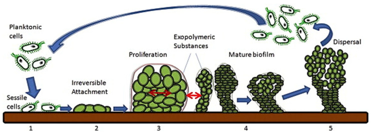
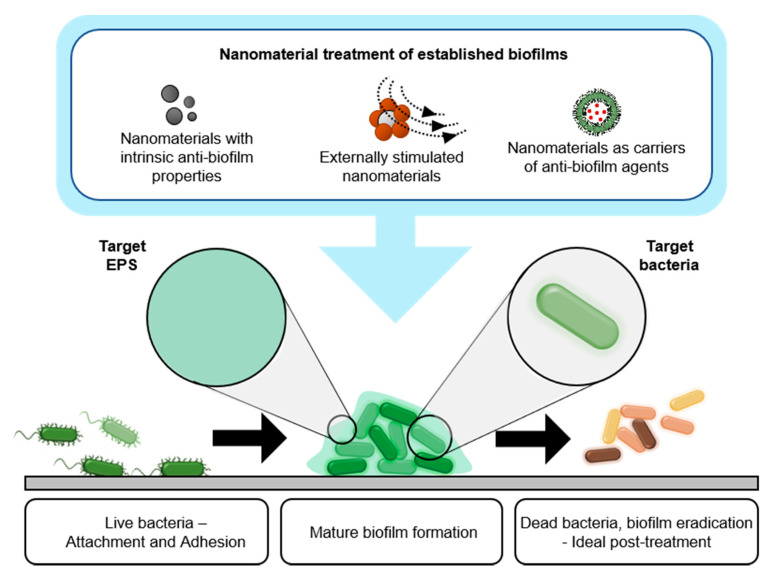
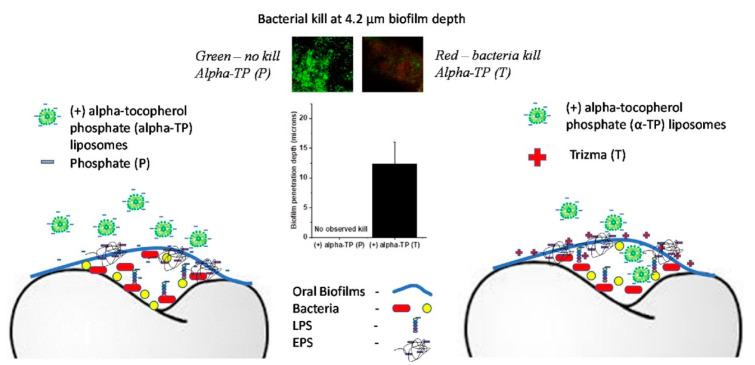
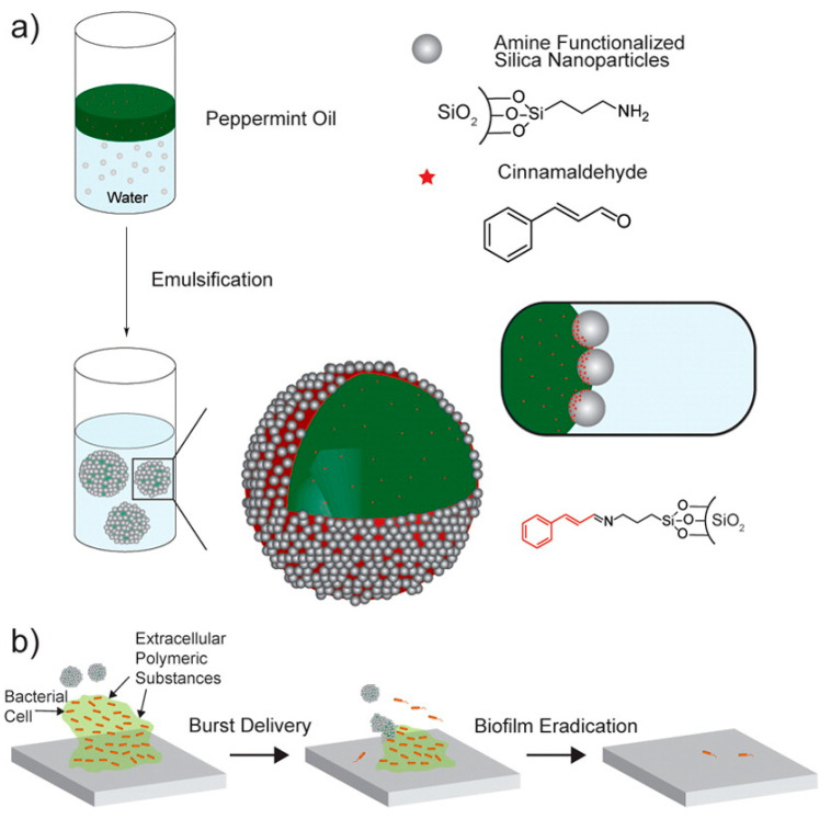
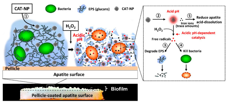
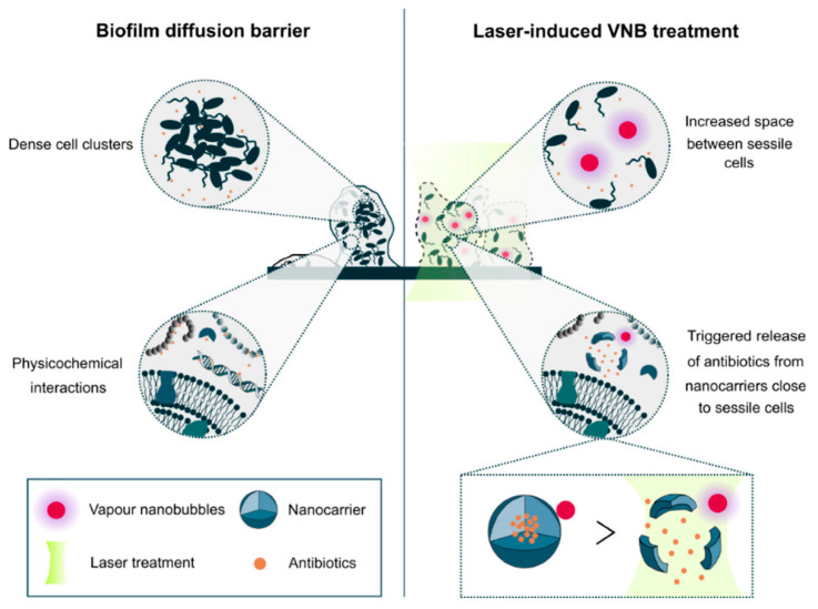
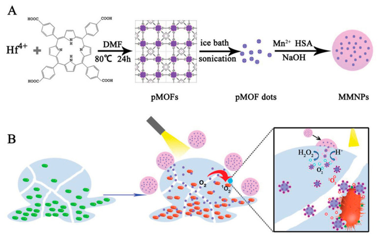
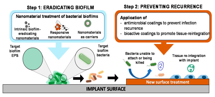
Bacterial biofilms are involved in most device-associated infections and remain a challenge for modern medicine. One major approach to addressing this problem is to prevent the formation of biofilms using novel antimicrobial materials, device surface modification or local drug delivery; however, successful preventive measures are still extremely limited. The other approach is concerned with treating biofilms that have already formed on the devices; this approach is the focus of our manuscript. Treating biofilms associated with medical devices has unique challenges due to the biofilm's extracellular polymer substance (EPS) and the biofilm bacteria's resistance to most conventional antimicrobial agents. The treatment is further complicated by the fact that the treatment must be suitable for applying on devices surrounded by host tissue in many cases. Nanomaterials have been extensively investigated for preventing biofilm formation on medical devices, yet their applications in treating bacterial biofilm remains to be further investigated due to the fact that treating the biofilm bacteria and destroying the EPS are much more challenging than preventing adhesion of planktonic bacteria or inhibiting their surface colonization. In this highly focused review, we examined only studies that demonstrated successful EPS destruction and biofilm bacteria killing and provided in-depth description of the nanomaterials and the biofilm eradication efficacy, followed by discussion of key issues in this topic and suggestion for future development.

Keywords: bacteria; biofilm; in situ; infection; nanomaterials; treatment.


Conflict of interest statement

The authors declare no conflict of interest.

Figures

Figure 1
Figure 1
Biofilm formation and dispersion. (1) Bacterial cell attachment onto the surface. (2) Bacterial cells becoming irreversibly attached. (3) Bacterial proliferation and EPS secretion. (4) Biofilm formation and maturation. (5) Biofilm dispersal and mobility of planktonic cells (reproduced from [12] with permission from Elsevier, 2015).
Figure 2
Figure 2
Schematic indicating the growth of mature bacterial biofilm and three major types of nanomaterials for biofilm treatment: (i) intrinsically biofilm-eradicating nanomaterials, (ii) nanocarriers of biofilm-eradicating agents, and (iii) responsive biofilm-eradicating nanomaterials.
Figure 3
Figure 3
Schematic of biofilm elimination by combination of charge screening and alpha-tocopherol phosphate (α-TP) charged liposome nanoparticles. The penetration of negatively charged α-TP liposomes were enhanced in the Tris buffer (positively charged) causing bacteria death as compared to poor penetration and ineffective in phosphate buffer saline (PBS) (reproduced from [47] with permission from Elsevier, 2015).
Figure 4
Figure 4
(a) Schematic describing the preparation of nanocarriers loaded with antimicrobial agents by an emulsification method, and (b) illustration of functionalized silica nanocarriers for bacterial biofilm treatment (reproduced from [57] with permission from American Chemical Society, 2015).
Figure 5
Figure 5
Schematic of biofilm disruption using hydrogen peroxide and iron oxide nanoparticles (ION). (1) The CAT-NP distributed in 3D biofilm structure after a brief topical exposure. (2) The CAT-NP catalyzed H2O2 via the Fenton reaction under acidic condition to produce free radicals. The generated free radicals (3) degraded EPS and (4) killed bacteria inside the biofilm. (5) CAT-NP were also able to release iron ions that can reduce acid dissolution of hydroxyapatite (reproduced from [70] with permission from Elsevier, 2016).
Figure 6
Figure 6
Laser-induced vapor nanobubbles (VNB) improved antibiotic delivery to biofilms. Impaired biofilm diffusion is caused by the fact that sessile cells cluster together into dense aggregates of hundreds of micrometers in size and because of the multi-component nature of the biofilm matrix which can trap molecules in their passage through biofilms. The mechanical impact of laser-induced VNB could increase the space between sessile cells, leading to a better flux and the effectivity of antimicrobial agents and their mechanical force can trigger antibiotic release from nanocarriers close to sessile bacteria (reproduced from [72] with permission from MDPI, 2019).
Figure 7
Figure 7
(A) Schematic illustration of nanomaterial fabrication for a multi-component nanoplatform (MMNPs) containing porphyrin-containing metal–organic framework (pMOF) dots. (B) Schematic of the mechanism of bacterial biofilm treatment by the nanoplatform (MMNPs) and external activation by NIR light. The MMNPs decomposed under an acidic environment, causing MnO2 degradation and pMOF dot release. The pMOF dots were then activated by NIR light to produce the reactive oxygen species. Additionally, in the presence of H2O2, MnO2 catalyzed H2O2 to form O2, enhancing the photodynamic therapy (reproduced from [75] with permission from John Wiley and Sons, 2019).
Figure 8
Figure 8
‘Eradicating-and-preventing’ approach to medical device-associated biofilms. After the biofilm has been eradicated (step 1), the surface of the implant should be further treated (step 2, for example by coating with antimicrobial agents or bioactive coating or with bioactive agents to enable rapid tissue-implant reintegration) to prevent infection recurrence. This proposed approach is essentially a combination of treating biofilms and preventing biofilm formation.

References

    1. Tran N., Tran P.A. Nanomaterial-Based Treatments for Medical Device-Associated Infections. ChemPhysChem. 2012;13:2481–2494. doi: 10.1002/cphc.201200091. - DOI - PubMed
    1. Hetrick E.M., Schoenfisch M.H. Reducing implant-related infections: Active release strategies. Chem. Soc. Rev. 2006;35:780–789. doi: 10.1039/b515219b. - DOI - PubMed
    1. Gnanadhas D.P., Elango M., Janardhanraj S., Srinandan C.S., Datey A., Strugnell R.A., Gopalan J., Chakravortty D. Successful treatment of biofilm infections using shock waves combined with antibiotic therapy. Sci. Rep. 2015;5:17440. doi: 10.1038/srep17440. - DOI - PMC - PubMed
    1. Donlan R.M. Biofilm Formation: A Clinically Relevant Microbiological Process. Clin. Infect. Dis. 2001;33:1387–1392. doi: 10.1086/322972. - DOI - PubMed
    1. Ikuma K., Decho A.W., Lau B.L.T. When nanoparticles meet biofilms—Interactions guiding the environmental fate and accumulation of nanoparticles. Front. Microbiol. 2015;6:591. doi: 10.3389/fmicb.2015.00591. - DOI - PMC - PubMed

LinkOut - more resources