Effect of Nardostachys jatamansi DC. on Apoptosis, Inflammation and Oxidative Stress Induced by Doxorubicin in Wistar Rats
- PMID: 33203171
- PMCID: PMC7734586
- DOI: 10.3390/plants9111579
Effect of Nardostachys jatamansi DC. on Apoptosis, Inflammation and Oxidative Stress Induced by Doxorubicin in Wistar Rats
Abstract
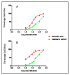
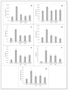
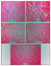
The study aimed to investigate the protective action of jatamansi (Nardostachys jatamansi DC.) against doxorubicin cardiotoxicity. Methanolic extract of jatamansi (MEJ) was prepared and standardized using HPTLC fingerprinting, GC-MS chemoprofiling, total phenolic content, and antioxidant activity in vitro. Further in vivo activity was evaluated using rodent model. Animals were divided into five groups (n = 6) namely control (CNT) (Normal saline), toxicant (TOX, without any treatment), MEJ at low dose (JAT1), MEJ at high dose (JAT2), and standard desferrioxamine (STD). All groups except control received doxorubicin 2.5 mg per Kg intra-peritoneally for 3 weeks in twice a week regimen. After 3 weeks, the blood samples and cardiac tissues were collected from all groups for biochemical and histopathological evaluation. Treatment with MEJ at both dose levels exhibited significant reduction (p < 0.001 vs. toxicant) of serum CK-MB (heart creatine kinase), LDH (Lactate dehydrogenase) & HMG-CoA (3-hydroxy-3-methylglutaryl-coenzyme A) levels, and tissue MDA (melondialdehyde) level; insignificant difference was observed (p > 0.05) in TNF-alpha (tumour necrosis factor), IL-6 (interleukine-6) levels and caspase activity as compared to TOX. Histopathological evaluation of cardiac tissues of different treatment groups further reinforced the findings of biochemical estimation. This study concludes that jatamansi can protect cardiac tissues from oxidative stress-induced cell injury and lipid peroxidation as well as against inflammatory and apoptotic effects on cardiac tissues.
Keywords: Nardostachys jatamansi DC.; biochemical estimation; cardioprotection; cardiotoxicity; doxorubicin; lipid peroxidation.
Conflict of interest statement
The authors have declared that there are no conflicts of interest.
Figures


References
-
- Lee K., Sung R.Y., Huang W.Z., Yang M., Pong N.H., Lee S.M., Chan W.Y., Zhao H., To M.Y., Fok T.F., et al. Thrombopoietin protects against in vitro and in vivo cardiotoxicity induced by DOX. Circulation. 2006;113:2211–2220. - PubMed
-
- Neilan T.G., Blake S.L., Ichinose F., Raher M.J., Buys E.S., Jassal D.S., Furutani E., Perez-Sanz T.M., Graveline A., Janssens S.P., et al. Disruption of nitric oxide synthase 3 protects against the cardiac injury, dysfunction, and mortality induced by DOX. Circulation. 2007;116:506–514. doi: 10.1161/CIRCULATIONAHA.106.652339. - DOI - PubMed
-
- Arola O.J., Saraste A., Pulkki K., Kallajoki M., Parvinen M., Voipio-Pulkki L.M. Acute DOX cardiotoxicity involves cardiomyocyte apoptosis. Cancer Res. 2000;60:1789–1792. - PubMed
Grants and funding
LinkOut - more resources
Full Text Sources
Research Materials
Miscellaneous

