. 2020 Nov 17;17(1):343.
doi: 10.1186/s12974-020-02018-6.

Sodium para-aminosalicylic acid inhibits manganese-induced NLRP3 inflammasome-dependent pyroptosis by inhibiting NF-κB pathway activation and oxidative stress

Affiliations

Sodium para-aminosalicylic acid inhibits manganese-induced NLRP3 inflammasome-dependent pyroptosis by inhibiting NF-κB pathway activation and oxidative stress

Dongjie Peng et al. J Neuroinflammation. .

Abstract

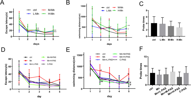
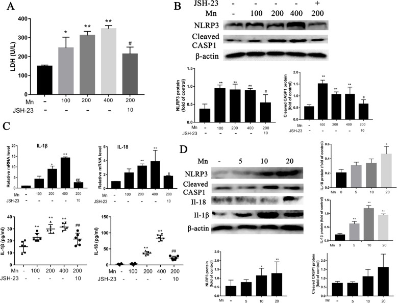
Background: The activation of NOD-like receptor protein 3 (NLRP3) inflammasome-dependent pyroptosis has been shown to play a vital role in the pathology of manganese (Mn)-induced neurotoxicity. Sodium para-aminosalicylic acid (PAS-Na) has a positive effect on the treatment of manganism. However, the mechanism is still unclear. We hypothesized that PAS-Na might act through NLRP3.

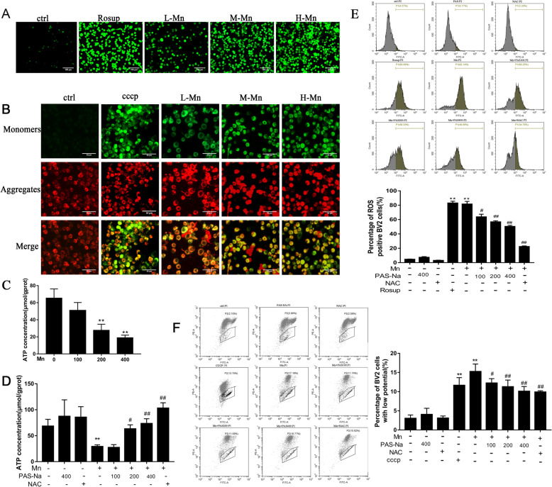
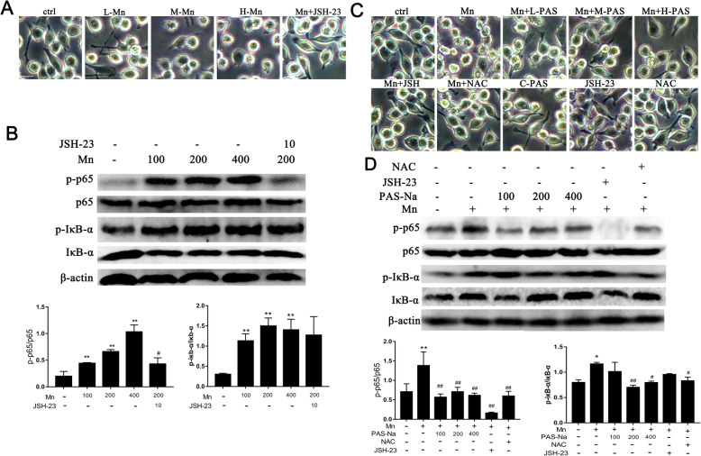
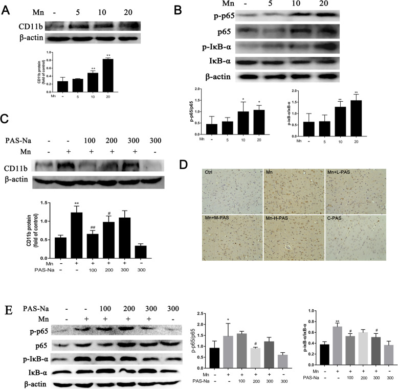
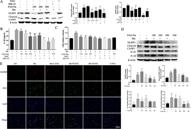
Methods: The microglial cell line BV2 and male Sprague-Dawley rats were used to investigate the impacts of PAS-Na on Mn-induced NLRP3 inflammasome-dependent pyroptosis. The related protein of the NF-κB pathway and NLRP3-inflammasome-dependent pyroptosis was detected by western blot. The reactive oxygen species and mitochondrial membrane potential were detected by immunofluorescence staining and flow cytometry. The activation of microglia and the gasdermin D (GSDMD) were detected by immunofluorescence staining.

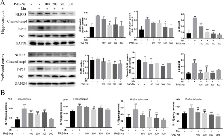
Results: Our results showed that Mn treatment induced oxidative stress and activated the NF-κB pathway by increasing the phosphorylation of p65 and IkB-α in BV2 cells and in the basal ganglia of rats. PAS-Na could alleviate Mn-induced oxidative stress damage by inhibiting ROS generation, increasing mitochondrial membrane potential and ATP levels, thereby reducing the phosphorylation of p65 and IkB-α. Besides, Mn treatment could activate the NLRP3 pathway and promote the secretion of IL-18 and IL-1β, mediating pyroptosis in BV2 cells and in the basal ganglia and hippocampus of rats. But an inhibitor of NF-κb (JSH-23) treatment could significantly reduce LDH release, the expression of NLRP3 and Cleaved CASP1 protein and IL-1β and IL-18 mRNA level in BV2 cells. Interestingly, the effect of PAS-Na treatment in Mn-treated BV2 cells is similar to those of JSH-23. Besides, immunofluorescence results showed that PAS-Na reduced the increase number of activated microglia, which stained positively for GSDMD.

Conclusion: PAS-Na antagonized Mn-induced NLRP3 inflammasome dependent pyroptosis through inhibiting NF-κB pathway activation and oxidative stress.

Keywords: Mn; NF-κB pathway; NLRP3 inflammasome; Oxidative stress; PAS-Na; pyroptosis.


Conflict of interest statement

There is no conflict of interest with any of the authors. This manuscript has not been published before and is not currently submitted for review to any other journal.

Figures

Fig. 1
Fig. 1
PAS-Na enhanced the learning ability of Mn-exposed rats. Experimental period of 8 weeks: the rats were treated with 5, 10, and 20 mg/kg MnCl2 for 8 weeks (ac). Experimental period of 14 weeks: the rats were treated with 20 mg/kg MnCl2 for 8 weeks and then treated with 100, 200, and 300 mg/kg PAS-Na for an additional 6 weeks (df). Escape latency (a, d), swimming distance (b, e), and number of platform crosses (Probe times) (c, f) of rats were tested by Morris water maze. Data are presented as mean ± SD (n = 10 per group). *p < 0.05: significant as compared to the control group; #p < 0.05 and ##p < 0.01: significant as compared to Mn-treated group
Fig. 2
Fig. 2
PAS-Na inhibited Mn-induced oxidative stress in BV2 cells. ac BV2 cells were treated with 100, 200, and 400 μmol/L MnCl2 for 24 h, respectively. Cells in positive control group were treated with rosup or CCCP (a, e) (a compound mixture that was provided by the manufacturer) for 1 h after normal medium cultured for 23 h. a ROS in BV2 cells was detected by DCFH-DA kit. After DCFH-DA treatment, ROS shows green fluorescence. Scale bars: 100 μm. b Mt∆Ψm of BV2 cells was determined by the lipophilic cationic probe JC-1. When the mt∆Ψm is high, JC-1 gathers in the mitochondrial matrix to form J-aggregates and produces red fluorescence. On the contrary, JC-1 is presented as a monomer and produces green fluorescence. Scale bars: 50 μm. c Mn decreased intracellular ATP concentration in BV2 cells. df BV2 cells were treated with 200 μmol/L MnCl2 for 24 h, following treatment with 100, 200, and 400 μmol/L PAS-Na and 50 umol/L NAC for 24 h, respectively. d PAS-Na recovered the ATP levels in Mn-treated BV2 cells. e Flow cytometry analysis of intracellular ROS in BV2 cells. f Flow cytometry analysis of intracellular the mt∆Ψm change of BV2 cells. All tests were repeated independently three times. Data are presented as mean ± SD. *p < 0.05 and **p < 0.01: significant as compared to the control group; #p < 0.05 and ##p < 0.01: significant as compared to Mn-treated group
Fig. 3
Fig. 3
PAS-Na inhibited Mn-induced NF-κb pathway activation by suppressing oxidative stress in BV2 cells. a, b BV2 cells were treated with 100, 200, and 400 μmol/L MnCl2 or 200 μmol/L MnCl2 + 10 μmol/L JSH-23 (L-, M-, H-Mn, Mn + JSH-23) for 24 h, respectively. a Morphology of BV2 cell. Scale bars: 20μm. b The protein expressions of p-p65, p65, p-IκB-α, and IκB-α in BV2 cells were detected by western blot. c, d BV2 cells were treated with 200 μmol/L MnCl2 or 200 μmol/L MnCl2 + 10 μmol/L JSH-23 for 24 h, following treatment with 100, 200, and 400 μmol/L PAS-Na and 50 μmol/L NAC for 24 h, respectively. c Morphology of BV2 cell. Scale bars: 20μm. (D) The protein expressions of p-p65, p65, p-IκB-α, and IκB-α in BV2 cells were detected by western blot. The protein expression was normalized by β-actin or corresponding total protein content. All tests were repeated independently three times. Data are presented as mean ± SD.*p < 0.05 and **p < 0.01: significant as compared to the control group; #p < 0.05 and ##p < 0.01: significant as compared to corresponding Mn-treated group
Fig. 4
Fig. 4
PAS-Na mitigates Mn-induced BV2 cells activation and microglia proliferation in basal ganglia of rats by inhibited NF-κB pathway activation. a, b The rats were treated with 5, 10, and 20 mg/kg MnCl2 for 8 weeks. a The protein expressions of CD11b in the basal ganglia of rats was analyzed by western blot (n = 4 per group). b The protein expressions of p-p65, p65, p-IκB-α, and IκB-α in the basal ganglia of rats was detected by western blot (n = 4 per group). cd The rats were treated with 20 mg/kg MnCl2 for 8 weeks and then treated with 100, 200, and 300 mg/kg PAS-Na for an additional 6 weeks. c The protein expression of CD11b in the basal ganglia of rats were analyzed by western blot (n = 4 per group). d Immunohistochemical results of CD11b in the basal ganglia of rats (n = 3 per group). Scale bars: 100 μm. e The protein expressions of p-p65, p65, p-IκB-α, and IκB-α in the basal ganglia of rats were detected by western blot (n = 4 per group). The protein expression was normalized by β-actin or corresponding total protein content. Data are presented as mean ± SD.*p < 0.05 and **p < 0.01: significant as compared to the control group; #p < 0.05 and ##p < 0.01: significant as compared to corresponding Mn-treated group
Fig. 5
Fig. 5
Mn activated the NLRP3-CASP1 inflammasome-dependent pyroptosis both in BV2 cell and in the basal ganglia of rats. ac BV2 cells were treated with 100, 200, and 400 μmol/L MnCl2 or 200 μmol/L MnCl2 + 10 μmol/L JSH-23 for 24 h. a Relative release of LDH in BV2 cells culture medium. b The protein expressions of NLRP3, cleaved-caspase1 in BV2 cells were detected by western blot. c The IL-1β and IL-18 mRNA expression in BV2 cells were detected by qPCR. The IL-1β and IL-18 level in the culture medium were detected by ELISA. d The rats were treated with 5, 10, and 20 mg/kg MnCl2 for 8 weeks. The protein expressions of NLRP3, cleaved-caspase1, IL-1β, and IL-18 in the basal ganglia of rats were detected by western blot (n = 4 per group). The protein expression was normalized by β-actin. All tests were repeated independently three times. Data are presented as mean ± SD.*p < 0.05 and **p < 0.01: significant as compared to the control group; #p < 0.05 and ##p < 0.01: significant as compared to corresponding Mn-treated group
Fig. 6
Fig. 6
PAS-Na inhibited Mn-induced NLRP3 inflammasome-dependent pyroptosis by inhibited NF-κB pathway activation and anti-oxidative stress. ac BV2 cells were treated with 200 μmol/L MnCl2 or 200 μmol/L MnCl2 + 10 μmol/L JSH-23 for 24 h, following treatment with 100, 200, and 400 μmol/L PAS-Na and 50 μmol/L NAC for 24 h, respectively. a The protein expressions of NLRP3, cleaved-caspase1 in BV2 cells were detected by western blot. b Levels of IL1β in the BV2 cell culture supernatants were measured by ELISA. c Relative release of LDH in BV2 cells culture medium. d, e the rats were treated with 20 mg/kg MnCl2 for 8 weeks and then treated with 100, 200, and 300 mg/kg PAS-Na for an additional 6 weeks. d The protein expressions of NLRP3, cleaved-caspase1, IL-1β, and IL-18 in the basal ganglia of rats were detected by western blot (n = 4 per group). e The basal ganglia sections of rats were stained for IBA1 as the microglia marker (green staining), GSDMD (red staining). Nuclei were stained with DAPI. Bar: 100 μm. The protein expression was normalized by β-actin. All tests were repeated independently three times. Data are presented as mean ± SD.*p < 0.05 and **p < 0.01: significant as compared to the control group; #p < 0.05 and ##p < 0.01: significant as compared to corresponding Mn-treated group
Fig. 7
Fig. 7
PAS-Na inhibited Mn-induced NLRP3 inflammasome-dependent pyroptosis. a The protein expressions of NLRP3, cleaved-caspase1, P-P65, and P65 in the prefrontal cortex and hippocampus of rats were detected by western blot. b The IL-1β and IL-18 level in the prefrontal cortex and hippocampus of rats were measured by ELISA. The protein expression was normalized by GAPDH. All tests were repeated independently three times. Data are presented as mean ± SD. **p < 0.01 and ***p < 0.001: significant as compared to the control group; #p < 0.05, ##p < 0.01, and ###p < 0.001: significant as compared to corresponding Mn-treated group

Similar articles

Cited by

References

    1. Du S, Wu X, Han T, Duan W, Liu L, Qi J, Niu Y, Na L, Sun C. Dietary manganese and type 2 diabetes mellitus: two prospective cohort studies in China. Diabetologia. 2018;61:1985–1995. doi: 10.1007/s00125-018-4674-3. - DOI - PubMed
    1. Lu L, Zhang LL, Li GJ, Guo W, Liang W, Zheng W. Alteration of serum concentrations of manganese, iron, ferritin, and transferrin receptor following exposure to welding fumes among career welders. Neurotoxicology. 2005;26:257–265. doi: 10.1016/j.neuro.2004.09.001. - DOI - PMC - PubMed
    1. Jiang YM, Mo XA, Du FQ, Fu X, Zhu XY, Gao HY, Xie JL, Liao FL, Pira E, Zheng W. Effective treatment of manganese-induced occupational Parkinsonism with p-aminosalicylic acid: a case of 17-year follow-up study. J Occup Environ Med. 2006;48:644–649. doi: 10.1097/01.jom.0000204114.01893.3e. - DOI - PMC - PubMed
    1. Proudfoot O. Manganese in manganism, Parkinson's disease, Huntington's disease, amyotrophic lateral sclerosis, and Batten disease: A narrative review. Neurol India. 2017;65:1241–1247. doi: 10.4103/0028-3886.217949. - DOI - PubMed
    1. Guilarte TR. Manganese and Parkinson's disease: a critical review and new findings. Environ Health Perspect. 2010;118:1071–1080. doi: 10.1289/ehp.0901748. - DOI - PMC - PubMed

MeSH terms

LinkOut - more resources