. 2021 Feb 15;27(4):1200-1213.
doi: 10.1158/1078-0432.CCR-18-3683. Epub 2020 Nov 17.

Somatic Epigenetic Silencing of RIPK3 Inactivates Necroptosis and Contributes to Chemoresistance in Malignant Mesothelioma

Affiliations

Somatic Epigenetic Silencing of RIPK3 Inactivates Necroptosis and Contributes to Chemoresistance in Malignant Mesothelioma

Yinfei Tan et al. Clin Cancer Res. .

Abstract

Purpose: Receptor-interacting protein kinase 3 (RIPK3) phosphorylates effector molecule MLKL to trigger necroptosis. Although RIPK3 loss is seen in several human cancers, its role in malignant mesothelioma is unknown. This study aimed to determine whether RIPK3 functions as a potential tumor suppressor to limit development of malignant mesothelioma.

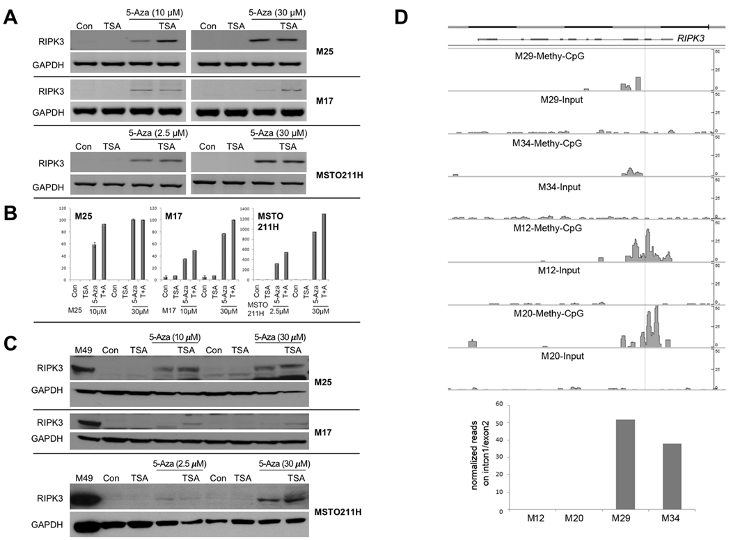
Experimental design: RIPK3 expression was examined in 66 malignant mesothelioma tumors and cell lines. Promoter methylation and DNMT1 siRNA studies were performed to assess the mode of RIPK3 silencing in RIPK3-deficient malignant mesothelioma cells. Restoration of RIPK3 expression in RIPK3-negative malignant mesothelioma cells, either by treatment with 5-aza-2'-deoxycytidine or lentiviral expression of cDNA, was performed to assess effects on cell viability, necrosis, and chemosensitization.

Results: Loss of RIPK3 expression was observed in 42/66 (63%) primary malignant mesotheliomas and malignant mesothelioma cell lines, and RT-PCR analysis demonstrated that downregulation occurs at the transcriptional level, consistent with epigenetic silencing. RIPK3-negative malignant mesothelioma cells treated with 5-aza-2'-deoxycytidine resulted in reexpression of RIPK3 and chemosensitization. Ectopic expression of RIPK3 also resulted in chemosensitization and led to necroptosis, the latter demonstrated by phosphorylation of downstream target MLKL and confirmed by rescue experiments. Mining of RIPK3 expression and survival outcomes among patients with malignant mesothelioma available from The Cancer Genome Atlas repository revealed that promoter methylation of RIPK3 is associated with reduced RIPK3 expression and poor prognosis.

Conclusions: These data suggest that RIPK3 acts as a tumor suppressor in malignant mesothelioma by triggering necroptosis and that epigenetic silencing of RIPK3 by DNA methylation impairs necroptosis and contributes to chemoresistance and poor survival in this incurable disease.


Conflict of interest statement

Disclosure of Potential Conflicts of Interest

The authors declare no potential conflicts of interest with regard to this work.

Figures

Figure 1.
Figure 1.
RIPK3 expression is frequently down regulated or undetectable in human MM cells, primary tumors and PDXs from pleural MM patients. A, Immunoblot analysis of RIPK1, RIPK3, MLKL, β-actin, FADD, caspase 8, NF2/Merlin, BAP1 and p16INK4A protein expression in a panel of human pleural MM cell lines. B, Semi-quantitative RT-PCR analysis (ethidium bromide gel) demonstrating transcriptional down regulation of RIPK3 mRNA in human MM cell lines. In both immunoblot and semi-quantitative RT-PCR analyses, the human mesothelial cell line NM311A was used as a normal control. C, Immunoblot analysis of primary human pleural MM specimens showing down regulated or absent expression of RIPK3 in 13 of 27 (48%) samples and of RIPK1 in 10 of 27 (37%) samples. Separate blots shown below depict expression of BAP1 and NF2 tumor suppressors in the same set of tumors for comparison.
Figure 1.
Figure 1.
RIPK3 expression is frequently down regulated or undetectable in human MM cells, primary tumors and PDXs from pleural MM patients. A, Immunoblot analysis of RIPK1, RIPK3, MLKL, β-actin, FADD, caspase 8, NF2/Merlin, BAP1 and p16INK4A protein expression in a panel of human pleural MM cell lines. B, Semi-quantitative RT-PCR analysis (ethidium bromide gel) demonstrating transcriptional down regulation of RIPK3 mRNA in human MM cell lines. In both immunoblot and semi-quantitative RT-PCR analyses, the human mesothelial cell line NM311A was used as a normal control. C, Immunoblot analysis of primary human pleural MM specimens showing down regulated or absent expression of RIPK3 in 13 of 27 (48%) samples and of RIPK1 in 10 of 27 (37%) samples. Separate blots shown below depict expression of BAP1 and NF2 tumor suppressors in the same set of tumors for comparison.
Figure 2.
Figure 2.
Treatment of MM cells with DNA methylation and/or histone deacetylase inhibitors results in restoration of RIPK3 expression. A-C, The expression of RIPK3 was also determined after treatment of MM cell lines with 2.5 μM, 10 μM or 30 μM 5-aza-2’-deoxycyditine alone for 7 days or 2.5 μM, 10 μM or 30 μM 5-aza-2’-deoxycyditine for 6 days with TSA for the final 24 hours (7 days total), followed by semi-quantitative RT-PCR (A), Real-Time PCR (B) and immunoblot analysis (C). In panel B, the Y-axis refers to relative RIPK3 mRNA expression in treated samples compared to control. Note that the Y-axis in the two graphs on the left and the one on the right differ, because the RIPK3-negative MM cell lines we tested showed variable levels of re-expression of RIPK3 following treatment with 5-aza-2’-deoxycyditine, possibly related to differences in degree of global methylation and/or chromatin compaction. D, RIPK3-positive cell lines (M29 and M34) and RIPK3-negative cell lines (M12 and M20) were used to study DNA methylation. Genomic DNA fragments with methylated-CpG were enriched with MBD2b/MBD3L1 and subjected to NGS and analyzed using MACS2 peak calling pipeline. Both RIPK3-negative cell lines show strong methylated-CpG peaks in RIPK3 intron1 and exon2, whereas both RIPK3-positive cell lines do not. The pre-captured DNAs serve as negative controls (Input). Histograph depicts fold changes of the methylated-CpG peaks among the cell lines studied.
Figure 3.
Figure 3.
RIPK3 suppresses clonogenic growth of MM cells. A, RIPK3-negative cells were nucleofected with pEGFP, pEGFP-RIPK3-WT or pEGFP-RIPK3-KD, and clonogenic assays were then performed. B, Quantification of colonies was performed in triplicate with error bars indicated. C, Immunoblot analysis of RIPK3, p-MLKL, MLKL (54 kDa) and GAPDH in nucleofected MM cells. The cells used for immunoblotting in panel C were harvested 48 h after nucleofection; and the remaining cells were seeded on dishes and selected in medium containing G418 and allowed to form the colonies shown in panel B.
Figure 4.
Figure 4.
Restoration of RIPK3 expression triggers necroptosis in RIPK3-negative MM cells, and RIPK3 inhibition rescues the cell death caused by necroptosis. A, Lentivirus expressing RIPK3 WT, RIPK3-KD, or empty vector only was used to transduce RIPK3-negative MM cells at MOI=5, and light microscopy photographs depict representative transduced MM cells 48 h after transduction. B, RIPK3-negative MM cell line MSTO-211H was transduced with WPI lentivirus expressing RIPK3 WT or empty vector and treated with the apoptosis inhibitor zVAD and/or the RIPK3 inhibitor GSK’872 for 24 h, 48 h, and 4 d. Results of immunoblot analysis (24 h) and cell viability assays (48 h), and clonogenic growth (4 d) are shown. A hemocytometer and trypan blue dye exclusion test was used to determine cell viability. Expression of RIPK3 induced activation of MLKL loss of cell viability, which was rescued by treatment with GSK’872. C, Fluorescence microscopy of MSTO-211H cells demonstrating spatial activation of MLKL, which was rescued by the RIPK3 inhibitor (R3i) GSK’872. D, Transduction of MSTO-211H cells with lentivirus expressing three different shMLKL or shGFP and then selected for 72 h with puromycin. Stable cells expressing shMLKL or shGFP were then infected with WPI or WPI expressing RIPK3. Cell viability assays and immunoblotting were performed after 48 h to detect the activation of MLKL and proportions of alive vs. dead cells. Expression of RIPK3 induced necroptosis, which was rescued by knockdown of MLKL.
Figure 5.
Figure 5.
Re-expression of RIPK3 induces necrosis and sensitizes RIPK3-negative, RIPK3 (–), MM cells to chemotherapeutic drugs. A, B, RIPK3 (–) MSTO-211H and M12 and RIPK3-positive, RIPK3 (+), cell lines M49 and M217 were treated with 10 μM of 5-Aza-dC for 4 days followed by treatment for 48 hours with 5-Aza-dC plus two different doses each of doxorubicin (0 μM, 0.625 μM, 1.25 μM) (A) or cisplatin (0 μM, 6.25 μM, 12.5 μM) (B), and MTS assays were performed to assess cell viability. Treatment with 5-Aza-dC sensitized RIPK3 (–) MM cell lines to both cisplatin and doxorubicin, whereas treatment with 5-Aza-dC showed little or no effect on RIPK3 (+) cells. Similar differences between RIPK3 (–) and RIPK3 (+) MM cell lines were observed when a lower dose (5 μM) of 5-Aza-dC was used (not shown). C, Semi-quantitative RT-PCR analysis of RIPK3 RNA expression in MM cell lines treated with or without 10 μM 5-Aza-dC for 6 days (end date of the chemosensitization assays). Expression of GAPDH was used as a loading control. D, Immunoblot analysis of RIPK3 expression 24 post-infection with control WPI or WPI-RIP3K expressing lentivirus. M8, M12, M17 and MSTO-211H cells were transduced with control WPI or WPI-RIPK3 lentivirus for 24 hours before being harvested for immunoblot analysis as well as seeding for chemosensitization and cell viability assays. RIPK3, P-MLKL, and total MLKL levels were determined to demonstrate re-expression of RIP3K and induction of necrosis. GAPDH levels are shown as a loading control. E, Re-expression of RIP3K sensitizes MM cells to cisplatin. RIPK3-negative MM cell lines were transduced as above for 24 hour and then seeded on a 96-well plate; 24 hours later the cells were treated with 0, 12.5 or 25 μM cisplatin for 48 hours. Cell viability was determined using MTS assay.
Figure 6.
Figure 6.
Loss of RIPK3 mRNA is associated with promoter methylation and poor prognosis in pleural MM patients. Data mining was performed using TCGA pleural MM dataset. A, Box plot showing RIPK3 promoter methylation for various degrees of RIPK3 expression. X-axis shows RIPK3 expression categories partitioned into three groups: Down (lower quartile), No-change (inter quartile range, designated as No Change in expression), and Up (upper quartile); Y-axis shows methylation β-values of promoter probes. White bars in the box plot indicate median β-value, while each color dot indicates an MM patient in the respective category. B, Dot plot showing negative correlation between RIPK3 promoter methylation and RIPK3 mRNA expression (Pearson correlation coefficient r = −0.59, p-value < 0.001). Red line indicates regression, and blue dots represent individual cases. X- and Y-axes indicate log-transformed RSEM expression values and median of CpG probe β-values in promoter regions, respectively. C, Box plot of mRNA expression for RIPK3 where Y-axis indicates the log-transformed RSEM mRNA expression values for different expression categories partitioned into three groups: Down (lower quartile), No-change (inter quartile range), and Up (upper quartile). D, Box plot showing RIPK3 expression for different histologic subtypes of MM. E,. Kaplan-Meier curves indicating OS based on expression categories as shown in panel C for RIPK3 (logrank test, p = 0.01) of MM patients based on RIPK3 mRNA expression, with poor survival of MM patients having low expression of RIPK3 (blue line) compared to those with high levels (orange) or No-change levels (gray) of RIPK3 expression. F, Kaplan-Meier curves indicating OS of expression categories of MM patients based on RIPK1 mRNA expression (p = 0.001), with poor survival of MM patients having low expression of RIPK1 (blue line) compared to those with high levels (red) or No-change levels (gray) of RIPK1 expression.

References

    1. Kadariya Y, Menges CW, Talarchek J, Cai KQ, Klein-Szanto AJ, Pietrofesa RA, et al. Inflammation-related IL1beta/IL1R signaling promotes the development of asbestos-induced malignant mesothelioma. Cancer Prev Res 2016;9:406–14. - PMC - PubMed
    1. Cheng JQ, Jhanwar SC, Klein WM, Bell DW, Lee W-C, Altomare DA, et al. p16 alterations and deletion mapping of 9p21-p22 in malignant mesothelioma. Cancer Res 1994;54:5547–51. - PubMed
    1. Bianchi AB, Mitsunaga S-I, Cheng JQ, Klein WM, Jhanwar SC, Seizinger B, et al. High frequency of inactivating mutations in the neurofibromatosis type 2 gene (NF2) in primary malignant mesotheliomas. Proc Natl Acad Sci U S A 1995;92:10854–8. - PMC - PubMed
    1. Bott M, Brevet M, Taylor BS, Shimizu S, Ito T, Wang L, et al. The nuclear deubiquitinase BAP1 is commonly inactivated by somatic mutations and 3p21.1 losses in malignant pleural mesothelioma. Nat Genet 2011;43:668–72. - PMC - PubMed
    1. Testa JR, Cheung M, Pei J, Below JE, Tan Y, Sementino E, et al. Germline BAP1 mutations predispose to malignant mesothelioma. Nat Genet 2011;43:1022–5. - PMC - PubMed

Publication types

MeSH terms

Substances