. 2020 Nov 17;11(6):e02540-20.
doi: 10.1128/mBio.02540-20.

The TLR3/IRF1/Type III IFN Axis Facilitates Antiviral Responses against Enterovirus Infections in the Intestine

Affiliations

The TLR3/IRF1/Type III IFN Axis Facilitates Antiviral Responses against Enterovirus Infections in the Intestine

Rui Su et al. mBio. .

Abstract

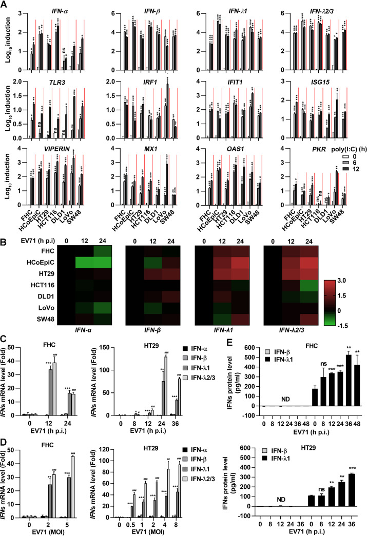
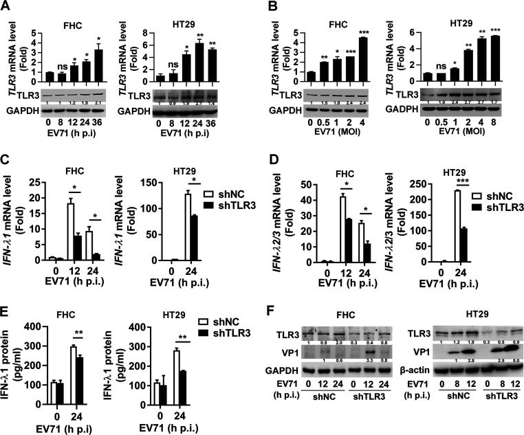
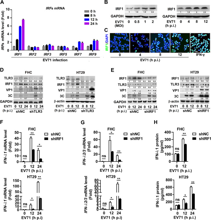
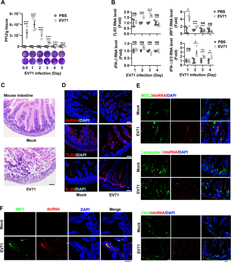
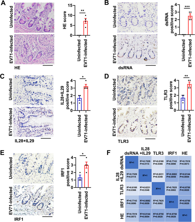
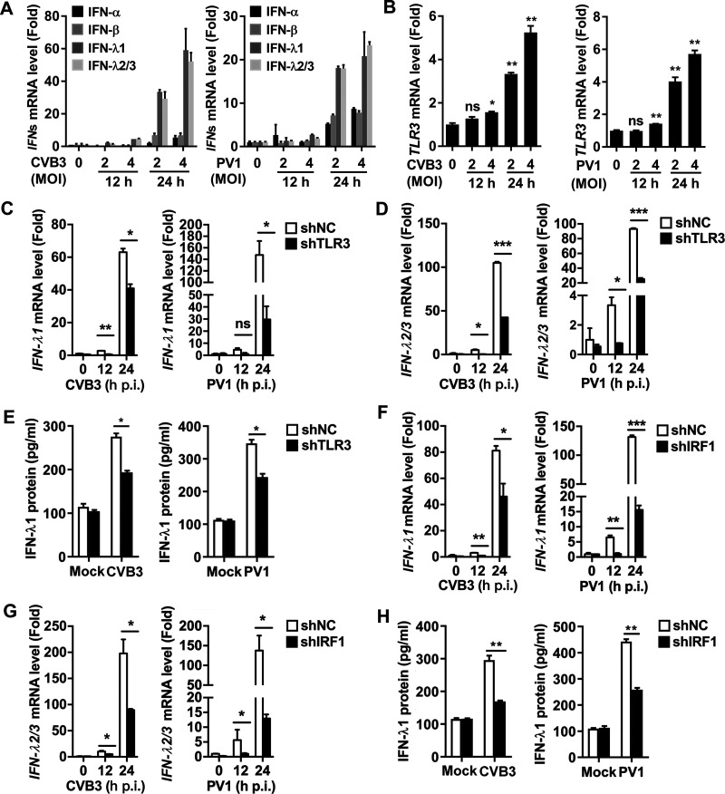
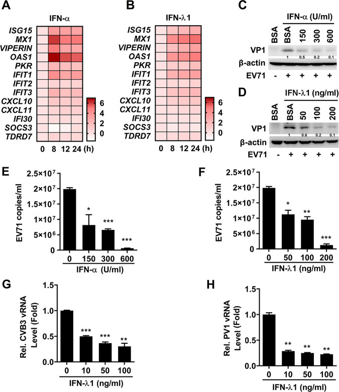
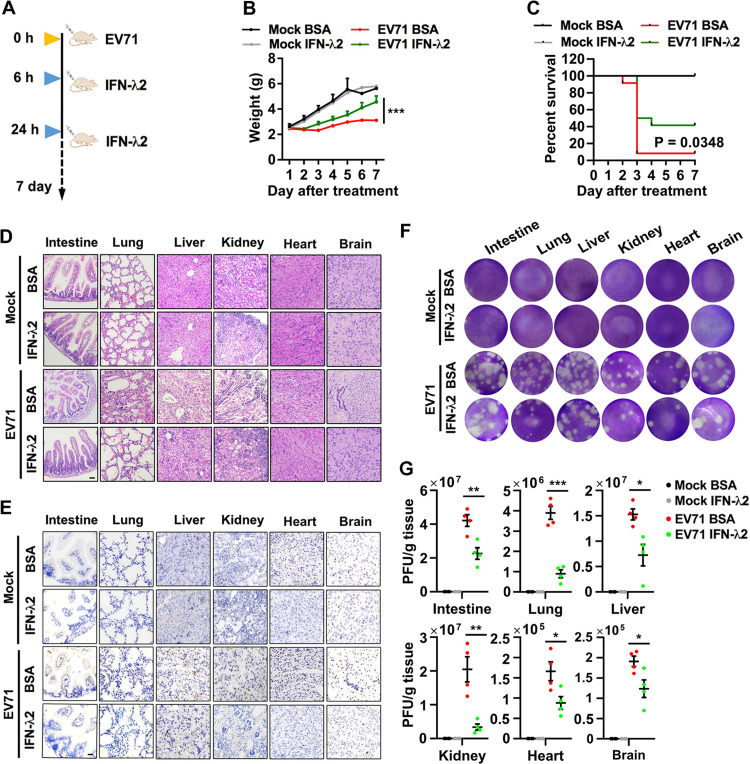
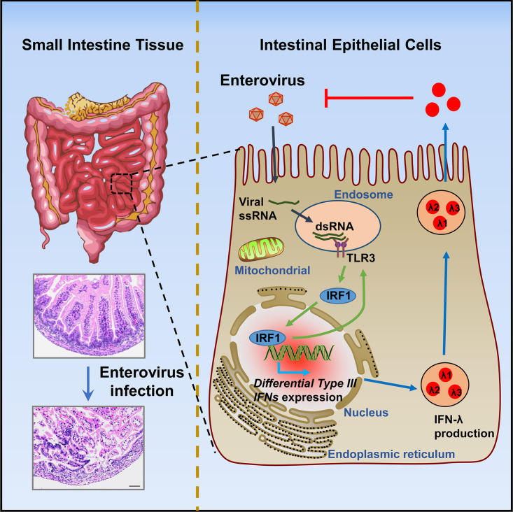
Enteroviruses infect gastrointestinal epithelium cells, cause multiple human diseases, and present public health risks worldwide. However, the mechanisms underlying host immune responses in intestinal mucosa against the early enterovirus infections remain elusive. Here, we showed that human enteroviruses including enterovirus 71 (EV71), coxsackievirus B3 (CVB3), and poliovirus 1 (PV1) predominantly induce type III interferons (IFN-λ1 and IFN-λ2/3), rather than type I interferons (IFN-α and IFN-β), in cultured human normal and cancerous intestine epithelial cells (IECs), mouse intestine tissues, and human clinical intestine specimens. Mechanistic studies demonstrated that IFN-λ production is induced upon enterovirus infection through the Toll-like receptor 3/interferon regulatory factor 1 (TLR3/IRF1) signaling pathway in IECs. In turn, the supplementation of IFN-λ subsequently induces intrinsically antiviral responses against enterovirus replication. Notably, intraperitoneal injection in neonatal C57BL/6J mice with mouse recombinant IFN-λ2 protein represses EV71 replication and protects mice from viral lethal effects. Altogether, these results revealed a distinct mechanism by which the host elicited immune responses against enterovirus infections in intestine through activating the TLR3/IRF1/type III IFN axis. The new findings would provide an antiviral strategy for the prevention and treatment of enterovirus infections and associated diseases.IMPORTANCE Enterovirus infections are significant sources of human diseases and public health risks worldwide, but little is known about the mechanism of innate immune response in host intestine epithelial surface during the viral replication. We reported the epithelial immune response in cultured human normal and cancerous cells (IECs), mouse tissues, and human clinical intestine specimens following infection with enterovirus 71. The results mechanistically revealed type III interferons (IFN-λ1 and IFN-λ2/3), rather than type I interferons (IFN-α and IFN-β), as the dominant production through TLR3/IRF1 signaling upon multiple human enterovirus infection, including enterovirus 71 (EV71), coxsackievirus B3 (CVB3), and poliovirus 1 (PV1). IFN-λ subsequently induced antiviral activity against enterovirus replication in vitro and in vivo. These studies uncovered the role of the novel process of type III IFN production involved in the TLR3/IRF1 pathway in host intestine upon enterovirus infection, which highlighted a regulatory manner of antiviral defense in intestine during enterovirus infection.

Keywords: Toll-like receptor 3; coxsackievirus B3; enterovirus; enterovirus 71; enterovirus infection; interferon regulatory factor 1; interferon-stimulated genes; intestine; intestine epithelial cells; neonatal C57BL/6J mice; poliovirus 1; type III interferons.


Figures

FIG 1
FIG 1
EV71 infection mainly induces type III interferon in IECs. (A) The human normal (FHC and HCoEpiC) and cancerous (HT29, HCT116, DLD1, LoVo, and SW48) intestinal cell lines were treated with poly(I·C) at a dose of 3 μg/ml for 6 h or 12 h. Human IFN and ISG gene expression was detected by qPCR analysis. Data are shown as a fold change (log10) relative to control (0-h group). (B) Seven human intestine cell lines were incubated with EV71 (MOI = 1) for 12 h or 24 h. The total mRNA of treated cells was extracted. Type I IFN (IFN-α [specific for IFN-α1 and IFN-α13] and IFN-β) and type III IFN (IFN-λ1 and IFN-λ2/3) mRNA levels were assessed by qPCR and visualized in a heatmap of expression values (log10 [fold change]). Data are representative of at least two independent experiments. (C and D) FHC cells were treated with EV71 at an MOI of 2 for 12 and 24 h (C) or at MOI of 2 and 5 for 12 h (D). HT29 cells were infected with EV71 at an MOI of 1 for 0, 8, 12, 24, and 36 h (C) or at MOI of 0, 0.5, 1, 2, 4, and 8 for 24 h (D). Type I IFN (IFN-α and IFN-β) and type III IFN (IFN-λ1 and IFN-λ2/3) mRNA levels were detected by qPCR and normalized to the GAPDH mRNA level, and results are expressed as fold induction relative to control. * and # indicate the statistical significance for IFN-λ1 and IFN-λ2/3 mRNA levels relative to each control, respectively. (E) ELISA of IFN-β and IFN-λ1 production in the supernatant of FHC (MOI = 2) and HT29 (MOI = 1) cells treated with EV71 for the indicated time. Data are shown as mean ± SD and correspond to a representative experiment out of three performed. ns, nonsignificant; **, P < 0.01; ***, P < 0.001. ND, not detected. Statistical significance was determined by Student’s t test.
FIG 2
FIG 2
TLR3 is important for type III IFN production in human IECs upon EV71 infection. (A and B) EV71 infected FHC (MOI = 2) and HT29 (MOI = 1) cells for 8, 12, 24, or 36 h, respectively (A). FHC (MOI = 0, 0.5, 1, 2, and 4) and HT29 (MOI = 0, 0.5, 1, 2, 4, and 8) cells were treated with EV71 for 24 h (B). TLR3 mRNA and TLR3 protein levels were measured by qPCR and Western blot analysis, respectively. (C and D) FHC cells stably expressing shNC or shTLR3 were infected with EV71 at an MOI of 2 for 12 or 24 h. HT29 cells stably expressing shNC or shTLR3 were infected with EV71 (MOI = 1) for 24 h. IFN-λ1 (C) and IFN-λ2/3 (D) mRNA levels were detected by qPCR. (E) FHC (MOI = 2) and HT29 (MOI = 1) cells stably expressing shNC or shTLR3 were treated with EV71 for 24 h. IFN-λ1 protein secreted in the cell supernatants was analyzed by ELISA. (F) EV71 infected FHC (0, 12, and 24 h, MOI = 2) and HT29 (0, 8, and 12 h, MOI = 1) shNC/shTLR3 stably expressing cells. TLR3, VP1, and GAPDH expressions were analyzed by Western blotting. Protein expression relative to internal control is quantified using Image J software. Data are shown as mean ± SD and correspond to a representative experiment out of three performed. ns, nonsignificant; *, P < 0.05; **, P < 0.01; ***, P < 0.001. Statistical significance was determined by Student’s t test.
FIG 3
FIG 3
IRF1 is important for TLR3-mediated type III IFN production upon EV71 infection. (A) HT29 cells were infected with EV71 (MOI = 1) for the indicated time. Total RNA was extracted from the cells, and the level of IRF1, IRF2, IRF3, IRF5, IRF7, and IRF9 mRNA was determined by qPCR. (B) HT29 cells were infected with EV71 at indicated MOI for 8 h or infected with EV71 at an MOI of 1 for 0, 4, 8, and 12 h. The cells were lysed, and IRF1 and GAPDH proteins were detected by Western blotting analyses. (C) HT29 cells were infected with EV71 at an MOI of 1 for 0, 4, 8, and 12 h or treated with 100 ng/ml of IFN-γ for 4 h, used as a positive control. The localization of IRF1 (green) and DAPI (blue) was analyzed by confocal microscopy. Bar = 20 μm. (D) FHC (MOI = 2) and HT29 (MOI = 1) stably expressing shNC or shTLR3 cells were infected with EV71 for the indicated time. Cell lysates were prepared, and the TLR3, IRF1, VP1, 3C, and GAPDH/β-actin proteins in the cell lysates were detected by Western blot analyses. (E to H) FHC and HT29 cells stably expressing shNC or shIRF1 were generated and then infected with EV71 at an MOI of 2 and an MOI of 1 for indicated times, respectively. Cell lysates were prepared, and the TLR3, IRF1, VP1, 3C, and GAPDH/β-actin proteins in the cell lysates were detected by Western blot analyses. The levels of IFN-λ1 mRNA (F) and IFN-λ2/3 mRNA (G) were detected by qPCR. The levels of IFN-λ1 protein secreted in the cell culture supernatants were determined by ELISA (H). All qPCR assays used GAPDH mRNA as an internal control. Results are expressed as fold induction relative to control. Protein expression relative to internal control is quantified using Image J software. Data are shown as mean ± SD and correspond to a representative experiment out of three performed. ns, nonsignificant; *, P < 0.05; **, P < 0.01. Statistical significance was determined by Student’s t test.
FIG 4
FIG 4
EV71 infection induces an antiviral response in mouse intestine. Three-day-old C57BL/6J mice were infected by intraperitoneal injection of 1 × 107 PFU of EV71 (n = 20) or mock infected with PBS (n = 20). At 0.5, 1, 2, 3, 4, and 5 days after infection, animals were euthanized, and the small intestine tissues were collected. Each group, n = 3 to 4. (A) The EV71 titers of mice were quantified by plaque assay shown as PFU/g tissue. The representative images of viral plaque assay were displayed. (B) The levels of TLR3, IRF1, IFN-β, and IFN-λ2/3 mRNAs were detected by qPCR. Data are shown as fold changes in RNA expression compared to the control group at 1 day to 4 days postinfection. Graphs represent the mean ± SEM. ns, nonsignificant; *, P < 0.05; **, P < 0.01; ***, P < 0.001. Statistical significance was determined by Student’s t test. (C and D) Sections of the mouse small intestine at 3 days postinfection were stained by H&E (C) or fluorescently labeled using antibodies against TLR3, IL-28, or dsRNA (red) and DAPI to label the nuclei (blue) (D). (E) Immunofluorescence images from intestine were immunostained with DAPI (blue), intestine cell markers (MUC2, goblet cell marker; lysozyme C, Paneth cell marker; ChrA, enteroendocrine cell marker) (green), and dsRNA (red). (F) The small intestine sections of mice on day 3 postinfection from PBS and EV71 groups were immunostained with IRF1 (green), dsRNA (red), and DAPI (blue). All images are representative of each group (n = 3) with similar results. Bar = 100 μm.
FIG 5
FIG 5
EV71 infection triggers the TLR3/IRF1/type III IFN signaling in human intestines. (A to E) Uninfected intestinal tissue samples (n = 3) and EV71-infected intestinal tissue specimens (n = 4) were collected from deceased human individuals. The small intestine tissue sections were analyzed by H&E staining (A) or immunohistochemistry using antibodies against dsRNA (B), IL-28+IL-29 proteins (C), TLR3 protein (D), and IRF1 protein (E). Positive staining is represented by brown coloration in these representative images. Light microscopy, bar = 100 μm. The relative levels of proteins were quantified with Image J software and are expressed as a positive score. Graphs show mean ± SEM. *, P < 0.05; **, P < 0.01; ***, P < 0.001. Statistical significance was determined by Student’s t test. (F) Pearson’s correlation analyses of H&E scores, the viral dsRNA expression, IL-28+IL-29 production, TLR3 protein, and IRF1 protein in the 7 human intestine specimens. Statistical significance was determined by two-tailed t test.
FIG 6
FIG 6
CVB3 and PV1 induce type III IFN production through the TLR3/IRF1 signaling in IECs. (A and B) HT29 cells were infected with CVB3 or PV1 at MOI of 0, 2, and 4 for 12 and 24 h. The levels of type I IFN (IFN-α and IFN-β) and type III IFN (IFN-λ1 and IFN-λ2/3) mRNAs (A) and TLR3 mRNA (B) were measured by qPCR. (C to E) HT29 cell lines stably expressing shNC or shTLR3 were infected with CVB3 and PV1 at an MOI of 2 for 12 h and 24 h. The levels of IFN-λ1 mRNA (C) and IFN-λ2/3 mRNA (D) were measured by qPCR. The levels of IFN-λ1 protein were determined by ELISA (E). (F to H) HT29 cell lines stably expressing shNC or shIRF1 were infected with CVB3 and PV1 at an MOI of 2 for 12 h and 24 h. The levels of IFN-λ1 mRNA (F) and IFN-λ2/3 mRNA (G) were measured by qPCR. The levels of IFN-λ1 protein were determined by ELISA (H). All qPCR assays used GAPDH mRNA as an internal control. Results are expressed as fold induction relative to control. Data are shown as mean ± SD and correspond to a representative experiment out of three performed. ns, nonsignificant; *, P < 0.05; **, P < 0.01; ***, P < 0.001. Statistical significance was determined by Student’s t test.
FIG 7
FIG 7
IFN-λ1 exerts antiviral activity against enterovirus replication in IECs. (A and B) HT29 cells were incubated with IFN-α (subtype IFN-α2b) (150 U/ml) (A) or IFN-λ1 (50 ng/ml) (B) for the indicated times. The mRNA levels of 13 IFN-inducible stimulated genes (ISGs) were assessed by qPCR. The data for all genes are normalized to GAPDH and visualized in a heatmap of expression values (log2 [fold change]). Data are representative of at least two independent experiments. (C to F) HT29 cells were plated in 12-well plates; treated with BSA, IFN-α (C and E), or IFN-λ1 (D and F) at different concentrations as indicated for 8 h; and infected with EV71 at an MOI of 1 for 16 h. The levels of EV71 VP1 protein and β-actin protein in the cell lysates were determined by Western blot analysis (C and D). EV71 copy numbers in the supernatants were determined by absolute qPCR analysis (E and F). (G and H) HT29 cells were seeded in 12-well plates, treated with IFN-λ1 at different concentrations for 24 h, and then infected with CVB3 (G) or infected with PV1 (H) at an MOI of 1 for 24 h. The levels of viral RNAs in the cell lysates were measured by qPCR. Protein expression relative to internal control is quantified using Image J software. Data are shown as mean ± SD and correspond to a representative experiment out of three. ns, nonsignificant; *, P < 0.05; **, P < 0.01; ***, P < 0.001. Statistical significance was determined by Student’s t test.
FIG 8
FIG 8
IFN-λ2 suppresses EV71 replication in vivo. (A) Schematic of mouse experiment. (B and C) Three-day-old suckling mice were injected with EV71 (intraperitoneally), 1 × 107 PFU in 20 μl PBS, and then injected with IFN-λ2 (0.5 μg per mouse) at 6 and 24 h postinfection. The weight (B) and survival (C) were monitored for 7 days after infection (Mock, n = 6 in each group; EV71, n = 12 in each group). Statistical significance in weight and survival was calculated via two-way ANOVA and log rank tests, respectively. (D to G) Mouse tissues were stained with H&E (D) and IHC for viral dsRNA (E). Representative light microscopy images are shown. Bar = 100 μm. The EV71 titers in the intestine, lung, liver, kidney, heart, and brain tissues (each group, n = 4) at 3 days after the infection were determined by plaque assay (F) and quantified (G). Graphs show the mean ± SEM. *, P < 0.05; **, P < 0.01; ***, P < 0.001. Statistical significance was determined by Student’s t test.
FIG 9
FIG 9
A proposed model underlying the TLR3/IRF1 signaling pathway. TLR3/IRF1 signaling regulates type III interferon production and antiviral activity in intestinal epithelial cells upon enterovirus infection. Upon infection by enterovirus in human or murine intestine tissues, intestinal epithelial cells are the first line of defense against virus infection. Viral dsRNA is recognized by TLR3 to trigger signaling events, and then TLR3-IRF1 signaling is activated. The IRF1 is translocated into the nucleus to induce the differential accumulation of type III interferon production, resulting in the activation of the innate immune antiviral response in intestinal epithelial cells.

References

    1. Pons-Salort M, Grassly NC. 2018. Serotype-specific immunity explains the incidence of diseases caused by human enteroviruses. Science 361:800–803. doi: 10.1126/science.aat6777. - DOI - PMC - PubMed
    1. Black S, Bradley C, Lai FY, Shenoy S, Bandi S, Allen DJ, Tang JW. 2019. Comparing the clinical severity of disease caused by enteroviruses and human parechoviruses in neonates and infants. Pediatr Infect Dis J 38:e36–e38. doi: 10.1097/INF.0000000000002145. - DOI - PubMed
    1. Wells AI, Coyne CB. 2019. Enteroviruses: a gut-wrenching game of entry, detection, and evasion. Viruses 11:460. doi: 10.3390/v11050460. - DOI - PMC - PubMed
    1. Huang PN, Shih SR. 2014. Update on enterovirus 71 infection. Curr Opin Virol 5:98–104. doi: 10.1016/j.coviro.2014.03.007. - DOI - PubMed
    1. Li Y, Chang Z, Wu P, Liao Q, Liu F, Zheng Y, Luo L, Zhou Y, Chen Q, Yu S, Guo C, Chen Z, Long L, Zhao S, Yang B, Yu H, Cowling BJ. 2018. Emerging enteroviruses causing hand, foot and mouth disease, China, 2010–2016. Emerg Infect Dis 24:1902–1906. doi: 10.3201/eid2410.171953. - DOI - PMC - PubMed

Publication types

MeSH terms

LinkOut - more resources