. 2020 Nov 17;12(1):31.
doi: 10.1038/s41368-020-00098-x.

Hollow silica reinforced magnesium nanocomposites with enhanced mechanical and biological properties with computational modeling analysis for mandibular reconstruction

Affiliations

Hollow silica reinforced magnesium nanocomposites with enhanced mechanical and biological properties with computational modeling analysis for mandibular reconstruction

Somasundaram Prasadh et al. Int J Oral Sci. .

Abstract

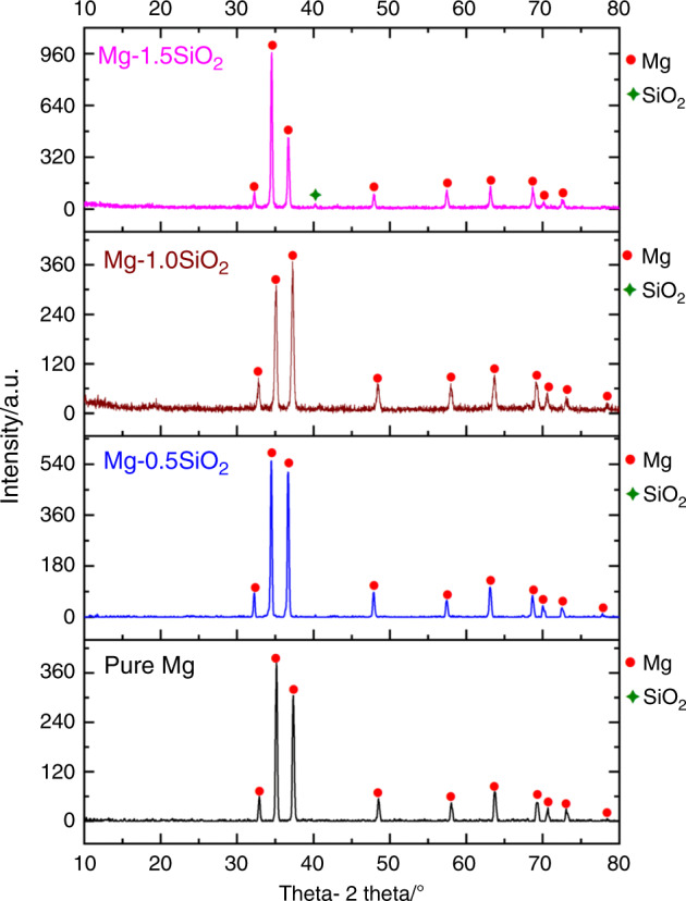
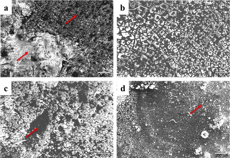
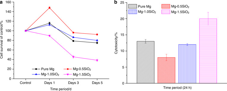
The present study investigates Mg-SiO2 nanocomposites as biodegradable implants for orthopedic and maxillofacial applications. The effect of presence and progressive addition of hollow silica nanoparticles (0.5, 1, and 1.5) vol.% on the microstructural, mechanical, degradation, and biocompatibility response of pure Mg were investigated. Results suggest that the increased addition of hollow silica nanoparticles resulted in a progressive increase in yield strength and ultimate compressive strength with Mg-1.5 vol.% SiO2 exhibiting superior enhancement. The response of Mg-SiO2 nanocomposites under the influence of Hanks' balanced salt solution revealed that the synthesized composites revealed lower corrosion rates, indicating rapid dynamic passivation when compared with pure Mg. Furthermore, cell adhesion and proliferation of osteoblast cells were noticeably higher than pure Mg with the addition of 1 vol.% SiO2 nanoparticle. The biocompatibility and the in vitro biodegradation of the Mg-SiO2 nanocomposites were influenced by the SiO2 content in pure Mg with Mg-0.5 vol.% SiO2 nanocomposite exhibiting the best corrosion resistance and biocompatibility when compared with other nanocomposites. Enhancement in mechanical, corrosion, and biocompatibility characteristics of Mg-SiO2 nanocomposites developed in this study are also compared with properties of other metallic biomaterials used in alloplastic mandibular reconstruction in a computational model.


Conflict of interest statement

The authors declare no competing interests.

Figures

Fig. 1
Fig. 1
X-ray diffractograms of pure Mg and Mg-SiO2 nanocomposites taken along the longitudinal direction of hot extruded samples. X, Y, Z represent 2θ = 32°, 34°, and 36° corresponding to (10–10) prism, (0002) basal, and (10–11) pyramidal planes, respectively
Fig. 2
Fig. 2
The immersion response of the synthesized samples. a Calculated corrosion rates based on weight loss and b pH measurements from immersion testing
Fig. 3
Fig. 3
Scanning electron microscope images of a pure Mg, b Mg-0.5 SiO2, c Mg-1.0 SiO2, and d Mg-1.5 SiO2 nanocomposites after 7 days of immersion (red arrows indicating the corroded and non-corroded areas). Magnification, ×75. Scale bars are 200 μm
Fig. 4
Fig. 4. Cytotoxicity in vitro for pure Mg and Mg-SiO2 nanocomposites.
a Remaining cellular activity of and b relative cytotoxicity of MC3T3-E1 cells upon exposure to sample discs, as measured by MTS assay and LDH, respectively
Fig. 5
Fig. 5
LIVE/DEAD staining of primary MC3T3-E1 cells cultured on samples for a Day 1, b Day 3, c Day 5, respectively, and d Percentage of living cells. Viable cells are labeled in green, the nucleus of live cells (blue), dead cells (red) and merge images of live and dead cells. d Percentage of living cells. Magnification: ×10 and scale bar are 200 µm
Fig. 6
Fig. 6
Scanning microscope images of MC3T3-E1 cultured on Mg-SiO2 nanocomposite samples for a Day 1, b Day 3, and c Day 7. Arrows show the cell attachment. Scale bars are 400 µm
Fig. 7
Fig. 7
Von Misses stress values (MPa): a Pure magnesium, b Mg/1 SiO2, c stress concentration areas. Magnitude of displacement: d Pure magnesium, € Mg/1 SiO2, f Stress concentration areas
Fig. 8
Fig. 8
Von Misses stress values of mandible (MPa): a Pure magnesium, b Mg/1 SiO2, c stress concentration areas. Magnitude of displacement of mandible: d pure magnesium, e Mg/1 SiO2, f stress concentration areas

References

    1. Prasadh S, et al. The potential of magnesium based materials in mandibular reconstruction. Metals. 2019;9:302. doi: 10.3390/met9030302. - DOI
    1. Angrisani N, et al. Biocompatibility and degradation of LAE442-based magnesium alloys after implantation of up to 3.5 years in a rabbit model. Acta Biomater. 2016;44:355–365. doi: 10.1016/j.actbio.2016.08.002. - DOI - PubMed
    1. Ali M, Hussein MA, Al-Aqeeli N. Magnesium-based composites and alloys for medical applications: a review of mechanical and corrosion properties. J. Alloy. Compd. 2019;792:1162–1190. doi: 10.1016/j.jallcom.2019.04.080. - DOI
    1. Liu C, et al. Biodegradable magnesium alloys developed as bone repair materials: a review. Scanning. 2018;2018:9216314. - PMC - PubMed
    1. Dziuba D, et al. Long-term in vivo degradation behaviour and biocompatibility of the magnesium alloy ZEK100 for use as a biodegradable bone implant. Acta Biomater. 2013;9:8548–8560. doi: 10.1016/j.actbio.2012.08.028. - DOI - PubMed

Publication types