. 2020 Nov 17;11(1):5834.
doi: 10.1038/s41467-020-19633-9.

XPC deficiency increases risk of hematologic malignancies through mutator phenotype and characteristic mutational signature

Affiliations

XPC deficiency increases risk of hematologic malignancies through mutator phenotype and characteristic mutational signature

Andrey A Yurchenko et al. Nat Commun. .

Abstract

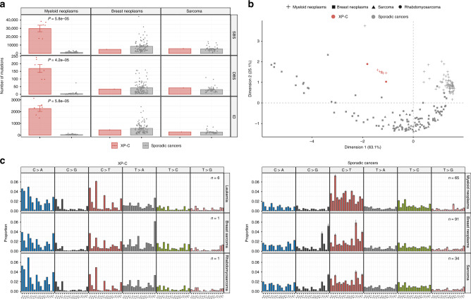
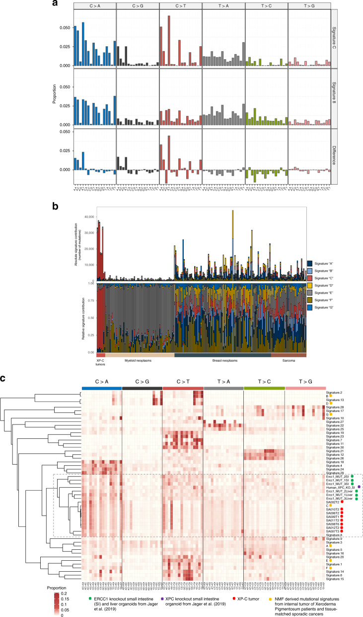
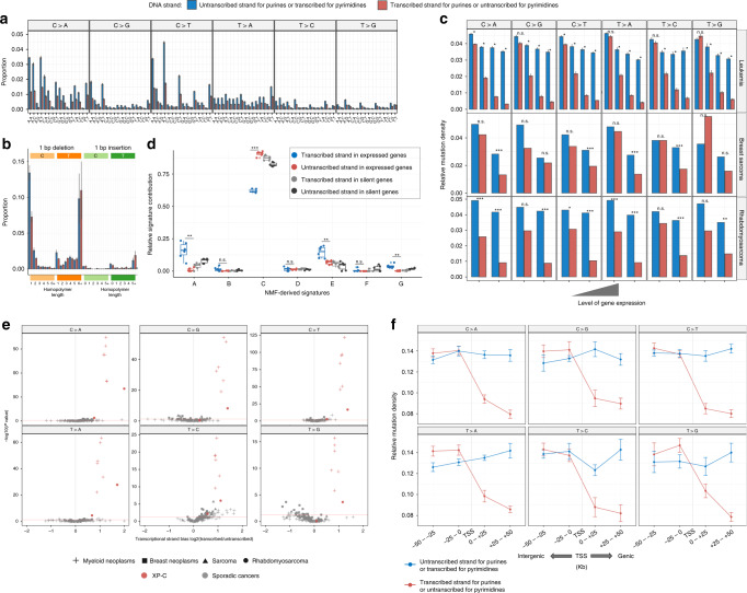
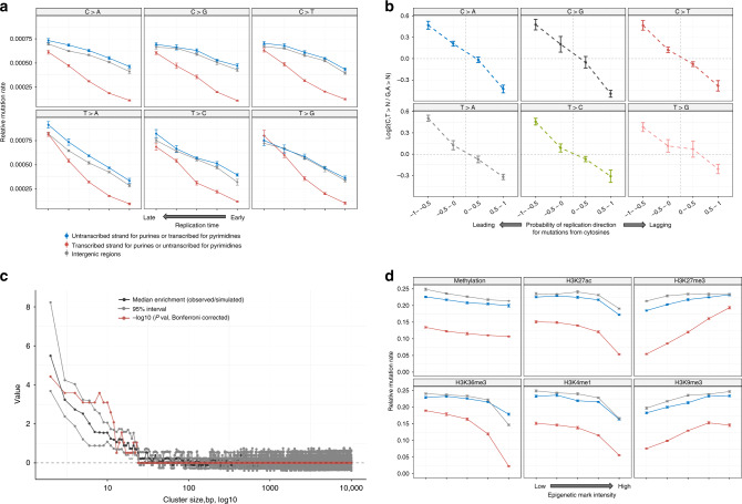
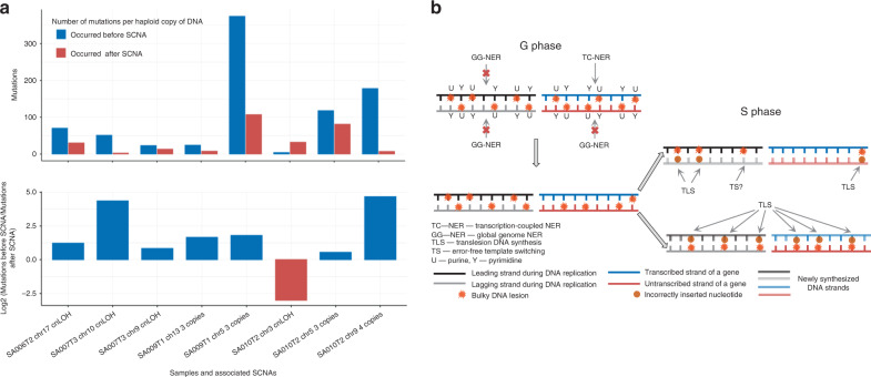
Recent studies demonstrated a dramatically increased risk of leukemia in patients with a rare genetic disorder, Xeroderma Pigmentosum group C (XP-C), characterized by constitutive deficiency of global genome nucleotide excision repair (GG-NER). The genetic mechanisms of non-skin cancers in XP-C patients remain unexplored. In this study, we analyze a unique collection of internal XP-C tumor genomes including 6 leukemias and 2 sarcomas. We observe a specific mutational pattern and an average of 25-fold increase of mutation rates in XP-C versus sporadic leukemia which we presume leads to its elevated incidence and early appearance. We describe a strong mutational asymmetry with respect to transcription and the direction of replication in XP-C tumors suggesting association of mutagenesis with bulky purine DNA lesions of probably endogenous origin. These findings suggest existence of a balance between formation and repair of bulky DNA lesions by GG-NER in human body cells which is disrupted in XP-C patients.


Conflict of interest statement

The authors declare no competing interests.

Figures

Fig. 1
Fig. 1. Mutational load and profiles of XP-C and 190 tissue-matched sporadic cancers.
a Number of SBS (single base substitutions), DBS (double base substitutions) and ID (indels) in XP-C and sporadic cancers with indicated SEM intervals. The difference is highly significant for myeloid neoplasms (Mann–Whitney U test, two-sided, n = 6 for XP-C and n = 65 for myeloid neoplasms), but number of mutations in breast sarcoma (n = 1) and rhabdomyosarcoma (n = 1) are in the range of sporadic tumors (n = 91 and n = 34 for breast cancer and sarcoma respectively). b Multidimension scaling plot based on the Cosine similarity distance between the mutational profiles of the samples. XP-C tumors clearly groups together and are distant from tissue-matched sporadic cancers. c Trinucleotide-context mutational profiles (SEM intervals are shown in case of multiple samples, n represents the number of independent cancer samples). An x-axis represents the nucleotides upstream and downstream of mutation. XP-C tumors demonstrate high similarity with each other (left panel), but profiles of sporadic cancers (right panel) are different from them.
Fig. 2
Fig. 2. Mutational profiles of XP-C tumors in the context of known mutational signatures.
a NMF-derived mutational Signature “C” from XP-C tumors and tissue-matched sporadic cancers in comparison with COSMIC Signature 8  (Cosine similarity = 0.86). b Relative contribution of NMF-derived mutational signatures in XP-C and tissue-matched sporadic cancers (NMF approach). XP-C tumor mutational profiles are dominated by Signature “C”, while sporadic cancers by other signatures with relatively small proportion of Signature “C”. c Unsupervised hierarchical clustering based on the Cosine similarity distances between the XP-C tumors mutational profiles, NMF-derived mutational signatures from XPC tumors and tissue-matched sporadic cancers, COSMIC mutational signatures (Signatures 1−30), and XPC and Ercc1 organoid knockouts. XP-C tumors cluster with each other and COSMIC Signature 8 forming a larger cluster with Ercc1 and XPC organoid knockouts.
Fig. 3
Fig. 3. Strong transcriptional bias (TRB) is a specific feature of XP-C tumors.
a TRB is observed in the majority of trinucleotide contexts of XP-C leukemia samples (n = 6, SEMs are indicated). b TRB is highly pronounced for specific single nucleotide C:G deletions in XP-C leukemia samples (n = 6, SEMs are indicated). c TRB strength depends on the level of gene expression and is most pronounced in highly expressed genes (SEMs are indicated for leukemia; Poisson, two-sided test used for breast sarcoma (n = 1) and rhabdomyosarcoma (n = 1); Wilcoxon signed-rank, two-sided test for leukemia (n = 6), P: ns—nonsignificant, *<0.5, **<0.01, ***<0.001). d Relative mutational signature contribution for mutations separated by transcribed and untranscribed strands in transcriptionally active (FPKM > 2) and silent genes (FPKM < 0.05) of XP-C leukemia. Boxes depict the interquartile range (25–75% percentile), lines—the median, whiskers—1.5× the IQR below the first quartile and above the third quartile. Predominant in XP-C leukemia Signature “C” is depleted on the transcribed strands with functional TC-NER, but relative contribution of signatures “A” and “E” typical for sporadic leukemia is enriched on the transcribed strand (t test, two-sided, paired between transcribed and untranscribed strands in expressed genes (n = 6), P: ns—nonsignificant, *<0.5, **<0.01, ***<0.001). e TRB is highly significant and pronounced in XP-C samples for all six substitution classes in comparison with sporadic cancers (Poisson two-sided test). f The strong TRB observed in XP-C leukemia (n = 6) is caused by transcriptional-coupled repair (TC-NER) but not transcriptional-associated damage. Strong decrease of mutation rate is observed on the genic untranscibed strand for pyrimidines (transcribed for purines, red; right side of transcription start site, TSS), but not on the transcribed strand for pyrimidines (untranscribed for purines, blue) as compared to neighboring intergenic regions (±50 kbp from transcription start site, SEMs are indicated).
Fig. 4
Fig. 4. Genomic landscape of mutagenesis in XP-C internal tumors.
a Mutational density on the transcribed, untranscribed DNA strands of genes as well in intergenic regions presented as a function of replication timing in XP-C leukemia (n = 6, SEMs are indicated). Replication time is split onto five quantiles. Mutation rate for pyrimidines on the transcribed strand (or purines on the untranscribed, blue) is not different from intergenic regions within the same bin, which is compatible with the absence of GG-NER. b Pyrimidine/purine ratios of mutation rates for regions of the genome grouped by propensity of reference DNA strand to be replicated as leading (left) or lagging (right) strand during DNA synthesis for XP-C leukemia genomes (n = 6, SEMs are indicated). Strong enrichment of mutagenesis on the leading strand from pyrimidines (C and T) (lagging strands from purines (G and A)) is observed for all six classes of mutations. Mutations from purines on the lagging DNA strand may result from error-prone Translesion Synthesis. c The assessment of the length of clustered mutation events on the distances ranging between 2 and 10,000 bp. Sliding window of 5 bp was used to estimate median effect size (black) and its 95% confidence interval (gray) as well as Bonferroni-corrected −log10 (P value) (red, Wilcoxon signed-rank test, two-sided) for different length of clusters in real data (XP-C leukemia, n = 6) against simulations (see “Methods” section). The highest and significant enrichment of clustered mutations was observed for short clusters with lengths between 2 and 16 bp. d Intensity of epigenetic marks (5 quantiles) and relative mutation load for XP-C leukemia (n = 6, SEMs are indicated). Mutational density on the transcribed, untranscribed DNA strands of genes as well in intergenic regions positively correlated with repressive histone marks H3K27me3 and H3K9me3, and inversely correlated with active chromatin marks (H3K27ac, H3K36me3, H3K4me1). For all three genomic categories, effect of the majority of epigenetic marks was similar. At the same time correlations of untranscribed strand for pyrimidines (or transcribed for purines, red) with H3K27me3 and H34M36me3 were more important than for the other two categories.
Fig. 5
Fig. 5. Accumulation of DNA lesions and mutations observed in XP-C tumors.
a Relative number of mutations that occurred before and after SCNAs in XP-C cancer genomes (normalized per haploid DNA copy number). The majority of events demonstrate an excess of mutations that were accumulated before the SCNA and may have occurred in tumor-progenitor cells or at early stages of carcinogenesis. b A model of DNA lesion accumulation and mutagenesis in XP-C cells. In XP-C cells where GG-NER is dysfunctional, bulky lesions cannot be efficiently repaired and persist everywhere in the genome except transcribed strands of active genes where TC-NER is operative. During the S-phase a part of bulky lesions on the leading strand may be removed by error-free template switching (TS) mechanisms while on the lagging strand they are converted to mutations by error-prone translesion synthesis (TLS) more frequently, causing mutation accumulation with cell divisions and observed transcriptional and replication biases.

References

    1. Lehmann AR, McGibbon D, Stefanini M. Xeroderma pigmentosum. Orphanet J. Rare Dis. 2011;6:1–6. doi: 10.1186/1750-1172-6-70. - DOI - PMC - PubMed
    1. Kraemer, K. H., Lee, M. M., Andrews, A. D. & Lambert, W. C. The role of sunlight and DNA repair in melanoma and nonmelanoma skin cancer: the Xeroderma Pigmentosum paradigm. Arch. Dermatol. 10.1001/archderm.1994.01690080084012 (1994). - PubMed
    1. Bradford, P. T. et al. Cancer and neurologic degeneration in xeroderma pigmentosum: long term follow-up characterises the role of DNA repair. J. Med. Genet. 10.1136/jmg.2010.083022 (2011). - PMC - PubMed
    1. Sethi M, et al. Patients with xeroderma pigmentosum complementation groups C, e and v do not have abnormal sunburn reactions. Br. J. Dermatol. 2013;169:1279–1287. doi: 10.1111/bjd.12523. - DOI - PubMed
    1. Kraemer, K. H. Xeroderma pigmentosum. Cutaneous, ocular, and neurologic abnormalities in 830 published cases. Arch. Dermatol.10.1136/jmg.2010.083022 (1987). - PubMed

Publication types

Supplementary concepts