. 2021 Sep;18(9):2262-2274.
doi: 10.1038/s41423-020-00583-7. Epub 2020 Nov 17.

TRAF2 regulates T cell immunity by maintaining a Tpl2-ERK survival signaling axis in effector and memory CD8 T cells

Affiliations

TRAF2 regulates T cell immunity by maintaining a Tpl2-ERK survival signaling axis in effector and memory CD8 T cells

Xiaoping Xie et al. Cell Mol Immunol. 2021 Sep.

Abstract

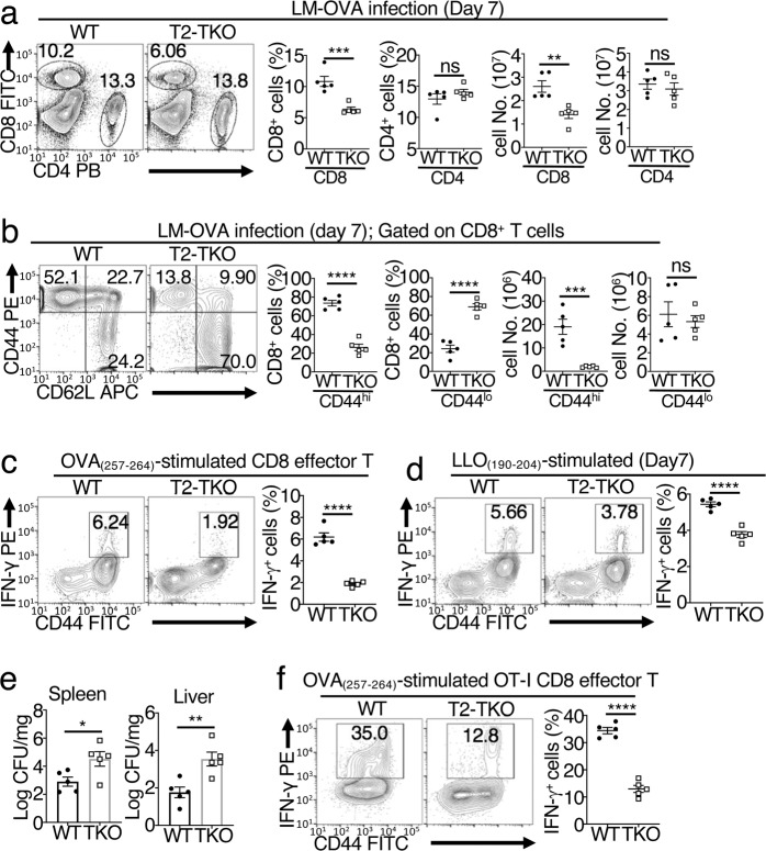
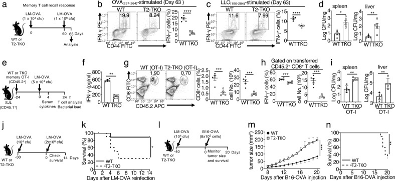
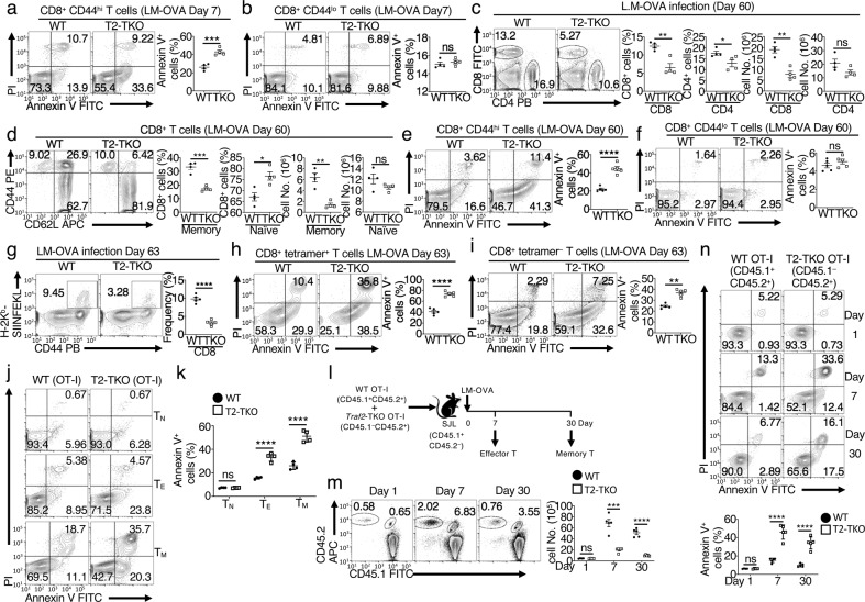
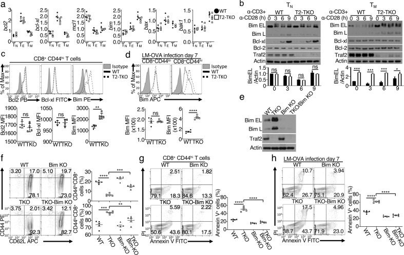
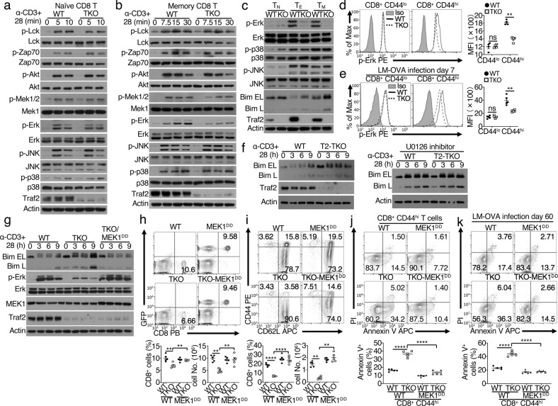
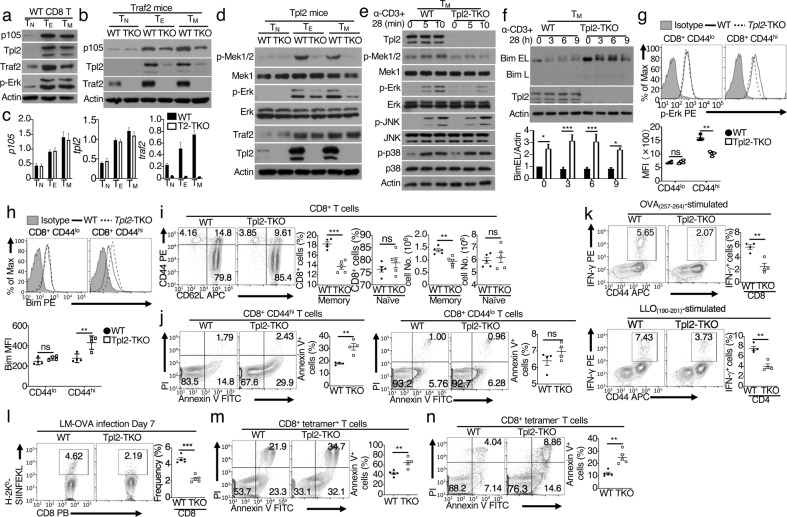
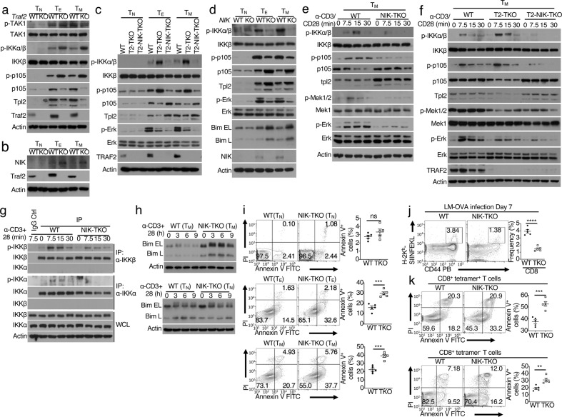
Generation and maintenance of antigen-specific effector and memory T cells are central events in immune responses against infections. We show that TNF receptor-associated factor 2 (TRAF2) maintains a survival signaling axis in effector and memory CD8 T cells required for immune responses against infections. This signaling axis involves activation of Tpl2 and its downstream kinase ERK by NF-κB-inducing kinase (NIK) and degradation of the proapoptotic factor Bim. NIK mediates Tpl2 activation by stimulating the phosphorylation and degradation of the Tpl2 inhibitor p105. Interestingly, while NIK is required for Tpl2-ERK signaling under normal conditions, uncontrolled NIK activation due to loss of its negative regulator, TRAF2, causes constitutive degradation of p105 and Tpl2, leading to severe defects in ERK activation and effector/memory CD8 T cell survival. Thus, TRAF2 controls a previously unappreciated signaling axis mediating effector/memory CD8 T cell survival and protective immunity.

Keywords: Bacterial infection; Effector and memory CD8 T cells; NIK; Protective immunity; T cell survival; TRAF2; Tpl2.


Conflict of interest statement

The authors declare no competing interests.

Figures

Fig. 1
Fig. 1
TRAF2 deficiency dampens antibacterial immune responses. ad Measuring primary T cell responses 7 days after infection of wild-type (WT) and Traf2-TKO mice with LM-OVA (n = 5). Flow cytometric analysis of the percentages and absolute numbers of splenic CD4 and CD8 T cells (a) or naïve (CD44lo) and memory (CD44hi) CD8 T cells (b). ICS and flow cytometric analysis of IFN-γ-producing CD8 (c) and CD4 (d) T cells in splenocytes restimulated in vitro for 6 h with OVA257–264 (c) or LLO190–201 (d) peptide in the presence of monensin. e Bacterial load in the spleen and liver in wild-type and Traf2-TKO mice 5 days after infection with LM-OVA (n = 5). f Flow cytometric analysis of IFN-γ-producing CD8 T cells in the spleens of wild-type and Traf2-TKO OT-I mice 7 days after infection with LM-OVA (n = 5). Data are representative of three independent experiments. Summary data are shown as the mean ± SEM values, and p values were determined by unpaired Student’s t test. *p < 0.05; **p < 0.01; ***p < 0.001; ****p < 0.0001; ns not significant
Fig. 2
Fig. 2
TRAF2 deficiency impairs memory CD8 T cell responses and protective immunity. a Schematic of the experimental design, in which wild-type (WT) and Traf2-TKO (T2-TKO) mice were intravenously infected with a low dose (5 × 104 cfu/mouse) of LM-OVA, reinfected on day 60 with a higher dose (1 × 106 cfu/mouse) of LM-OVA, and analyzed after 3 days (n = 5). ICS and flow cytometric analyses of IFN-γ-producing CD8 (b) and CD4 (c) T cells among splenocytes after in vitro restimulation with the indicated peptides; analysis of the bacterial load in the spleen and liver (d) in the LM-OVA-infected mice described in a. ei Study of memory CD8 T cell responses by adoptive transfer of wild-type or Traf2-TKO memory OT-I CD8 T cells (CD45.2+, 5 × 105) into wild-type WT B6.SJL (CD45.1+) mice prior to LM-OVA infection (n = 5). Schematic of the experimental design (e), ELISA of serum IFN-γ 4 h after infection (f), flow cytometric analysis of donor (CD45.2+) splenic OT-I CD8 T cells (g) or IFN-γ-producing OT-I CD8 T cells (h) 24 h after infection, and bacterial load 24 h after infection (i). Schematic of the experimental design (j) and survival curves (k) of WT and Traf2-TKO mice infected with a low dose (1 × 104 cfu/mice) of LM-OVA 30 days before rechallenge with a lethal dose (2 × 106 cfu/mice) of LM-OVA (n = 10, p = 0.0133 by the log-rank test). Schematic of the experimental design (l), tumor size data (m), and survival curves (n) of WT and Traf2-TKO mice infected with a low dose (1 × 104 cfu/mice) of LM-OVA 40 days before injection (s.c.) with B16-OVA melanoma cells (8 × 105 cells/mouse) (n = 10). Data are representative of two (en) or three (ad) independent experiments. Summary data are shown as the mean ± SEM values, and p values were determined by unpaired Student’s t test (af, bd, fi), two-way ANOVA with the Bonferroni correction (m), or log-rank test (k, n). *p < 0.05; **p < 0.01; ***p < 0.001; ****p < 0.0001
Fig. 3
Fig. 3
TRAF2 is required for the survival of effector and memory but not naïve CD8 T cells. a, b Flow cytometric analysis of apoptotic memory (CD44hi) and naïve (CD44lo) CD8 T cells in the spleens of wild-type (WT) and Traf2-TKO mice 7 days after infection with LM-OVA (n = 5). Flow cytometric analysis of the percentages and absolute numbers of CD4 and CD8 T cells (c), the percentages and absolute numbers of naïve (CD44lo) and memory (CD44hi) CD8 T cells (d), and the percentages of apoptotic memory (CD44hi, e), and naïve (CD44lo, f) CD8 T cells in the spleens of WT and Traf2-TKO mice 60 days after infection with LM-OVA (n = 5). Detection of memory responses to LM-OVA infection. Flow cytometric analysis of antigen-specific CD8 T cells using the H-2Kb OVA (SIINFKL) tetramer (g); flow cytometric analysis of apoptosis within the OVA tetramer-positive (h), and negative (i) CD8 T cell populations in the spleens of WT and Traf2-TKO mice infected with LM-OVA for 60 days and reinfected with LM-OVA for 3 additional days (n = 5). j, k Flow cytometric analysis of the percentage of apoptotic cells among naïve (TN) or in vitro-differentiated effector (TE), and memory (TM) WT and Traf2-TKO OT-I CD8 T cells (n = 4), based on annexin V and PI staining. Data are presented as a representative plot (j) and summary graph based on multiple mice (k). ln Mixed T cell transfer to study the cell-intrinsic function of TRAF2 in regulating CD8 T cell survival. l Schematic of the experimental design. WT B6.SJL (CD45.1+, CD45.2) mice were adoptively transferred with a mixture of WT (CD45.1+, CD45.2+) and Traf2-TKO (CD45.1, CD45.2+) naïve OT-I T cells (1 × 106 each). After 24 h, recipient mice were infected with 5 × 104 cfu of LM-OVA, and flow cytometric analysis of the donor T cells in the spleen at the indicated time points was performed. m Flow cytometric analysis of the percentages and absolute numbers of adoptively transferred WT (CD45.1+, CD45.2+) and Traf2-TKO (CD45.1, CD45.2+) OT-I CD8 T cells (n = 5). n Flow cytometric analysis of the apoptosis rate of adoptively transferred WT or Traf2-TKO T cells (n = 5). Data are representative of two (ln) or three (ak) independent experiments. Summary data are presented as the mean ± SEM values based on multiple animals (each symbol represents a mouse). p values were determined by unpaired Student’s t test. *p < 0.05; **p < 0.01; ***p < 0.001; ****p < 0.0001; ns not significant
Fig. 4
Fig. 4
TRAF2 controls the fate of Bim and supports the survival of effector and memory CD8 T cells. a QPCR analysis of the indicated genes in naïve (TN) or in vitro-differentiated effector (TE), and memory (TM) CD8 T cells (n = 4). b Immunoblot analysis of the indicated proteins in whole-cell lysates of naïve (TN) or in vitro-differentiated memory (TM) CD8 T cells stimulated with plate-bound anti-CD3 plus anti-CD28 antibodies for the indicated durations. Quantification of Bim EL bands is presented as the ratio of the Bim EL level to the Actin level (lower). c Flow cytometric analysis, based on intracellular staining, of the indicated proteins in memory-like (CD44hi) CD8 T cells in the spleens of 8-week-old wild-type (WT) and Traf2-TKO mice (n = 4). d Flow cytometric analysis of intracellular Bim in naïve (CD44lo) or memory (CD44hi) CD8 T cells from the spleens of WT and Traf2-TKO mice 7 days after infection with LM-OVA (n = 5). e Immunoblot analysis of Traf2 and Bim isoforms in whole-cell lysates of in vitro-differentiated memory CD8 T cells from wild-type (WT), Traf2-TKO (TKO), Bim KO, and Traf2-TKO/Bim KO mice. Flow cytometric analysis of the percentages of naïve (CD44lo) and memory (CD44hi) CD8 T cells (f) and the percentage of apoptotic cells within the memory (CD44hi) CD8 T cell population (g) in the spleens of 8-week-old WT, Traf2-TKO (TKO), Bim KO (WT-Bim KO), and Traf2-TKO/Bim KO mice (n = 5). h Flow cytometric analysis of apoptosis in splenic memory (CD44hi) CD8 T cells from the indicated mice 7 days after infection with LM-OVA (n = 5). Data are representative of two (e, h) or three (ad, f, g) independent experiments. Data in summary graphs are presented as the mean ± SEM values based on multiple mice (each symbol represents a mouse), and p values were determined by unpaired Student’s t test. *p < 0.05; **p < 0.01; ***p < 0.001; ****p < 0.0001; ns not significant
Fig. 5
Fig. 5
TRAF2 is required for ERK activation. Immunoblot analysis of the indicated phosphorylated (p-) and total proteins in whole-cell lysates of wild-type (WT) and Traf2-TKO naïve (a) or memory (b) CD8 T cells stimulated for the indicated durations with anti-CD3 plus anti-CD28 antibodies. c Immunoblot analysis of the indicated phosphorylated (p-) or total proteins in whole-cell lysates of naïve (TN) or in vitro-differentiated effector (TE), and memory (TM) WT and Traf2-TKO CD8 T cells. Flow cytometric analysis of phosphorylated ERK (p-ERK) in naïve (CD44lo) or memory-like (CD44hi) CD8 T cells in WT or Traf2-TKO mice that were either uninfected (d) or 7 days after infection with LM-OVA (n = 5) (e). f Immunoblot analysis of Bim isoforms in WT and Traf2-TKO memory CD8 T cells stimulated with plate-bound anti-CD3 plus anti-CD28 antibodies in the absence (left) or presence (right) of the MEK1 inhibitor U0126. g Immunoblot analysis of phosphorylated (p-) and total protein in whole-cell lysates of WT, Traf2-TKO (TKO), and Traf2-TKO/MEK1DD (TKO/MEK1DD) memory CD8 T cells stimulated for the indicated durations with anti-CD3 plus anti-CD28 antibodies. Flow cytometric analysis of the percentages and absolute numbers of CD8 T cells (h) and naïve (CD44lo) and memory-like (CD44hi) CD8 T cells (i) in the spleens of 8-week-old WT, Traf2-TKO (TKO), MEK1DD, and Traf2-TKO/MEK1DD (TKO-MEK1DD) mice (n = 5). Flow cytometric analysis of apoptosis in splenic memory-like (CD44hi) CD8 T cells from uninfected mice (j) and splenic memory (CD44hi) CD8 T cells in mice of the indicated genotypes 60 days after infection with LM-OVA (k) (n = 5). Data are representative of two (f, k) or three (ae, gj) independent experiments. Summary data are presented as the mean ± SEM values based on multiple mice, and p values were determined by unpaired Student’s t test. **p < 0.01; ****p < 0.0001; ns not significant
Fig. 6
Fig. 6
TRAF2 regulates ERK signaling by stabilizing the p105/Tpl2 complex. Immunoblot analysis of the indicated proteins in whole-cell lysates of naïve (TN) or in vitro-differentiated effector (TE), and memory (TM) CD8 T cells derived from WT mice (a) and from wild-type (WT) and Traf2-TKO (TKO) mice (b). c QPCR analysis of the indicated genes in naïve (TN) and in vitro-differentiated effector (TE) and memory (TM) CD8 T cells (n = 4). d Immunoblot analysis of the indicated proteins in whole-cell lysates of naïve (TN) and in vitro-differentiated effector (TE), and memory (TM) CD8 T cells from WT or Tpl2-TKO mice. e, f Immunoblot analysis of phosphorylated (p-) and total proteins in whole-cell lysates of WT and Tpl2-TKO memory CD8 T cells stimulated for the indicated durations with anti-CD3 plus anti-CD28 antibodies. The Bim EL band was quantified, and the level is presented as the ratio of Bim EL to Actin (f, lower). Flow cytometric analysis of p-ERK (g) and Bim (h) levels in naïve (CD44lo) and memory (CD44hi) CD8 T cells from the spleens of 8-week-old WT and Tpl2-TKO mice (n = 4). i Flow cytometric analysis of the percentages and absolute numbers of naïve (CD44lo) and memory-like (CD44hi) CD8 T cells in the spleens of 8-week-old WT and Tpl2-TKO mice (n = 5). j Flow cytometric analysis of apoptotic cells within the memory-like (CD44hi) and naïve (CD44lo) CD8 T cell populations in the spleens of 8-week-old WT and Tpl2-TKO mice (n = 4). k Intracellular cytokine staining (ICS) and flow cytometric analysis of IFN-γ-producing CD8 and CD4 effector T cells in the spleens of WT and Tpl2-TKO mice 7 days after infection with LM-OVA (n = 4). Splenocytes were restimulated for 6 h with OVA257–264 (for CD8 T cell stimulation) or LLO190–204 (for CD4 T cell stimulation) peptide in the presence of monensin prior to analysis. l Flow cytometric analysis, using H-2Kb OVA (SIINFKL) tetramers, of the percentage of antigen-specific CD8 T cells in the spleens of WT and Tpl2-TKO mice 7 days after infection with LM-OVA (n = 5). m, n Flow cytometric analysis of apoptotic cells within the H-2Kb OVA tetramer-positive and H-2Kb OVA tetramer-negative CD8 T cell populations in the spleens of WT and Tpl2-TKO mice 7 days after infection with LM-OVA (n = 5). Data are representative of two (c) or three (a, b, dn) independent experiments. Summary data are presented as the mean ± SEM values based on multiple mice. Summary data are presented as the mean ± SEM values based on multiple mice, and p values were calculated by Student’s t test. *p < 0.05; **p < 0.01; ***p < 0.001; ns not significant
Fig. 7
Fig. 7
NIK is a key regulator of Tpl2-ERK signaling in effector and memory CD8 T cells. Immunoblot analysis of the indicated phosphorylated (p-) and total proteins in whole-cell lysates of naïve (TN) and in vitro-differentiated effector (TE) and memory (TM) CD8 T cells from wild-type (WT) and Traf2-TKO (KO) mice (a, b) and from WT, Traf2-TKO (T2-TKO), and Traf2-NIK double TKO (T2-NIK-TKO) mice (c). d Immunoblot analysis of phosphorylated (p-) and total proteins in whole-cell lysates of naïve (TN) and in vitro-differentiated effector (TE) and memory (TM) CD8 T cells from WT and NIK-TKO (KO) mice. Immunoblot analysis of phosphorylated (p-) and total proteins in whole-cell lysates of in vitro-differentiated memory (TM) CD8 T cells derived from WT and NIK-TKO mice (e) and from WT, Traf2-TKO (T2-TKO), and Traf2-NIK double TKO (T2-NIK-TKO) mice (f), stimulated with anti-CD3 plus anti-CD28 antibodies for the indicated durations. g Immunoblot analyses of phosphorylated (p-) and total IKK protein isolated by immunoprecipitation under denaturing conditions with IKKα or IKKβ antibodies or directly from whole-cell lysates (WCL) of WT and NIK-TKO memory (TM) CD8 T cells stimulated with anti-CD3 plus anti-CD28 antibodies. IgG immunoprecipitation was included as a negative control. h Immunoblot analysis of the indicated proteins in whole-cell lysates of WT and NIK-TKO memory (TM) and naïve (TN) CD8 T cells stimulated for the indicated durations with anti-CD3 plus anti-CD28 antibodies. i Flow cytometric analysis of apoptotic cells within the naïve (TN) and in vitro-differentiated effector (TE) and memory (TM) CD8 T cell populations derived from WT and NIK-TKO mice (n = 5). Flow cytometric analysis of the percentage of antigen (OVA)-specific CD8 T cells using H-2Kb OVA (SIINFKL) tetramers (j) and apoptotic cells within the tetramer+ and tetramer CD8 T cell populations (k) in the spleens of WT and NIK-TKO mice 7 days after infection with LM-OVA (n = 5). Data are representative of two (f, j, k) or three (ae, gi) independent experiments. Summary data are presented as the mean ± SEM values based on multiple mice. p values were determined by unpaired Student’s t test (ik). **p < 0.01; ***p < 0.001; ****p < 0.0001; ns not significant

References

    1. Durgeau A, Virk Y, Corgnac S, Mami-Chouaib F. Recent advances in targeting CD8 T-Cell immunity for more effective cancer immunotherapy. Front. Immunol. 2018;9:14. - PMC - PubMed
    1. Glimcher LH, Townsend MJ, Sullivan BM, Lord GM. Recent developments in the transcriptional regulation of cytolytic effector cells. Nat. Rev. Immunol. 2004;4:900–911. - PubMed
    1. Cui W, Kaech SM. Generation of effector CD8+ T cells and their conversion to memory T cells. Immunol. Rev. 2010;236:151–166. - PMC - PubMed
    1. Marsden VS, Strasser A. Control of apoptosis in the immune system: Bcl-2, BH3-only proteins and more. Annu. Rev. Immunol. 2003;21:71–105. - PubMed
    1. Wojciechowski S, et al. Bim/Bcl-2 balance is critical for maintaining naive and memory T cell homeostasis. J. Exp. Med. 2007;204:1665–1675. - PMC - PubMed

Publication types

MeSH terms

Substances