Review
. 2020 Oct 26;5(10):691-698.
doi: 10.1302/2058-5241.5.190081. eCollection 2020 Oct.

A painful unknown: sacroiliac joint diagnosis and treatment

Affiliations
Review

A painful unknown: sacroiliac joint diagnosis and treatment

Jean-Charles Le Huec et al. EFORT Open Rev. .

Abstract

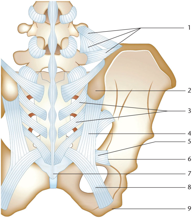
The sacroiliac joint (SIJ) is a complex anatomical structure located near the centre of gravity of the body.Micro-traumatic SIJ disorders are very difficult to diagnose and require a complete clinical and radiological examination.To diagnose micro-trauma SIJ pain it is recommended to have at least three positive provocative specific manoeuvres and then a radiologically controlled infiltration test.Conservative treatment combining physiotherapy and steroid injections is the most common therapy but has a low level of efficiency. SIJ thermolysis is the most efficient non-invasive therapy.SIJ fusion using a percutaneous technique is a solution that has yet to be confirmed on a large cohort of patients resistant to other therapies. Cite this article: EFORT Open Rev 2020;5:691-698. DOI: 10.1302/2058-5241.5.190081.

Keywords: diagnosis; dysfunction; micro-traumatic pains; minimally invasive fusion surgery; sacro-iliac joint (SIJ).


Conflict of interest statement

ICMJE Conflict of interest statement: J-CLH reports travel/accommodation/meeting expenses from Medtronic, Safeorthopaedics and Seaspine, outside the submitted work. The other authors declare no conflict of interest relevant to this work.

Figures

Fig. 1
Fig. 1
Anatomy of the SIJ. (A) Top view of the pelvic region. Sacro-iliac joint (SIJ – red line) is located between the sacrum (1) and iliac crest (2). A slight spread between the iliac crest and the sacrum allows a superior view of the SIJ region (3). (B) Transverse view of the pelvic region. The Iliac crest is spread from the sacrum to properly describe the anatomy of the pelvic region and observe the SIJ plate: (1) body of the pelvic bone, (2) auricular surface of the sacrum, (3) sacrum, (4) antero-superior iliac spine, (5) iliac tuberosity, (6) auricular surface of the ilium, (7) anterior sacro-iliac ligament, (8) postero-superior iliac spine.
Fig. 2
Fig. 2
Posterior ligaments of the sacro-iliac joint (posterior view). (1) Ilio-lumbar ligaments, (2) postero-superior iliac spine, (3) Inter-osseous sacro-iliac ligaments, (4) posterior sacro-iliac ligaments, (5) sacro-spinous ligament, (6) ischial spine, (7) coccyx; (8) sacro-tuberous ligament, (9) ischial tuberosity.
Fig. 3
Fig. 3
Mobility of the sacro-iliac joint. (A) Inter-osseous sacro-iliac ligaments (red points) are considered as the axis of rotation of the sacrum. Two mains movements are permitted depending on the sense of rotation: nutation and counter-nutation (red arrows). In the case of nutation, the coccyx moves away from the pelvis which is responsible for the enlargement of the lower outlet. Counter-nutation is described by the narrowing of the lower outlet and the enlargement of the upper outlet due to the close position of the coccyx to the pelvis. (B) During the nutation movement, the upper plate of S1 moves forward and the iliac crests are getting closer. The ischia move away which is associated with the increasing diameter of the lower outlet.
Fig. 4
Fig. 4
Provocative manoeuvres to diagnose a sacro-iliac joint (SIJ) dysfunction. If three of five of the following provocative manoeuvres are positives, the clinician can suspect an SIJ dysfunction. The diagnosis has to be confirmed using a radiological test or injection test. (A) Östgaard test. (B) Faber test. (C) Compression test. (D) Gaenslen’s test. (E) ‘Finger sign’ test. (F) Lasègue test.
Fig. 5
Fig. 5
Previous approaches. (A) Unilateral fusion of the sacro-iliac joint (SIJ): radiograph and computed tomography scan control. (B) External frame for sacro-iliac fusion.
Fig. 6
Fig. 6
Minimally invasive surgery of the sacro-iliac joint (SIJ). (A) Patient is in the ventral position. C-arm-guided approach allows for precisely inserting the first guide pin and to measure the screw length. The guide is perpendicular to the plane of instability through the ilium and the sacrum. A larger guide pin allows the insertion of the screws. SIJ fusion can be obtained using three triangular screws. The good position of each screw has to be post-operatively confirmed by computed tomography scan (B and C).

References

    1. Le Huec JC, Tsoupras A, Leglise A, Heraudet P, Celarier G, Sturresson B. The sacro-iliac joint: a potentially painful enigma. Update on the diagnosis and treatment of pain from micro-trauma. Orthop Traumatol Surg Res 2019;105:S31–S42. - PubMed
    1. Vleeming A, Schuenke MD, Masi AT, Carreiro JE, Danneels L, Willard FH. The sacroiliac joint: an overview of its anatomy, function and potential clinical implications. J Anat 2012;221:537–567. - PMC - PubMed
    1. Paquin JD, van der Rest M, Marie PJ, et al. Biochemical and morphologic studies of cartilage from the adult human sacroiliac joint. Arthritis Rheum 1983;26:887–895. - PubMed
    1. Solonen KA. The sacroiliac joint in the light of anatomical, roentgenological and clinical studies. Acta Orthop Scand Suppl 1957;27:1–127. - PubMed
    1. Nakagawa T. [Study on the distribution of nerve filaments over the iliosacral joint and its adjacent regio n in the Japanese]. Nihon Seikeigeka Gakkai Zasshi 1966;40:419–430. - PubMed