Case Reports
. 2020 Nov 3:2020:8823428.
doi: 10.1155/2020/8823428. eCollection 2020.

Diagnosis of Leiomyosarcoma after Uterine Artery Embolization for Multiple Leiomyomas

Affiliations
Case Reports

Diagnosis of Leiomyosarcoma after Uterine Artery Embolization for Multiple Leiomyomas

Maako Tsuji et al. Case Rep Obstet Gynecol. .

Abstract

Uterine sarcoma is significantly rarer than leiomyoma and has poor prognosis. Moreover, the diagnosis of leiomyosarcoma is difficult because its symptoms, including pelvic pain, uterine mass, and/or uterine bleeding, are very similar to those of leiomyoma. There are a few cases of leiomyosarcoma wherein leiomyoma was treated with uterine artery embolization (UAE); these reports revealed that the symptoms of hypermenorrhea or/and pelvic pain persisted even after UAE. Symptoms persisting even after UAE treatment for leiomyomas, especially multiple leiomyomas, should be investigated to rule out leiomyosarcoma. Therefore, long-term follow-up is needed. Here, we describe a case of a 39-year-old woman diagnosed with leiomyosarcoma 3 years after undergoing UAE for multiple leiomyomas.


Conflict of interest statement

The authors declare that they have no conflicts of interest.

Figures

Figure 1
Figure 1
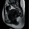
Magnetic resonance imaging was performed before uterine artery embolization. MR images show clearly visible fibroids and not tumors.
Figure 2
Figure 2
Magnetic resonance imaging was performed 2 years after uterine artery embolization. The leiomyomas grew smaller.
Figure 3
Figure 3
Magnetic resonance imaging was performed 3.5 years after uterine artery embolization. The leiomyomas were stable, and a new uterine mass was noted.

Similar articles

Cited by

References

    1. Norris H. J., Taylor H. B. Mesenchymal tumors of the uterus. I. A clinical and pathological study of 53 endometrial stromal tumors. Cancer. 1966;19(6):755–766. doi: 10.1002/1097-0142(196606)19:6<755::aid-cncr2820190604>3.0.co;2-u. - DOI - PubMed
    1. Ricci S., Stone R. L., Fader A. N. Uterine leiomyosarcoma: epidemiology, contemporary treatment strategies and the impact of uterine morcellation. Gynecologic Oncology. 2017;145(1):208–216. doi: 10.1016/j.ygyno.2017.02.019. - DOI - PubMed
    1. Papadia A., Salom E. M., Fulcheri E., Ragni N. Uterine sarcoma occurring in a premenopausal patient after uterine artery embolization: a case report and review of the literature. Gynecologic Oncology. 2007;104(1):260–263. doi: 10.1016/j.ygyno.2006.08.051. - DOI - PubMed
    1. Parker W. H., Fu Y. S., Berek J. S. Uterine sarcoma in patients operated on for presumed leiomyoma and rapidly growing leiomyoma. Obstetrics and Gynecology. 1994;83:p. 414. - PubMed
    1. Hinchcliff E. M., Esselen K. M., Watkins J. C., et al. The role of endometrial biopsy in the preoperative detection of uterine leiomyosarcoma. Journal of Minimally Invasive Gynecology. 2016;23(4):567–572. doi: 10.1016/j.jmig.2016.01.022. - DOI - PubMed

Publication types

LinkOut - more resources