Case Reports
. 2020 Oct 29:34:101472.
doi: 10.1016/j.eucr.2020.101472. eCollection 2021 Jan.

Needle in a haystack: Robotic removal of a retroperitoneal wire

Affiliations
Case Reports

Needle in a haystack: Robotic removal of a retroperitoneal wire

Max S Yudovich et al. Urol Case Rep. .

Abstract

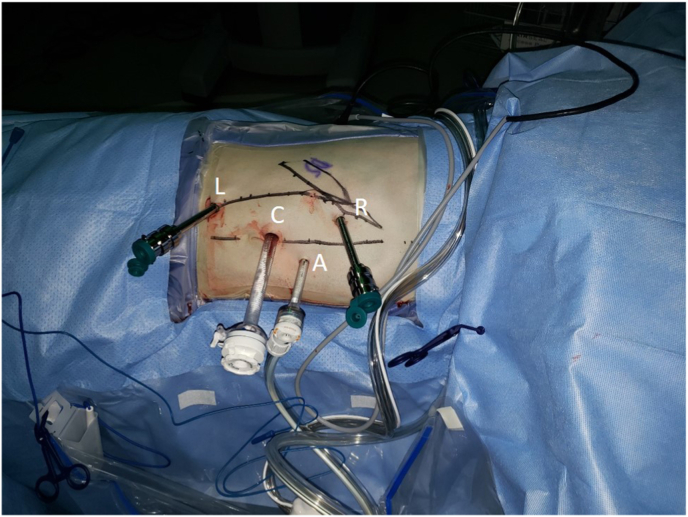
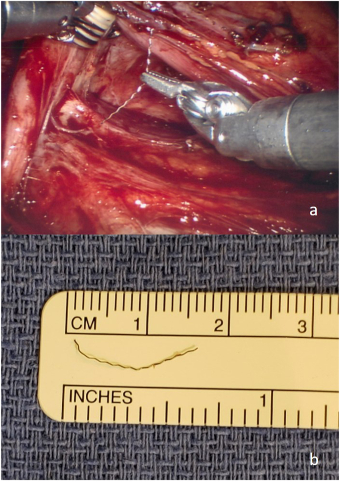
Retroperitoneal foreign bodies are rare indications for exploratory surgery. We present a case of a 19-year-old male with abdominal pain after a fall who was found to have a linear metallic object adjacent to the right ureter and inferior vena cava. Given the patient's pain and discomfort, he elected for robotic exploration of the retroperitoneum, which was carried out successfully with the Da Vinci Si® robot. This case demonstrates the feasibility of robotic retroperitoneal exploration and foreign body retrieval for a very small object.

Keywords: Robotic; foreign body; retroperitoneal.


Conflict of interest statement

The authors declare no conflicts of interest.

Figures

Fig. 1
Fig. 1
a. Initial axial CT image of the abdomen showing the metallic object (arrow) medial to the right lobe of the liver and posterior to the rectus musculature, and b. Subsequent axial CT image of the abdomen showing migration of the object (arrow) to be adjacent to the right ureter (U), inferior vena cava, and right lower renal pole (RP).
Fig. 2
Fig. 2
Robotic trocar placement for the procedure, including 5 mm left arm port for Maryland bipolar grasper (L), 12 mm camera port (C), 5 mm AirSeal® assistant port (A), and 5 mm right arm port for monopolar scissors (R).
Fig. 3
Fig. 3
a. Intraoperative image showing retrieval of the foreign body, and b. Foreign body with ruler to scale, demonstrating length consistent with estimates from pre-operative imaging.

References

    1. Mikhail D., Sarcona J., Mekhail M., Richstone L. Urologic robotic surgery. Surg Clin North Am. 2020;100(2):361–378. doi: 10.1016/j.suc.2019.12.003. - DOI - PubMed
    1. Corcione F., Esposito C., Cuccurullo D. Advantages and limits of robot-assisted laparoscopic surgery: preliminary experience. Surg Endosc. 2005;19(1):117–119. doi: 10.1007/s00464-004-9004-9. - DOI - PubMed
    1. Koutserimpas C., Ioannidis A., Konstantinidis M., Athanasopoulos P., Antonakopoulos F., Konstantinidis K. Robot-assisted removal of a broken scalpel blade following discectomy. Case Rep Surg. 2019;2019:8609246. doi: 10.1155/2019/8609246. - DOI - PMC - PubMed
    1. Grogan R.H. Current status of robotic adrenalectomy in the United States. Gland Surg. 2020;9(3):840–843. doi: 10.21037/gs.2020.03.39. - DOI - PMC - PubMed
    1. Reeves J.M., Wade M.D., Edwards J. Ingested foreign body mimicking acute appendicitis. Int J Surg Case Rep. 2018;46:66–68. doi: 10.1016/j.ijscr.2018.04.003. - DOI - PMC - PubMed

Publication types

LinkOut - more resources