

CT image evaluation of one-screw fixation in the Latarjet procedure
- PMID: 33204803
- PMCID: PMC7649353
- DOI: 10.1016/j.tcr.2020.100372
CT image evaluation of one-screw fixation in the Latarjet procedure
Erratum in
-
Erratum regarding missing patient consent statement in previously published articles.Trauma Case Rep. 2023 Mar 1;45:100809. doi: 10.1016/j.tcr.2023.100809. eCollection 2023 Jun. Trauma Case Rep. 2023. PMID: 37234577 Free PMC article.
Abstract
Background: In the Latarjet procedure, two screws are used for secure fixation. However, when the graft is small, two-screw fixation is technically difficult. The purpose of this study was to evaluate the bone union of one-screw fixation on CT images.
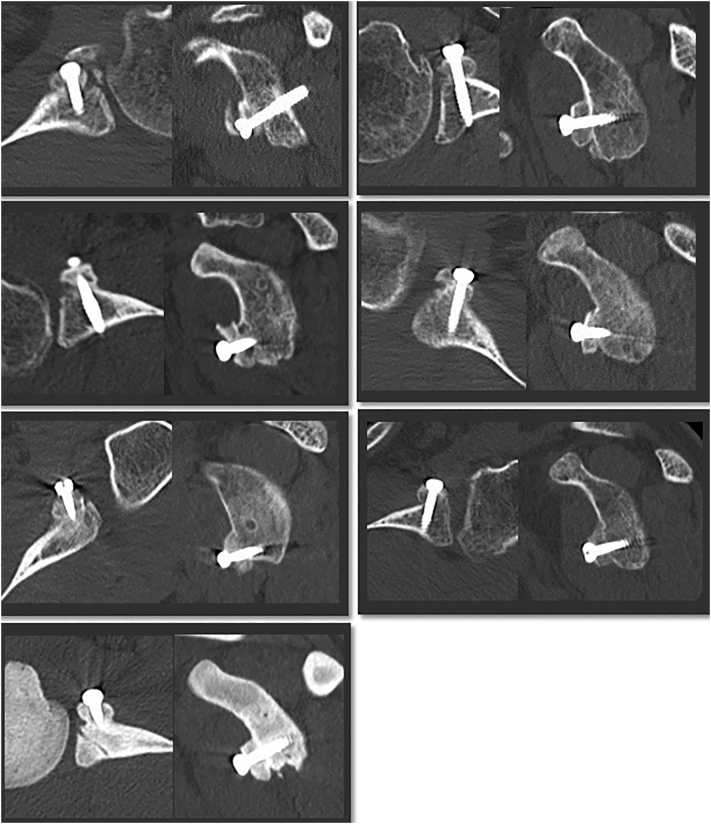
Methods: Ten shoulders with anterior recurrent dislocation underwent the open Latarjet procedure using one-screw fixation combined with arthroscopic Bankart repair. The bone union and the graft position were evaluated on CT images at 3, 6, and 12 months after the operation.
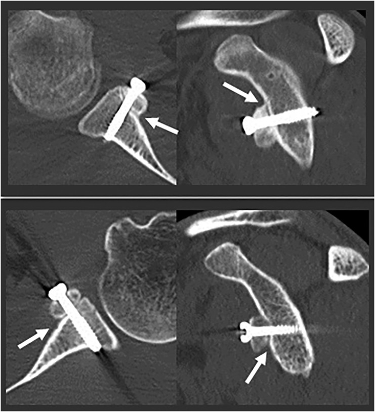
Results: Seven of 10 shoulders showed bone union at 3 months after the operation. In two shoulders, the removal of cortex was insufficient, and bone union was observed at 6 months. In one shoulder, the distal part of the graft was not in contact with the glenoid, and bone union was observed at 12 months. In one shoulder, the graft was healed in a 70-degree-rotated position. The average thickness of the graft was 7.2 ± 1.0 mm.
Conclusion: One-screw fixation in the Latarjet procedure did not show non-union. Sufficient removal of the graft cortex and good contact were needed for early union.
Keywords: Anterior instability; Bankart repair; Bone union; CT images; Latarjet procedure; One-screw fixation.
© 2020 The Author.
Conflict of interest statement
None.
Figures


References
-
- Skare Ø., Schrøder C.P., Mowinckel P., Reikerås O., Brox J.I. Reliability, agreement and validity of the 1988 version of the Rowe Score. J Shoulder Elbow Surg. 2011;20:1041–1049. - PubMed
-
- Chuang T.Y., Adams C.R., Burkhart S.S. Use of preoperative three-dimensional computed tomography to quantify glenoid bone loss in shoulder instability. Arthroscopy. 2008;24:376–382. - PubMed
-
- van der Linde J.A., van Wijngaarden R., Somford M.P., van Deurzen D.F., van den Bekerom M.P. The Bristow-Latarjet procedure, a historical note on a technique in comeback. Knee Surg Sports Traumatol Arthrosc. 2016;24:470–478. - PubMed
-
- Cowling P.D., Akhtar M.A., Liow R.Y. What is a Bristow-Latarjet procedure? A review of the described operative techniques and outcomes. Bone Joint J. 2016;98-B:1208–1214. - PubMed
-
- Mizuno N., Denard P.J., Raiss P., Melis B., Walch G. Long-term results of the Latarjet procedure for anterior instability of the shoulder. J Shoulder Elbow Surg. 2014;23:1691–1699. - PubMed
Publication types
LinkOut - more resources
Full Text Sources
