. 2020 Jun 8;6(6):3231-3257.
doi: 10.1021/acsbiomaterials.9b01667. Epub 2020 May 10.

Microphysiological Systems: Design, Fabrication, and Applications

Affiliations

Microphysiological Systems: Design, Fabrication, and Applications

Kai Wang et al. ACS Biomater Sci Eng. .

Abstract

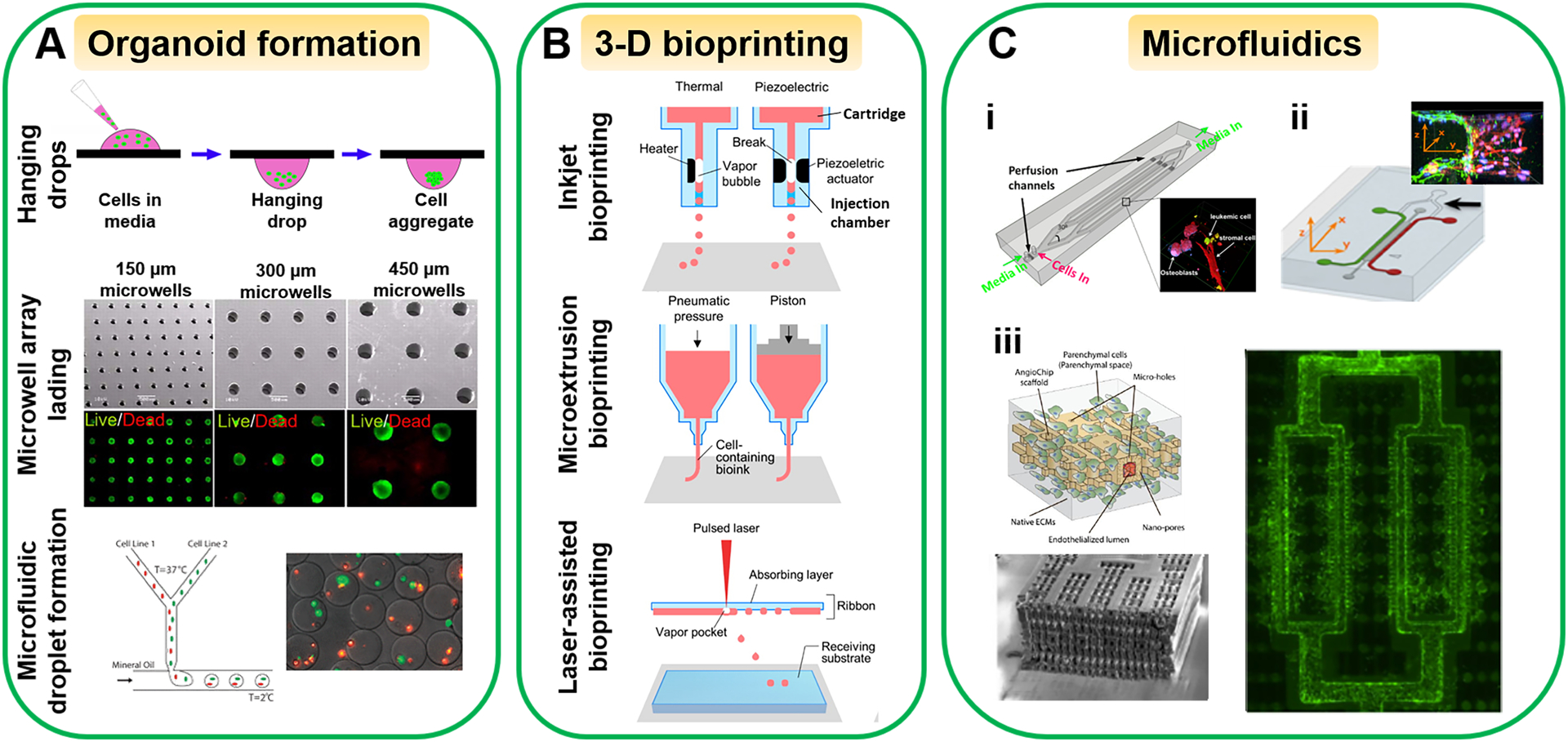
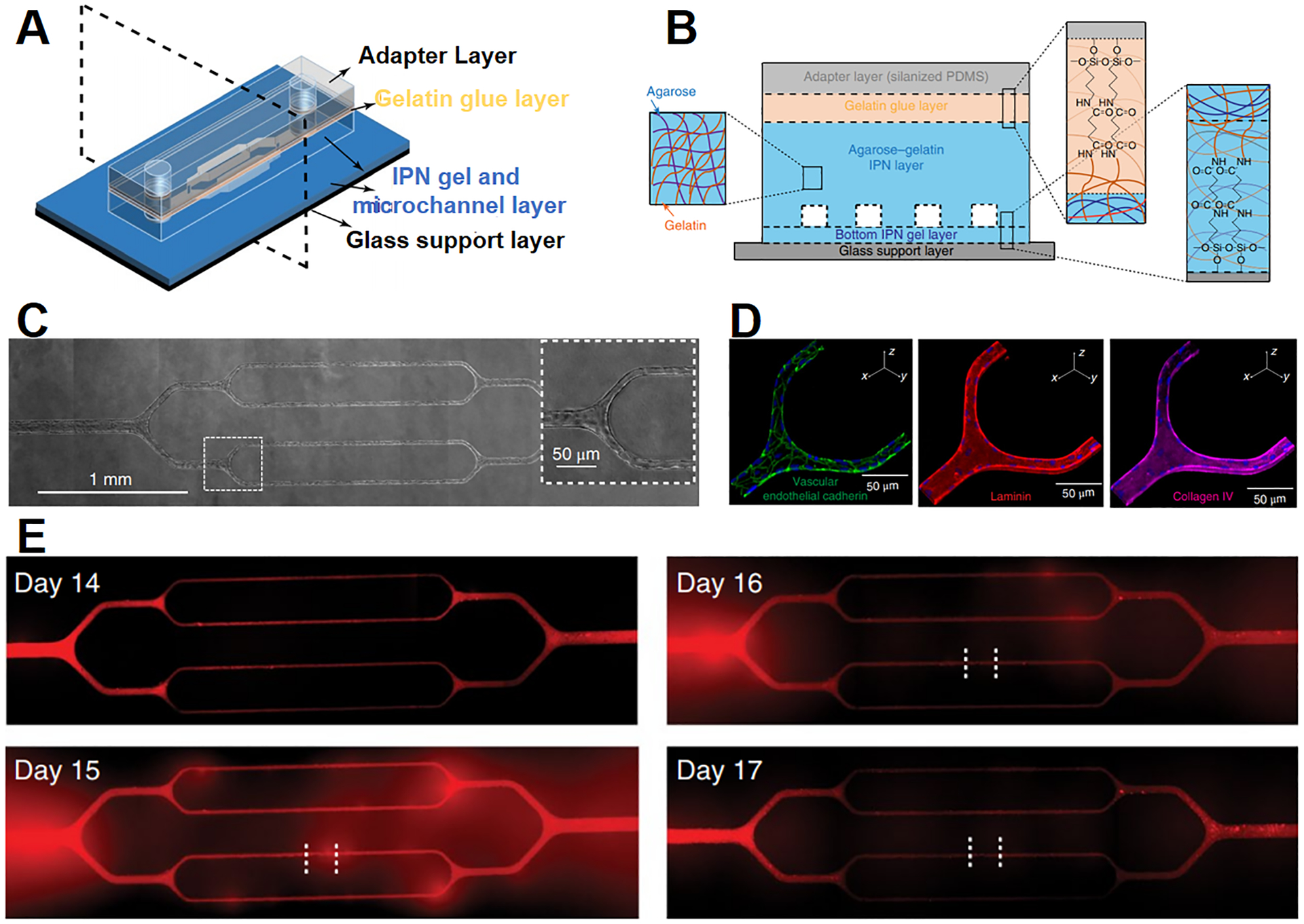
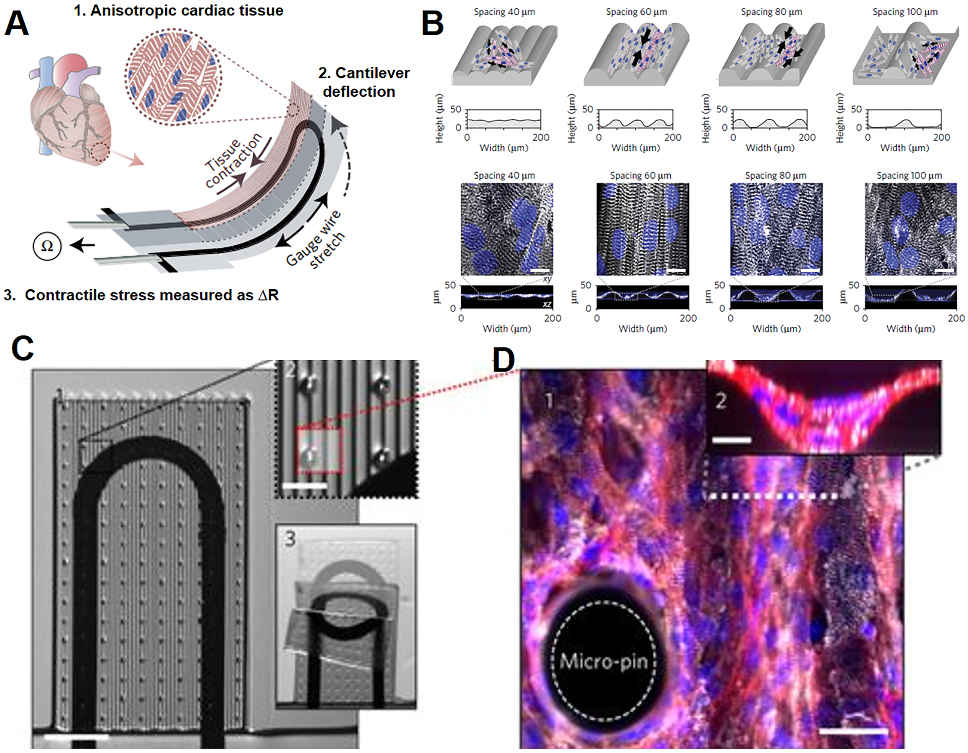
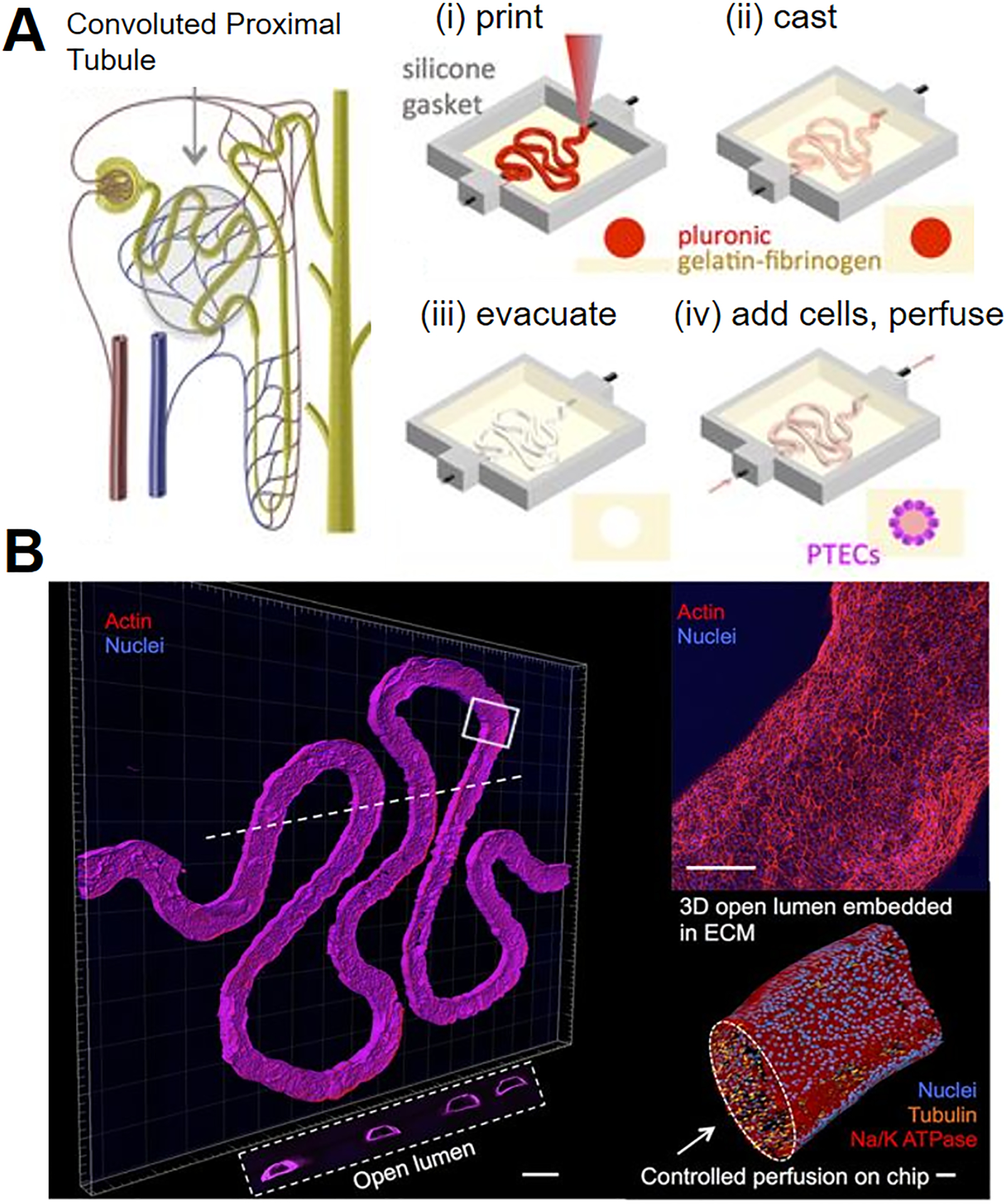
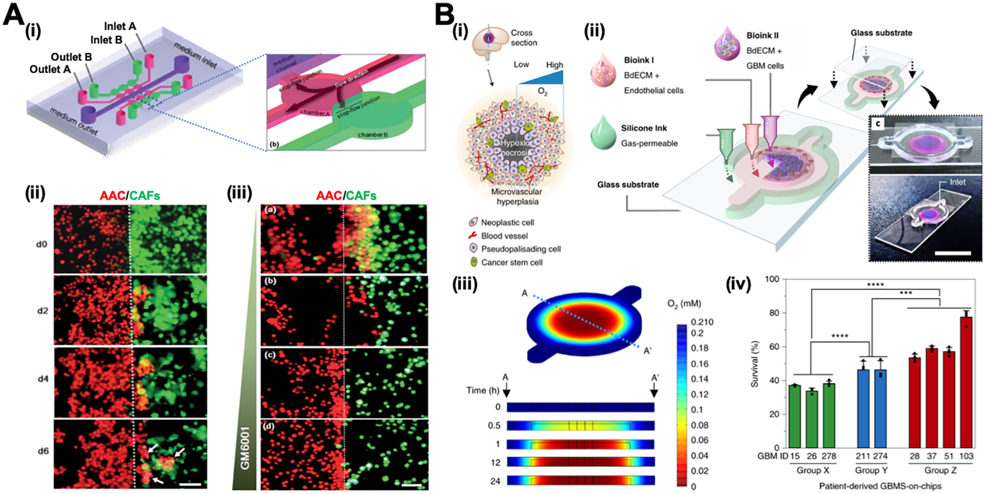
Microphysiological systems, including organoids, 3-D printed tissue constructs and organ-on-a-chips (organ chips), are physiologically relevant in vitro models and have experienced explosive growth in the past decades. Different from conventional, tissue culture plastic-based in vitro models or animal models, microphysiological systems recapitulate key microenvironmental characteristics of human organs and mimic their primary functions. The advent of microphysiological systems is attributed to evolving biomaterials, micro-/nanotechnologies and stem cell biology, which enable the precise control over the matrix properties and the interactions between cells, tissues and organs in physiological conditions. As such, microphysiological systems have been developed to model a broad spectrum of organs from microvasculature, eye, to lung and many others to understand human organ development and disease pathology and facilitate drug discovery. Multiorgans-on-a-chip systems have also been developed by integrating multiple associated organ chips in a single platform, which allows to study and employ the organ function in a systematic approach. Here we first discuss the design principles of microphysiological systems with a focus on the anatomy and physiology of organs, and then review the commonly used fabrication techniques and biomaterials for microphysiological systems. Subsequently, we discuss the recent development of microphysiological systems, and provide our perspectives on advancing microphysiological systems for preclinical investigation and drug discovery of human disease.

Keywords: 3-D printing; anatomy; microenvironment; microphysiological systems; organ chips; organoids; physiology.


Conflict of interest statement

Disclosure The authors declare no conflict of interest.

Figures

Figure 1.
Figure 1.. Overview of the design, fabrication and applications of microphysiological systems.
The figure is adapted with permissions from refs (Copyright 2010, The American Association for the Advancement of Science), (Copyright 2016, Dove Medical Press), (Copyright 2019, American Chemical Society), (Copyright 2018, American Chemical Society), (Copyright 2016, Elsevier), (Copyright 2016, Springer Nature), (Copyright 2016, American Chemical Society), (Copyright 2015, PLOS), (Copyright 2018, Royal Society of Chemistry), (Copyright 2017, Springer Nature), (Copyright 2018, Springer Nature), (Copyright 2010, Royal Society of Chemistry).
Figure 2.
Figure 2.. Exemplification of the design principles of microphysiological systems.
(A) Schematic and histological images of human cornea. Scale bar is 50 μm. Adapted with permission from ref . Copyright 2015, Mary Ann Liebert, Inc. (B) In vitro cornea model consisting of multiple epithelial layers, stroma and innervation. Adapted with permission from ref . Copyright 2016, Elsevier. (C) Illustration of the BBB. Adapted with permission from ref . Copyright 2016, Springer Nature. (D) A microfluidic chip recapitulating the physiological (stiffness, fluidic flows and cell-cell interactions) characteristics of the BBB. Adapted with permission from ref . Copyright 2015, AIP Publishing. (E) Illustration of the mechanical stretching of lung alveoli during breathing. Adapted with permission from ref . Copyright 2010, The American Association for the Advancement of Science. (F) A lung chip recreating the alveolar-capillary interface with fluidic flows and cyclic mechanical stretching. Adapted with permission from ref . Copyright 2010, The American Association for the Advancement of Science.
Figure 3.
Figure 3.. Commonly used fabrication techniques to engineer microphysiological systems.
(A) Organoid formation, (B) 3-D bioprinting, and (C) Microfluidic techniques. (C-i) An engineered 3-D bone marrow with leukemic cells in microchannels as a tumor-on-a-chip. Adapted with permission from ref . Copyright 2015, PLOS. (C-ii) The tumor-vascular interface on a 3-microchannel chip for tumor cell intravasation study. Adapted with permission from ref . Copyright 2012, National Academy of Sciences. (C-iii) An AngioChip with perfusable, branched, 3-D microchannel network. Adapted with permission from ref . Copyright 2016, Springer Nature. The microwell array lading images in (A) were adapted with permission from ref . Copyright 2009, National Academy of Sciences. The microfluidic droplet formation images in (A) were adapted with permission from ref . Copyright 2011, Oxford University Press. (B) was adapted with permission from ref . Copyright 2016, American Chemical Society.
Figure 4.
Figure 4.. A microvasculature-on-a-chip to study endothelial barrier dysfunction.
Schematic illustration of (A) the design and (B) the cross-sectional view of the microvasculature chip. (C) Brightfield images of the microvasculature chip with endothelial cells cultured for 14 days. The enlarged view of the boxed area shows the cells reach confluency in microchannels. (D) Confocal images of VE-cadherin (green), laminin (red) and collagen IV (purple) of HUVECs after 14 days of culture. The nuclei are in blue. (E) Fluorescent images demonstrate the change in endothelial permeability after the treatment of plasmodium falciparum-infected RBCs (iRBCs). Endothelial layer is treated with iRBCs for 4 h at day 14, fluorescent images show increased permeability of the cell layer at day 15 and 16, while the barrier function is recovered at day 17. Adapted with permission from ref . Copyright 2018, Springer Nature.
Figure 5.
Figure 5.. A heart-on-a-chip capable of monitoring cardiac contractility.
(A) Illustration of the heart chip design. Cardiomyocytes form anisotropic myocardium on the engineered cantilever, which is deflected by myocardial contraction and stretches the soft strain gauge. The resistance change is used to calculate the contractile stress of myocardium. (B) The microgrooves are engineered on the cantilever to guide the elongation of cardiomyocytes. White: α-actinin; Blue: DAPI. Scale bars are 10 μm. (C) 1. Modified cantilever containing micropins is used to support approximately four cardiomyocyte layers. 2. Close-up of the micropins. 3. A cantilever under deflecting. (D) 1. Fluorescent image of the myocardial tissue on the modified cantilever. Scale bar is 30 μm. 2. A cross-sectional image of the myocardial tissue. Scale bar is 10 μm. White: α-actinin; Red: actin; Blue: DAPI. Adapted with permission from ref . Copyright 2017, Springer Nature.
Figure 6.
Figure 6.. A liver-on-a-chip with perfusion culture of 3D hepatic spheroids.
(A) Schematic illustration of the hepatic sinusoid structure in vivo and the corresponding chip design. (B) A cross-sectional view of the multilayered liver chip. The lower microwell layer is used for hepatic spheroids culture, and the middle microporous membrane permits the nutrients and waste exchange yet serves as a barrier to protect the hepatic spheroids from the high fluid shear stress in the upper layer. (C) Fluorescent images of MRP-2 and ZO-1 in 2-D hepatic monolayer and 3-D hepatic spheroids. Adapted with permission from ref . Copyright 2018, Royal Society of Chemistry.
Figure 7.
Figure 7.. A renal proximal tubule-on-a-chip.
(A) Schematic illustration of a nephron containing proximal tubule and the fabrication process of the proximal tubule chip. (B) 3-D confocal images of the formed proximal tubule. Scale bar is 500 μm. Upper right image: the enlarged view of the proximal tubule. Scale bar is 200 μm. Lower right image: a 3-D rendering of proximal tubule to show the open lumen. Scale bar is 50 μm. The white dash line shows the location of the cross-sectional view in the white box. Adapted with permission from ref . Copyright 2016, Springer Nature.
Figure 8.
Figure 8.. Tumor-on-a-chip systems (A) to study tumor-CAF interactions and (B) to identify patient-specific responses to chemoradiotherapy.
(A) i: Schematic illustration of the chip design. ii: Invasion of AAC cells co-cultured with CAFs over time. White arrows show that AAC cells form spheroids in CAF-containing matrix. iii: Invasion of AAC cells co-cultured with CAFs under the treatment of GM6001 at the concentration of (a) 0 mg/mL; (b) 10 mg/mL; (c) 50 mg/mL; and (d) 100 mg/mL. Scale bars are 100 μm. Adapted with permission from ref . Copyright 2010, Royal Society of Chemistry. (B) i: Schematic illustration of the heterogeneous structure of the glioblastoma tissue. ii: Schematic illustration and photos of the glioblastoma chip. iii: Heatmap images of oxygen distribution in the glioblastoma chip along the crosssection A–A’. iv: The percentage survival after chemoradiation of the glioblastoma chip prepared by using patient-specific glioblastoma cells. The patients exhibited low-to-moderate resistance in Group X and high resistance in Group Y to chemoradiation treatment, while experienced extremely aggressive tumor progression in Group Z after the treatment. Adapted with permission from ref . Copyright 2019, Springer Nature.

References

    1. Greek R; Menache A, Systematic reviews of animal models: methodology versus epistemology. Int. J. Med. Sci 2013, 10 (3), 206–221. - PMC - PubMed
    1. Low L; Tagle D, Tissue chips–innovative tools for drug development and disease modeling. Lab Chip 2017, 17 (18), 3026–3036. - PMC - PubMed
    1. Park SE; Georgescu A; Huh D, Organoids-on-a-chip. Science 2019, 364 (6444), 960–965. - PMC - PubMed
    1. Huh D; Matthews BD; Mammoto A; Montoya-Zavala M; Yuan Hsin H; Ingber DE, Reconstituting organ-level lung functions on a chip. Science 2010, 328 (5986), 1662–1668. - PMC - PubMed
    1. Ronaldson-Bouchard K; Vunjak-Novakovic G, Organs-on-a-chip: a fast track for engineered human tissues in drug development. Cell Stem Cell 2018, 22 (3), 310–324. - PMC - PubMed

Publication types

LinkOut - more resources