. 2020 Oct 8:24:100824.
doi: 10.1016/j.bbrep.2020.100824. eCollection 2020 Dec.

Mitofusin-2 mediates doxorubicin sensitivity and acute resistance in Jurkat leukemia cells

Affiliations

Mitofusin-2 mediates doxorubicin sensitivity and acute resistance in Jurkat leukemia cells

Carl W Decker et al. Biochem Biophys Rep. .

Abstract

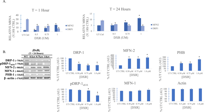
Mitochondria oscillate along a morphological continuum from fragmented individual units to hyperfused tubular networks. Their position at the junction of catabolic and anabolic metabolism couples this morphological plasticity, called mitochondrial dynamics, to larger cellular metabolic programs, which in turn implicate mitochondria in a number of disease states. In many cancers, fragmented mitochondria engage the cell with the biosynthetic capacity of aerobic glycolysis in service of proliferation and progression. Chemo-resistant cancers, however, favor remodeling dynamics that yield fused mitochondrial assemblies utilizing oxidative phosphorylation (OXPHOS) through the electron transport chain (ETC). In this study, expression of Mitofusin-2 (MFN-2), a GTPase protein mediator of mitochondrial fusion, was found to closely correlate to Jurkat leukemia cell survival post doxorubicin (DxR) assault. Moreover, this was accompanied by dramatically increased expression of OXPHOS respiratory complexes and ATP Synthase, as well as a commensurate escalation of state III respiration and respiratory control ratio (RCR). Importantly, CRISPR knockout of MFN-2 resulted in a considerable decrease of doxorubicin (DxR) median lethal dose compared to a treated wildtype control, suggesting an important role of mitochondrial fusion in chemotherapy sensitivity and acute resistance.

Keywords: Doxorubicin; MFN-2; Mitochondrial fusion; OXPHOS; Sensitivity.


Conflict of interest statement

The authors declare that they have no known competing financial interests or personal relationships that could have appeared to influence the work reported in this paper.

Figures

Image 1
Graphical abstract
Fig. 1
Fig. 1
A) Doxorubicin (DxR) cytotoxicity for suspended jukrat t-cell cultures over a treatment interval of 24 h determined through trypan exclusion. Viable population is reduced dose-dependently with increases to DxR treatment concentration, with approximate acute median lethal dose (LD50) achieved at 0.75 μM (53% of untreated control, ± 6.6%). B) Cell cycle phase analysis via flow cytometry. Fragmented DNA is used as indication of apoptosis, given DNA intercalation mechanism of DxR. DxR treatment at 0.75 μM increases DNA fragmented cell proportion from 15.8% ± 3.0% of population to 52.3% ± 6.4% versus untreated WT (wildtype). Additionally, the proportion of proliferative G2/M phase in treated cells is 4.5% ± 0.9, down from 12.1% ± 0.5% in control. Data represent the mean ± SD, n > 3.
Fig. 2
Fig. 2
A) Real-time quantitative PCR gene detection comparison measuring protein mediators of mitochondrial morphology across doxorubicin (DxR) treatment concentrations and untreated control (UT) at 1 and 24 h relative to an 18s control gene. At 1 h post-DxR treatment, transcript of dynamin related protein 1 (DRP-1), a GTPase mediator of mitochondrial fission, is shown to decrease. This is accompanied by a concurrent increase of MFN-2, a mitochondrial outer membrane fusion mediator, indicating hasty mitochondrial fusion in response to DxR; these same dynamics are observed at a 24 h endpoint. B) Immunoblot and densitometry (relative arbitrary units, AU) of mitochondrial dynamics mediators in DxR treated t-cells at 24 h. Corroborating RT-qPCR data, both DRP-1 and activated pDRP-1s616 are decreased in response to treatment. MFN-2 is likewise shown to increase as in RT-qPCR assay, though an increase of MFN-1 was determined to be statistically insignificant. Prohibitin 1 (PHB-1), a pleiotropic scaffolding protein known to stabilize mitochondrial inner membrane proteins and support cristae morphogenesis and maintenance, is upregulated in treated conditions compared to control. Taken together, data suggest MFN-2 mediated fusion in response to DxR, beginning at the latest 1 h after treatment, through to a 24 h interval. Data represent the mean ± SD, n > 3.
Fig. 3
Fig. 3
A) Immunoblot of electron transport chain respiratory proteins. Compared to untreated control (UT), complexes I, III, and IV are greatly upregulated, with the largest increase in expression occurring in complex V (ATP Synthase), indicating increased ATP production via oxidative phosphorylation. Interestingly, complex II (succinate dehydrogenase) is not increased, perhaps to further produce ATP through glutamate/malate-driven respiration via complex I, which is more efficient per molecule of glucose. Data are representative of n > 3. B) Oxygen consumption/mitochondrial respiration rates (states III and IV) and RCR measured using polarographic oxygen electrode. State III respiration, in which ADP and substrate (for complex I, glutamate) are saturated, is indicative of maximal electron transport chain (ETC) respiration. The increased expression of oxidative phosphorylation (OXPHOS) proteins observed in DxR treated jurkats is accompanied by a commensurate increase in glutamate/malate-driven state III respiration. State IV respiration, or minimal respiratory capacity, is only marginally altered, suggesting very little proton leak or mitochondrial damage. Respiratory control ratio (RCR), a measure of ATP production efficiency via mitochondrial coupling, is increased with doxorubicin (DxR) treatment concentration in a dose dependent manner. Data represent the mean ± SD, n > 3.
Fig. 4
Fig. 4
A) Validation immunoblot of clustered regularly interspaced short palindromic repeats. CRISPR MFN-2 knockout. Vector was shown to be effective in target gene knockout. Data is representative of n > 3. B) New acute median lethal dose established for MFN-2 KO jurkat t-cells. Compared to treated wildtype control (WT), MFN-2 KO cells demonstrate significantly lower LD50, decreasing from 0.81 μM to 0.49 μM, respectively. Data suggest that MFN-2 mediated mitochondrial fusion contributes to jurkat t-cell insensitivity and subsequent survival. Data represent the mean ± SD, n > 3.

References

    1. Vos T., Allen C., Arora M., Barber R.M., Bhutta Z.A., Brown A., Carter A., Casey D.C., Charlson F.J., Chen A.Z., Coggeshall M., Cornaby L., Dandona L., Dicker D.J., Dilegge T., Erskine H.E., Ferrari A.J., Fitzmaurice C., Fleming T.…Murray C.J.L. Global, regional, and national incidence, prevalence, and years lived with disability for 310 diseases and injuries, 1990–2015: a systematic analysis for the Global Burden of Disease Study 2015. Lancet. 2016;388(10053):1545–1602. doi: 10.1016/s0140-6736(16)31678-6. - DOI - PMC - PubMed
    1. Wang H., Naghavi M., Allen C., Barber R.M., Bhutta Z.A., Carter A., Casey D.C., Charlson F.J., Chen A.Z., Coates M.M., Coggeshall M., Dandona L., Dicker D.J., Erskine H.E., Ferrari A.J., Fitzmaurice C., Foreman K., Forouzanfar M.H., Fraser M.S.…Murray C.J.L. Global, regional, and national life expectancy, all-cause mortality, and cause-specific mortality for 249 causes of death, 1980–2015: a systematic analysis for the Global Burden of Disease Study 2015. Lancet. 2016;388(10053):1459–1544. doi: 10.1016/s0140-6736(16)31012-1. - DOI - PMC - PubMed
    1. Whitehead T.P., Metayer C., Wiemels J.L., Singer A.W., Miller M.D. Childhood leukemia and primary prevention. Curr. Probl. Pediatr. Adolesc. Health Care. 2016;46(10):317–352. doi: 10.1016/j.cppeds.2016.08.004. - DOI - PMC - PubMed
    1. Cai P., Hao J., Wang D., Xu J. Comparative efficacy of different chemotherapies for non-Hodgkin lymphoma: a network-meta analysis. Oncotarget. 2017;8(53):91238–91247. doi: 10.18632/oncotarget.20437. - DOI - PMC - PubMed
    1. Moskowitz A.J., Lunning M.A., Horwitz S.M. How I treat the peripheral T-cell lymphomas. Blood. 2014;123(17):2636–2644. doi: 10.1182/blood-2013-12-516245. - DOI - PMC - PubMed

LinkOut - more resources