Review
. 2020 Sep 19;2(11):668-679.
doi: 10.1096/fba.2020-00058. eCollection 2020 Nov.

Current advances in regulation of bone homeostasis

Affiliations
Review

Current advances in regulation of bone homeostasis

Abdul Alim Al-Bari et al. FASEB Bioadv. .

Abstract

Bone homeostasis is securely controlled by the dynamic well-balanced actions among osteoclasts, osteoblasts and osteocytes. Osteoclasts are large multinucleated cells that degrade bone matrix and involve in the bone remodelling in conjunction with other bone cells, osteoblasts and osteocytes, the completely matured form of osteoblasts. Disruption of this controlling balance among these cells or any disparity in bone remodelling caused by a higher rate of resorption by osteoclasts over construction of bone by osteoblasts results in a reduction of bone matrix including bone mineral density (BMD) and bone marrow cells (BMCs). The dominating effect of osteoclasts results in advanced risk of bone crack and joint destruction in several diseases including osteoporosis and rheumatoid arthritis (RA). However, the boosted osteoblastic activity produces osteosclerotic phenotype and weakened its action primes to osteomalacia or rickets. On the other hand, senescent osteocytes predominately progress the senescence associated secretory phenotype (SASP) and may contribute to age related bone loss. Here, we discuss an advanced level work on newly identified cellular mechanisms controlling the remodelling of bone and crosstalk among bone cells as these relate to the therapeutic targeting of the skeleton.

Keywords: RANKL‐RANK pathway; aging; bone remodelling; cellular senescence; osteocytes; osteoporosis.


Conflict of interest statement

All of the authors clearly declare that they have no competing and commercial interests.

Figures

Figure 1
Figure 1
The regulation of bone homeostasis by cellular signalling molecules. Osteoblasts and chondrocytes originate from common MSC lineage precursors. Runx2, Osx, ATF4 are considered master genes for osteoblast differentiation. Ihh and Wnt/β‐catenin are also supporting signalling molecules in osteoblastogenesis. In response to various stressors such as oxidative stress, genomic instability and telomere shortening, osteocytes can be converted into osteocyte senescent cells which have SASP factor. The SASP upregulates NF‐κB and increases pro‐inflammatory factors such as IL‐1α. As a result, senescent cells and the SASP contribute to age‐related frailty and a number of age‐associated chronic diseases including osteoporosis. During osteoclast differentiation, osteoclasts derive from by the fusion of mononuclear progenitors of the monocyte/macrophage family, and osteocytes are non‐proliferative differentiated cells of the osteoblast lineage. M‐CSF and RANKL are essential external stimuli for osteoclastogenesis. PU.1, MITF, NF‐κB, AP‐1 and NFATc1 are essential for differentiation of functional osteoclasts. CT negatively regulates the osteoclast expression of spinster 2 (Spns2), which encodes a transporter for the signalling lipid sphingosine 1‐phosphate (S1P). CT suppresses Spns2 expression and reduces S1P release from osteoclasts. Sphingosine, derived from the membrane lipid sphingomyelin through intermediate ceramide, is phosphorylated to S1P by sphingosine kinases (Sphk1 and 2). S1P is either degraded by sphingosine lyase or secreted through obligatory interaction with Spns2 which is reduced by calcitonin. S1P acts through receptor S1PR3 in the osteoblast to increase osteoblast differentiation and bone formation. The osteocyte (stellate shaped blue cells on the right) expresses several membrane receptors including receptors including PTH and others and controls both osteoblast and osteoclast functions though sclerostin and RANKL. Osteocytes also secrete factors involved in phosphate homeostasis. formula image 
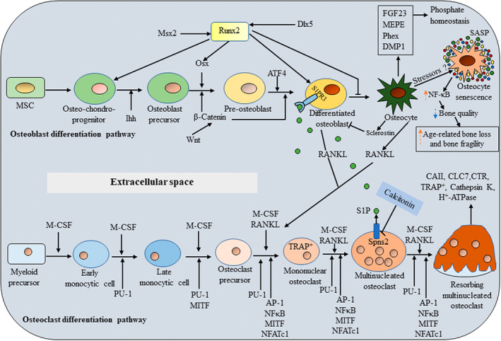
indicates upregulation and formula image indicates down‐regulation.
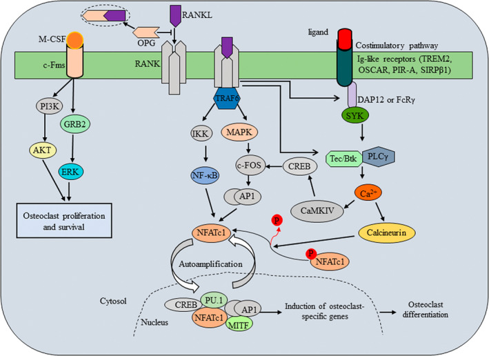
Figure 2
Figure 2
Major signaling pathways during the process of osteoclast differentiation. RANK and OPG are TNFR receptor‐related proteins, and RANKL is a TNF‐related cytokine that interacts specifically with either RANK or OPG. RANKL and M‐CSF regulate osteoclastogenesis through M‐CSF‐cFms, RANKL‐RANK as well as Ig‐like receptors associated with ITAM‐harbouring adaptor molecules such as DAP12 and Fc‐receptor common γ‐subunit (FcRγ). RANKL and its receptor RANK transduce a signal via the adaptor molecule TRAF6. TRAF6 recruits TAB2 and TAK1, which in turn activates the NF‐κB pathway and MAPK pathway. NF‐κB persuades c‐Fos expression via IKKs. Activation of MAPK pathway results in the activation of the Jun proteins. The c‐Fos and Jun proteins associate to form the complex AP‐1. The expression of the master regulator of osteoclastogenesis, NFATc1, is driven by AP‐1, NF‐κB and NFATc1 itself. The activation of NFATc1 is regulated by a co‐stimulatory signal pathway. FcRγ, DAP12 and their associating molecules activate Syk, which forms a complex with Btk/Tec and BLNK/SLP76. This complex further activates PLCγ, resulting in calcium signaling. The calcium signaling activates calcineurin, which dephosphorylates NFATc1, promoting its entry into the nucleus. Activated NFATc1 promotes its own expression making an autoamplification loop. The calcium signaling also induces c‐Fos expression via CAMKIV and CREB. NFATc1, together with other factors including PU.1 and MITF, promotes the expression of osteoclastogenic genes.

References

    1. Chotiyarnwong P, McCloskey EV. Pathogenesis of glucocorticoid‐induced osteoporosis and options for treatment. Nat Rev Endocrinol. 2020;16(8):437–447. - PubMed
    1. Karsenty G, Wagner EF. Reaching a genetic and molecular understanding of skeletal development. Dev Cell. 2002;2(4):389–406. - PubMed
    1. Takayanagi H. Osteoimmunology: Shared mechanisms and crosstalk between the immune and bone systems. Nat Rev Immunol. 2007;7(4):292–304. - PubMed
    1. Phan TCA, Xu J, Zheng MH. Interaction between osteoblast and osteoclast: Impact in bone disease. Histol Histopathol. 2004;19(4):1325–1344. - PubMed
    1. Crockett JC, Mellis DJ, Scott DI, Helfrich MH. New knowledge on critical osteoclast formation and activation pathways from study of rare genetic diseases of osteoclasts: focus on the RANK/RANKL axis. Osteoporos Int. 2011;22(1):1–20. - PubMed

LinkOut - more resources