. 2020 Aug 25;1(5):100058.
doi: 10.1016/j.xcrm.2020.100058.

Elevated Serum Amino Acids Induce a Subpopulation of Alpha Cells to Initiate Pancreatic Neuroendocrine Tumor Formation

Affiliations

Elevated Serum Amino Acids Induce a Subpopulation of Alpha Cells to Initiate Pancreatic Neuroendocrine Tumor Formation

Derek K Smith et al. Cell Rep Med. .

Abstract

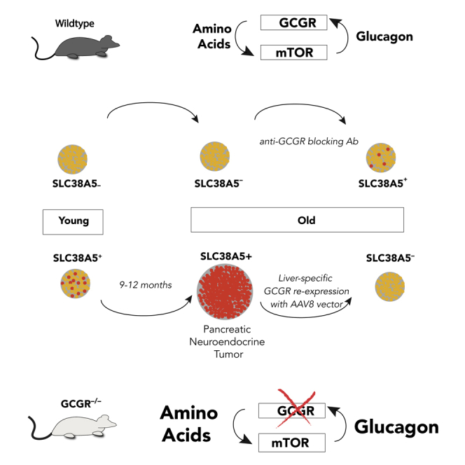
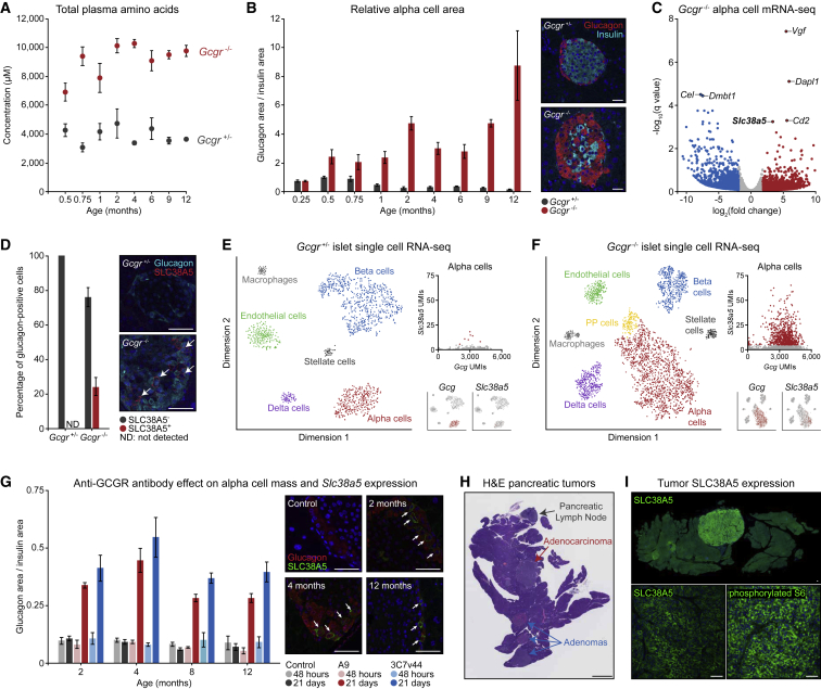
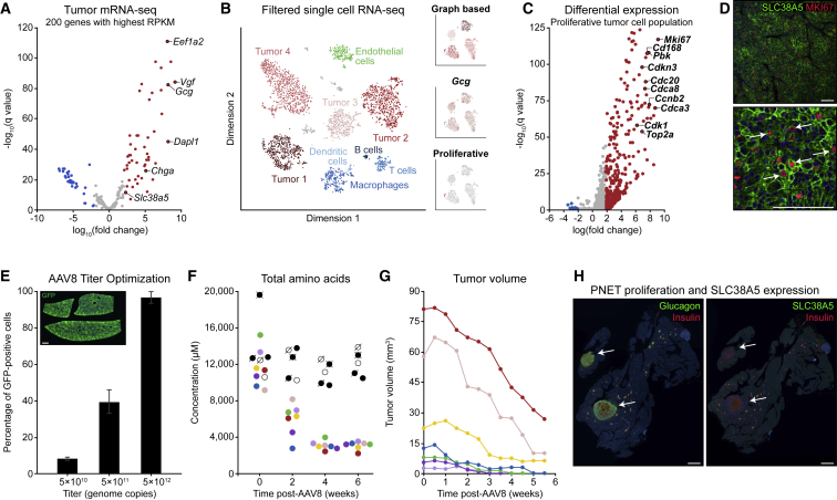
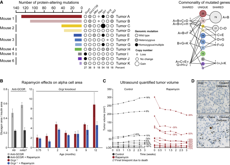
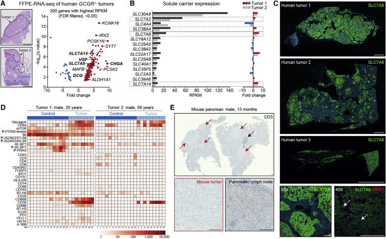
The cellular origin of sporadic pancreatic neuroendocrine tumors (PNETs) is obscure. Hormone expression suggests that these tumors arise from glucagon-producing alpha cells or insulin-producing β cells, but instability in hormone expression prevents linage determination. We utilize loss of hepatic glucagon receptor (GCGR) signaling to drive alpha cell hyperproliferation and tumor formation to identify a cell of origin and dissect mechanisms that drive progression. Using a combination of genetically engineered Gcgr knockout mice and GCGR-inhibiting antibodies, we show that elevated plasma amino acids drive the appearance of a proliferative population of SLC38A5+ embryonic progenitor-like alpha cells in mice. Further, we characterize tumors from patients with rare bi-allelic germline GCGR loss-of-function variants and find prominent tumor-cell-associated expression of the SLC38A5 paralog SLC7A8 as well as markers of active mTOR signaling. Thus, progenitor cells arise from adult alpha cells in response to metabolic signals and, when inductive signals are chronically present, drive tumor initiation.

Keywords: GCGR; Glucagon; PNET; SLC38A5; SLC38A7; mTOR; pancreatic neuroendocrine tumor; serum amino-acids; tumorigenesis.


Figures

None
Graphical abstract
Figure 1
Figure 1
Plasticity of Alpha Cell Identity in Aged Wild-Type and Gcgr Knockout Mice Reveals an SLC38A5+ Subpopulation Responsible for Tumor Initiation (A) Time-course analysis of blood amino acid concentrations in heterozygous and homozygous Gcgr knockout mice corresponding to the mice shown in (B). (B) Time-course analysis of relative alpha cell area in heterozygous and homozygous Gcgr knockout mice. Insets show representative islets from each genotype at 4 months. Five biological replicates per genotype at each time point. Scale bars, 50 μm. (C) Analysis of bulk RNA-seq data from FACS-isolated alpha cells from homozygous Gcgr knockout and heterozygous control mice. (D) Quantification of SLC38A5 immunostaining in 4-month-old mouse pancreas tissues. Five biological replicates per genotype. Scale bars, 50 μm (E) tSNE visualization of single-cell RNA-seq data from dispersed Gcgr heterozygous islets with alpha cell-specific Gcg and Slc38a5 expression plots. (F) T-distributed stochastic neighbor embedding (tSNE) visualization of single-cell RNA sequencing data from dispersed Gcgr homozygous knockout islets with alpha cell-specific Gcg and Slc38a5 expression plots. (G) Time-course analysis of alpha cell area and SLC38A5 expression in mice treated with mAb7 or A9 anti-GCGR antibodies for 2 or 21 days. Five biological replicates per condition at each time point. Scale bars, 50 μm. (H) Whole-slide H&E staining of pancreas tissue from a Gcgr homozygous knockout mouse with multiple PNETs. Scale bar, 2.5 mm. (I) SLC38A5 and phosphorylated S6 immunostaining of pancreatic tumor tissue from a Gcgr homozygous knockout mouse. Scale bars, 50 μm.
Figure 2
Figure 2
PNETs from Gcgr Knockout Mice Are Amino Acid Dependent and Dominantly Composed of Proliferative SLC38A5+ Tumor Cells (A) Analysis of bulk RNA-seq data from tumors and neighboring pancreatic control tissue from aged homozygous Gcgr knockout mice. (B) tSNE visualization of single-cell RNA-seq data from dispersed tumors, with insets showing graph-based clustering, Gcg UMI counts, and proliferation signatures for tumor cells. (C) Differential expression analysis of tumor cells classified as having a proliferative signature based on hallmark expression of MKI67 and cyclin-dependent kinases. (D) SLC38A5 and MKI67 co-immunostaining in mouse PNET tissue. Scale bars, 100 μm. (E) GFP immunostaining of liver tissues for quantification of AAV8 titer efficiency. The liver tissue section represents a 5 × 1012 unit titer. Three biological replicates per virus concentration. Scale bar, 1 mm. (F) Time-course analysis of blood amino acid concentrations in aged homozygous Gcgr knockout mice transduced with AAV8-Gcgr (colored symbols) and aged Gcgr knockout control mice (black and white symbols). (G) Ultrasound quantification of tumor volume in aged homozygous Gcgr knockout mice transduced with AAV8-Gcgr. Data point colors correspond to unique mice across (F) and (G). (H) Whole-slide glucagon, insulin, and SLC38A5 immunostaining of pancreas tissues from tumor-bearing Gcgr homozygous knockout mice that were transduced with AAV8 engineered for liver-specific expression of Gcgr. Scale bars, 1.5 mm.
Figure 3
Figure 3
Alpha Cell-Derived PNETs Are mTOR Dependent but Generally Harbor a Low Exomic Mutational Burden (A) Analysis of exome sequencing data generated from 11 pancreatic tumors isolated from six independent mice. Bar length represents the total number of protein-altering mutations per tumor, accompanied by mutation state and copy number, represented by circle size, for specific genes of interest. The rightmost network plot shows mutations shared across tumors, with node size representing the relative number of mutations. (B) Quantification of relative alpha cell area in wild-type mice treated with anti-GCGR antibody and homozygous Gcgr knockout mice treated with rapamycin. Five biological replicates per condition at each time point. (C) Ultrasound quantification of tumor volume over time in aged homozygous Gcgr knockout mice treated with vehicle or rapamycin. Relative volume change is shown as a percentage of the starting tumor volume. (D) Cleaved caspase-3 staining of tumor tissues isolated from Gcgr knockout mice treated with vehicle or rapamycin. Scale bars, 25 μm.
Figure 4
Figure 4
Human GCGR-Mutant, Alpha Cell-Derived PNETs Dominantly Express the Amino Acid Transporter SLC7A8+ and Exhibit an Immune Desert-like Phenotype (A) Analysis of bulk RNA-seq data from two human tumor biopsies obtained from patients harboring premature stop codons or missense mutations in both GCGR alleles. Scale bars, 5 mm. (B) Reads per kilobase of transcript, per million mapped reads (RPKM) expression analysis and fold change for human solute carrier genes. Amino acid transporters are highlighted by dashed boxes. (C) Whole-slide SLC7A8 immunostaining of three GCGR mutant human tumor biopsies. Scale bars, 2.5 mm and 250 μm. (D) Multiplexed digital spatial profiling of mTOR signaling-related and immune cell-related markers using two FFPE human tumor sections. (E) Whole-slide CD3 immunostaining of mouse pancreas tissue harboring multiple tumors with an in-tissue pancreatic lymph node as a positive control. Scale bars, 250 μm.

References

    1. Halfdanarson T.R., Rabe K.G., Rubin J., Petersen G.M. Pancreatic neuroendocrine tumors (PNETs): incidence, prognosis and recent trend toward improved survival. Ann. Oncol. 2008;19:1727–1733. - PMC - PubMed
    1. Ro C., Chai W., Yu V.E., Yu R. Pancreatic neuroendocrine tumors: biology, diagnosis,and treatment. Chin. J. Cancer. 2013;32:312–324. - PMC - PubMed
    1. Jiao Y., Shi C., Edil B.H., de Wilde R.F., Klimstra D.S., Maitra A., Schulick R.D., Tang L.H., Wolfgang C.L., Choti M.A. DAXX/ATRX, MEN1, and mTOR pathway genes are frequently altered in pancreatic neuroendocrine tumors. Science. 2011;331:1199–1203. - PMC - PubMed
    1. Bajetta E., Procopio G., Ferrari L., Catena L., Del Vecchio M., Bombardieri E. Update on the treatment of neuroendocrine tumors. Expert Rev. Anticancer Ther. 2003;3:631–642. - PubMed
    1. Sharma N., Naraev B.G., Engelman E.G., Zimmerman M.B., Bushnell D.L., Jr., OʼDorisio T.M., OʼDorisio M.S., Menda Y., Müller-Brand J., Howe J.R., Halfdanarson T.R. Peptide receptor radionuclide therapy outcomes in a North American cohort with metastatic well-differentiated neuroendocrine tumors. Pancreas. 2017;46:151–156. - PMC - PubMed

MeSH terms