. 2021 Feb;38(2):517-529.
doi: 10.1007/s10815-020-01998-x. Epub 2020 Nov 17.

Controlled hatching at the prescribed site using femtosecond laser for zona pellucida drilling at the early blastocyst stage

Affiliations

Controlled hatching at the prescribed site using femtosecond laser for zona pellucida drilling at the early blastocyst stage

Inna V Ilina et al. J Assist Reprod Genet. 2021 Feb.

Abstract

Purpose: To study whether the application of femtosecond laser pulses for zona pellucida (ZP) drilling of blastocysts at the embryonic or abembryonic poles can promote hatching to start immediately through the hole formed and ensure high hatching rates and embryo viability.

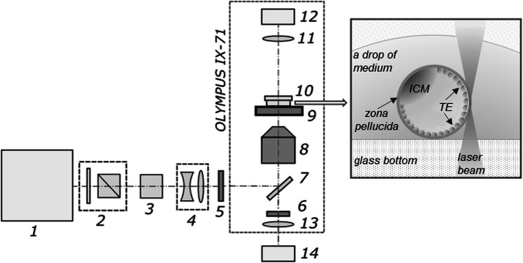
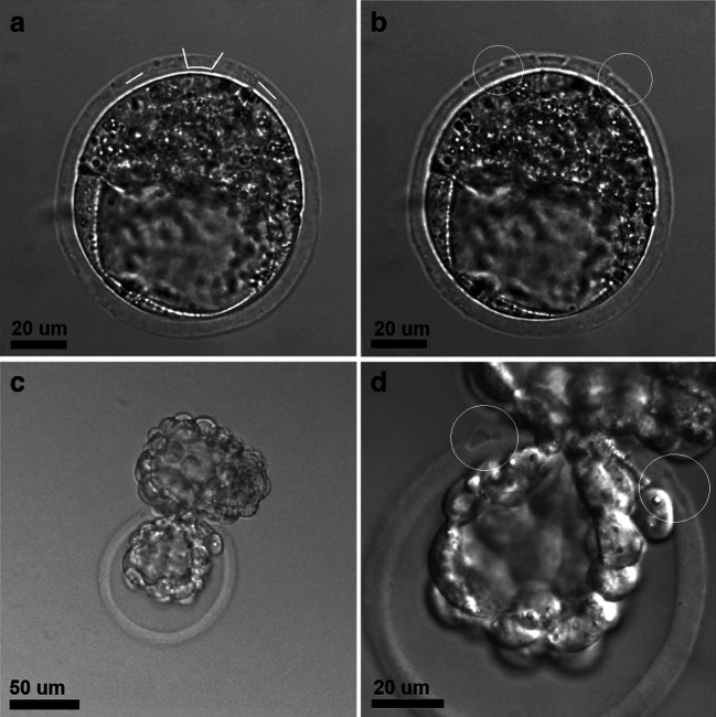
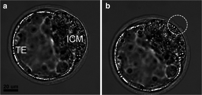
Methods: Mouse blastocyst (E3.5) ZP were microdissected with femtosecond laser pulses (514-nm wavelength, 280-fs pulse duration, 2.5-kHz repetition rate) close to the trophoblast or inner cell mass (ICM). The sizes of the holes formed were in the range of 4.5-8.5 μm. Additional longitudinal incisions (5-7-μm long) on either side of the hole were created to determine whether hatching had started at the correct position. Embryos post-laser-assisted ZP drilling and intact embryos were cultured under standard conditions for 2 days; embryo quality was assessed twice daily. The hatching rates and in vitro and in vivo implantation rates (only for embryos with ZP dissected close to the ICM) were estimated.

Results: Femtosecond laser-assisted ZP drilling at the early blastocyst stage facilitated embryo hatching to start at the artificial opening with probability approaching 100%. Despite the artificial opening's small size, no embryo trapping during hatching was observed. Both experimental groups had higher hatching rates than the control groups (93.3-94.7% vs. 83.3-85.7%, respectively). The in vitro implantation rate was comparable with that of the control group (92.3% vs. 95.4%). No statistically significant differences were obtained in the in vivo implantation rates between the experimental and control groups.

Conclusions: Blastocyst-stage femtosecond laser microsurgery of ZP is fast and delicate and enables the hatching process to be initiated in a controlled manner through a relatively small opening, with no embryo trapping.

Keywords: Blastocyst; Femtosecond laser microsurgery; Inner cell mass; Laser-assisted hatching; Trophectoderm cells; Zona pellucida drilling.


Conflict of interest statement

The authors declare that they have no conflict of interest.

Figures

Fig. 1
Fig. 1
Schematic setup of the system for femtosecond laser–assisted ZP drilling. 1, Femtosecond ytterbium laser; 2, beam attenuator; 3, second-harmonic generator; 4, telescope; 5, electro-mechanical shutter; 6, notch filter; 7, dichroic mirror; 8, microscope objective; 9, X-Y motorized stage; 10, Petri dish; 11, condenser lens; 12, microscope lamp; 13, tube lens; 14, CMOS camera. Inset: schematic presentation of glass-bottom Petri dish containing a blastocyst (in a fresh drop of embryo manipulation medium) during the process of laser-assisted ZP drilling in close proximity to TE cells
Fig. 2
Fig. 2
Femtosecond laser–assisted ZP drilling at the abembryonic pole. a Bright-field image of blastocyst with the ICM and TE clearly distinguishable. b The creation of primitive elements in the program interface at a position close to the TE cells. c Femtosecond laser–assisted ZP drilling (formation of an artificial opening in the ZP and two additional incisions [white circles]). d, e TE cells start herniating through the artificial opening in the ZP (5 h and 7 h post-ZP drilling, respectively); incisions on the ZP are blurred but nevertheless can be distinguished. f Embryo continues to hatch out of its shell at 24 h post-ZP drilling
Fig. 3
Fig. 3
Peri-implantation mouse embryos cultured on adhesive platform. а Bright-field image of mouse embryo on day 2 (6.5 days post-fertilization) of cultivation on the adhesive platform. b Immunofluorescence staining of embryo on day 2 (6.5 days post-fertilization) with otc4 for ICM visualization. c Embryo staining on day 2 (6.5 days post-fertilization) with Hoechst 33342 for nuclei detection and d merge. e, f Embryos on day 1 (5.5 days post-fertilization) and day 4 (8.5 days post-fertilization) of cultivation stained with calcein AM and Hoechst 33342; extensive TE growth is shown. g, h Embryos on day 1 (5.5 days post-fertilization) and day 4 (8.5 days post-fertilization) of cultivation stained with PI and Hoechst 33342
Fig. 4
Fig. 4
Femtosecond laser–assisted ZP drilling close to the embryonic pole. a The creation of primitive elements in the program interface at a position close to the ICM. b Femtosecond laser–assisted ZP microsurgery for forming an artificial opening and two additional incisions in the ZP (white circles). c, d The embryo hatching through the artificial opening; incisions on either side of the opening are clearly visualized and marked in white circles
Fig. 5
Fig. 5
Expanded blastocyst before (a) and immediately after (b) femtosecond laser–assisted ZP drilling at a position near the ICM. A tiny detached flap of the ZP is visible (white circle)

References

    1. Davidson LM, Liu Y, Griffiths T, Jones C, Coward K. Laser technology in the ART laboratory: a narrative review. Reprod BioMed Online. 2019;38(5):725–739. doi: 10.1016/j.rbmo.2018.12.011. - DOI - PubMed
    1. Bedient C, Khanna P, Desai N (2011) Laser pulse application in IVF. In: Jakubczak K (ed) Lasers - applications in science and industry. Rijeka, InTech, pp. 193–214. 10.5772/24024.
    1. Montag MHM, Klose R, Köster M, Rösing B, van der Ven K, Rink K, van der Ven H. Application of non-contact laser technology in assisted reproduction. Med Laser Appl. 2009;24(1):57–64. doi: 10.1016/j.mla.2008.11.001. - DOI
    1. Schopper B, Ludwig M, Edenfeld J, Al-Hasani S, Diedrich K. Possible applications of lasers in assisted reproductive technologies. Hum Reprod. 1999;14(Suppl 1):186–193. doi: 10.1093/humrep/14.suppl_1.186. - DOI - PubMed
    1. Ebner T, Yaman C, Moser M, Sommergruber M, Hartl J, Tews G. Laser assisted immobilization of spermatozoa prior to intracytoplasmic sperm injection in humans. Hum Reprod. 2001;16:2628–2631. doi: 10.1093/humrep/16.12.2628. - DOI - PubMed

MeSH terms