. 2021 Apr;23(2):e13514.
doi: 10.1111/tid.13514. Epub 2020 Dec 7.

Macrophage depletion of CMV latently infected donor hearts ameliorates recipient accelerated chronic rejection

Affiliations

Macrophage depletion of CMV latently infected donor hearts ameliorates recipient accelerated chronic rejection

Nicole N Haese et al. Transpl Infect Dis. 2021 Apr.

Abstract

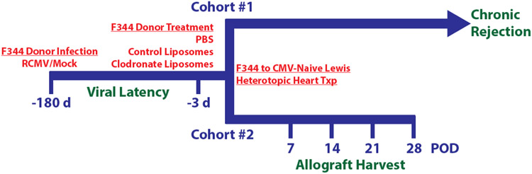
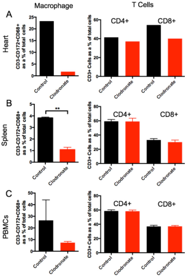
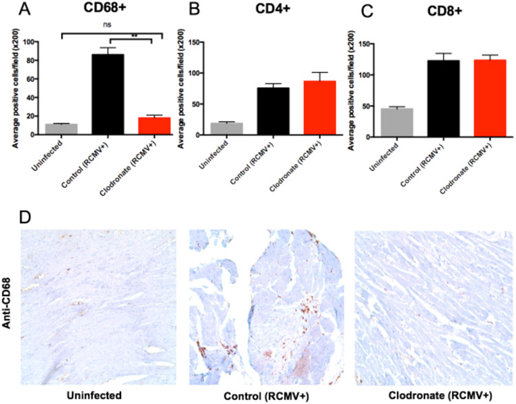
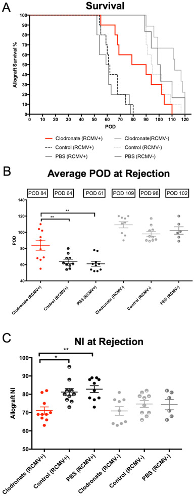
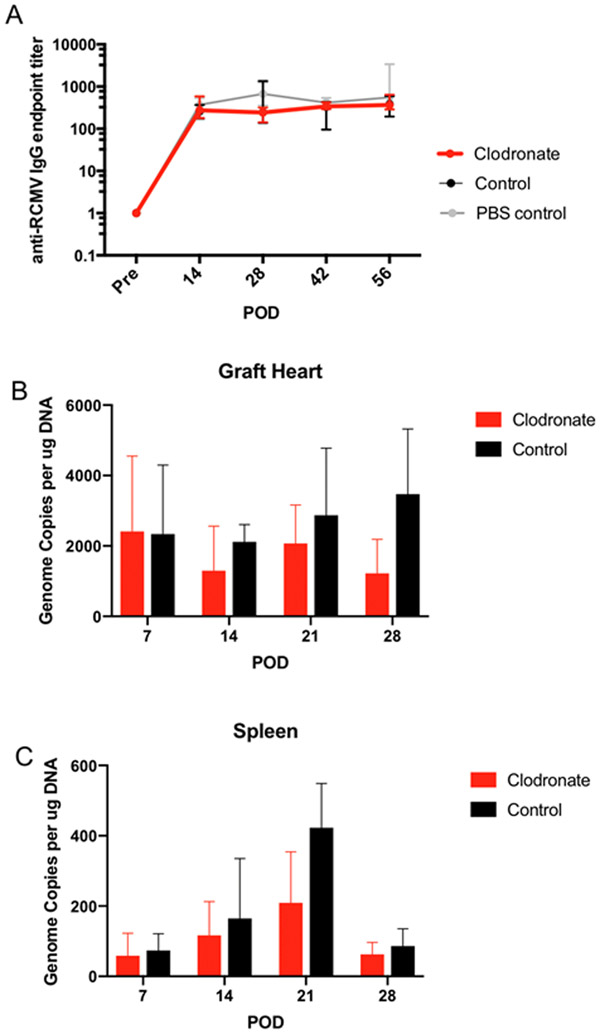
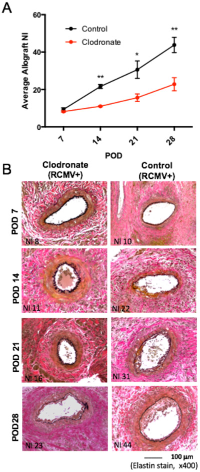
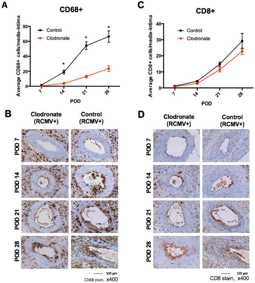
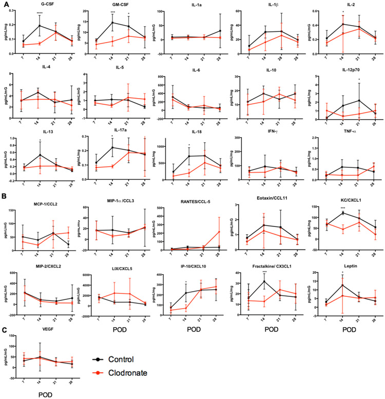
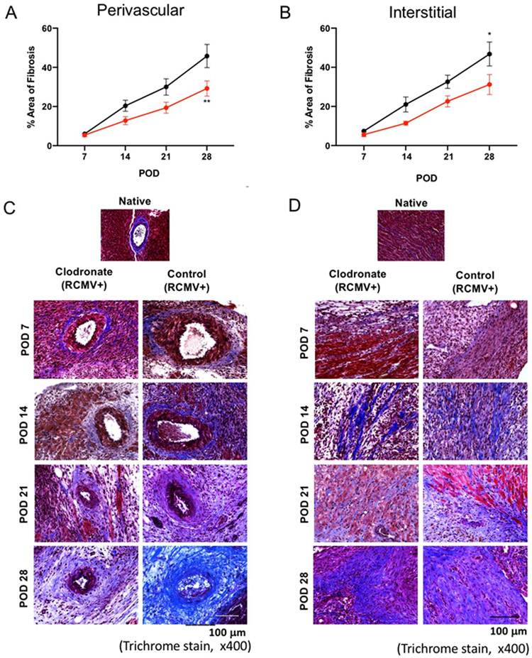
Cytomegalovirus (CMV) infection is linked to acceleration of solid organ transplant vascular sclerosis (TVS) and chronic rejection (CR). Donor latent CMV infection increases cardiac-resident macrophages and T cells leading to increased inflammation, promoting allograft rejection. To investigate the role of cardiac-resident passenger macrophages in CMV-mediated TVS/CR, macrophages were depleted from latently ratCMV (RCMV)-infected donor allografts prior to transplantation. Latently RCMV-infected donor F344 rats were treated with clodronate, PBS, or control liposomes 3 days prior to cardiac transplant into RCMV-naïve Lewis recipients. Clodronate treatment significantly increased graft survival from post-operative day (POD)61 to POD84 and decreased TVS at rejection. To determine the kinetics of the effect of clodronate treatment's effect, a time study revealed that clodronate treatment significantly decreased macrophage infiltration into allograft tissues as early as POD14; altered allograft cytokine/chemokine protein levels, fibrosis development, and inflammatory gene expression profiles. These findings support our hypothesis that increased graft survival as a result of allograft passenger macrophage depletion was in part a result of altered immune response kinetics. Depletion of donor macrophages prior to transplant is a strategy to modulate allograft rejection and reduce TVS in the setting of CMV + donors transplanted into CMV naïve recipients.

Keywords: chronic rejection; cytomegalovirus; latent infection; transplant vascular sclerosis.


Conflict of interest statement

Conflict of Interest

The authors do not have any conflicts of interest.

Figures

Figure 1.
Figure 1.. Experimental design.
F344 donor rats were latently infected with RCMV at least 180 days prior to transplant with 1x10⁵ pfu/rat salivary gland-derived RCMV or mock infected with PBS. At three days prior to transplant, donor rats were treated by intravenous infusion with PBS, control liposomes, or clodronate liposomes (10mL/kg). On experimental day 0, RCMV naïve recipient Lewis rats underwent heterotopic heart transplant. Cohort #1 (n=10 per treatment group) were monitored daily for signs of rejection by heartbeat palpation and harvested at the time of chronic rejection. Blood was drawn on post-operative days (POD) 14, 28, 42, and 56. A subgroup of donor heart, spleen, and PBMC samples were collected at day −3 from Cohort #1 animals to confirm macrophage depletion by clodronate using flow cytometry. Cohort #2 recipients were harvested at POD 7, 14, 21, and 28 (n=5 per time point for each experimental group) to monitor the effect of clodronate treatment over time. At CR (Cohort #1) or predetermined time points (Cohort #2), hearts (graft and native) spleen, SMG, and blood were collected for analysis.
Figure 2.
Figure 2.. Clodronate treatment depletes macrophages but not T cells from LDI organs and blood.
To confirm macrophage depletion by clodronate liposome treatment, RCMV latently-infected donor F344 rats were treated with control liposomes (n=3, black) or clodronate liposomes (n=3, red). At three days post treatment, tissues were harvested and leukocytes were prepared from heart, spleen, and peripheral blood (PBMC). The frequency of CD4+ and CD8+ T cells and macrophages in each of these samples was quantified via flow cytometry by cell surface staining using fluorescent antibodies directed against CD172 (myeloid lineage cells), CD68 (macrophage), CD3 (T cells), CD4 (CD4+ T cells), and CD8 (CD8+ T cells). Depicted is the frequency of macrophages (CD3CD172+CD68+) and CD4 (CD3+CD4+) and CD8 (CD3+CD8+) cells isolated from the heart (a), spleen (b), and PBMCs (c). **p≤0.01 as determined by Student’s t-test.
Figure 3.
Figure 3.. Clodronate treatment reduces allograft tissue macrophage levels to those observed in uninfected hearts.
Heart tissue sections from F344 rats that were either uninfected (gray bars), latently RCMV infected and treated with control liposomes (black bars) or latently RCMV infected and treated with clodronate liposome (red bars) were stained with antibodies directed against CD68 (macrophages) (a, d), CD4+ T cells (b) and CD8+ T cells (c). Immunohistochemical analysis was done in a blinded review and scored as follows: 0, no visible staining; 1, faint staining; 2, moderate intensity with multifocal staining; and 3, intense diffuse staining. CD4, CD8 and CD68 positively stained cells with scores 2 and 3 were counted in 10 fields from whole heart tissues at 200x magnification or in 6 fields from the coronary arteries at 400x magnification; and the mean number of positive cells per field was calculated. **p≤0.01 as determined by a One-way ANOVA with Tukey’s secondary testing.
Figure 4.
Figure 4.. Clodronate liposome treatment increases the time to allograft CR and reduces TVS.
Heterotopic heart allografts from LDI F344 rats or uninfected F344 donors treated three days prior to organ donation with clodronate liposomes, control liposomes, or PBS were transplanted into RCMV naïve Lewis recipients (n=10 for each treatment group). Allograft recipients were monitored daily for graft rejection. Kaplein-Meier graft survival curves are depicted in Panel A, and the average time of rejection reported as POD is shown in Panel B. At rejection, sections of allograft heart were stained with H&E and elastin to determine the degree of vascular disease in the coronary arteries. Average NI (a measure of TVS) for each group is reported along with individual allograft values (Panel C). Error bars represent standard deviation for each group. *p≤0.05, **p≤0.01 as determined by a One-way ANOVA with Tukey’s secondary testing.
Figure 5.
Figure 5.. Macrophage depletion does not alter recipient infection status.
Allograft hearts and spleen samples were collected from Cohort#2 recipients on POD7, 14, 21, and 28 to compare RCMV levels in between clodronate and control liposome treated animals. (n=5 per group/time point). LDI F344 rats were treated with either control liposomes (black) or clodronate liposomes (red) three days prior to heterotopic heart allograft transplant into RCMV naïve Lewis recipients. Recipients (n=5 per group/time point) were euthanized on POD 7, 14, 21, and 28 at which time recipient heart allografts) and spleen were collected in DNAzol. Total DNA was prepared using the DNAzol method and viral DNA loads were determined by qRT-PCR using virus specific primers and probes. Genome copies per μg total RNA were graphed for both graft heart in Panel A and spleen in Panel B. In Panel C serum was isolated from recipient rats in Cohort #1 on POD0, 14, 28, 42, and 56. RCMV-specific antibodies were quantified by limiting dilution ELISA for serum samples using high-binding 96-well plates coated with RCMV-infected cell lysates. Serum titers for anti-RCMV IgG were compared between LDI allograft recipients in all three treatment groups (clodronate liposomes – red, control liposomes – black, and PBS control – grey) but did not differ in their peak titers or kinetics of formation.
Figure 6.
Figure 6.. Transplant vascular sclerosis is decreased in clodronate-liposome treated LDI allografts.
TVS was quantified in Cohort #2 recipients to compare the degree of vascular disease present in allograft vessels between clodronate and control liposomes treated animals treated animals. LDI F344 rats were treated with either control liposomes (black) or clodronate liposomes (red) three days prior to heterotopic heart allograft transplant into RCMV naïve Lewis recipients. Recipients (n=5 per group/time point) were euthanized on POD 7, 14, 21, and 28 at which time rat tissues and heart allografts were collected for analysis. Embedded allograft heart tissues were sectioned and stained with H&E and elastin to visualize vessel neointimal formation. Panel A depicts graphical representation of TVS quantification as reported as the mean allograft NI. Images shown in Panel B shows representative stained graft heart tissue sections. Clodronate liposome treated allografts have reduced TVS relative to control liposome treated allografts starting at POD14 and persisting through POD28 *p≤0.05, **p≤0.01 as determined by Student’s t- test.
Figure 7.
Figure 7.. Macrophage and CD8+ T cell infiltration into the media/intima of allograft vessels is reduced in allografts derived from RCMV latently infected donors treated with clodronate.
Immunohistochemical staining for the presence of macrophages and T cells of heart allografts harvested from Cohort #2 recipients at POD7, 14, 21, and 28 (n=5 per group/time point). Embedded tissue sections were cut and stained using antibodies directed against CD68 (macrophages; panels a and b) and CD8+ T cells (panels B and C). The frequency of each cell population was measured over 10 serial tissue slides and averaged to determine the mean number of cells that infiltrated into the vessel wall media and intima. Results for clodronate liposome treated animals are depicted in red and control liposome treated animals are shown in black for panels A and C. Images are representative stained sections from each time point (panels B and D). *p≤0.05 as determined by Student’s t-test.
Figure 8.
Figure 8.. Macrophage depletion alters RCMV-infected allograft proinflammatory cytokine and chemokine profiles.
Rat allograft heart lysates from recipients in cohort 2 at POD 7, 14, 21, and 28 (n=5 per group/time point) were analyzed using a 27-plex cytokine magnetic bead assay to quantify the levels of cytokines (a), chemokines (b), and VEGF (c), clodronate liposome (red) and control liposome (black). *p≤0.05, **p≤0.01, ***p≤001, ****p<0.0001 as determined by Two-way ANOVA with Holm-Sidak correction for multiple comparisons.
Figure 9.
Figure 9.. LDI donor macrophage depletion alters the fibrosis pathway normally activated at POD14.
Ingenuity pathway core analysis was used to identify downstream effects in cardiac graft samples following transplantation (Clodronate-liposomes vs. Control-liposomes). Statistically significant alterations in gene expression from RNAseq analysis of graft tissues are represented by labeled shapes depending on their molecular function. Expression and predicted activation levels determined by fold-change between Clodronate-liposomes vs. Control-liposomes are presented using the following convention on color scales with increasing saturation associated with an greater absolute fold-change value: red = increased detection in Clodronate-Liposome treated donor hearts; green = decreased detection; orange=predicted activation; pink= fibrosis associated genes. Predicted relationships are indicated by arrow-heads and blue lines to indicate predicted inhibition; yellow lines indicate inconsistent findings that make it difficult to predict outcomes of interactions.
Figure 10.
Figure 10.. LDI macrophage depletion reduces cardiac allograft fibrosis.
Heart allografts harvested from Cohort #2 recipients at POD7, 14, 21, and 28 (n=5 per group/time point) were embedded and stained with Masson’s trichrome to identify levels of cardiac tissue fibrosis (healthy areas are red and collagen-rich areas are blue). Perivascular and interstial fibrosis was measured over 6 random vields of view of 10 serial tissue slides at a magnification of x400. Values were averaged and adjusted to a tooal tissue area in the image anlayzed to determine the mean percent area with fibrosis as depicted in panels A and B. Results for clodronate liposome treated animals are depicted in red and control liposome treated animals are shown in black. Images are representative stained sections from each time pointValues are presented as the means ± standard error of the mean (SEM) of each group and were analyzed using 2way ANOVA followed by Sidak’s multiple comparison test *p≤0.05, **p≤0.01.

References

    1. Ramanan P, Razonable RR. Cytomegalovirus infections in solid organ transplantation: a review. Infection & chemotherapy. 2013;45(3):260–271. - PMC - PubMed
    1. Hodson EM, Barclay PG, Craig JC, et al. Antiviral medications for preventing cytomegalovirus disease in solid organ transplant recipients. Cochrane Database Syst Rev. 2005(4):Cd003774. - PubMed
    1. Merigan TC, Renlund DG, Keay S, et al. A controlled trial of ganciclovir to prevent cytomegalovirus disease after heart transplantation. N Engl J Med. 1992;326(18):1182–1186. - PubMed
    1. Valentine VG, Weill D, Gupta MR, et al. Ganciclovir for cytomegalovirus: a call for indefinite prophylaxis in lung transplantation. The Journal of heart and lung transplantation : the official publication of the International Society for Heart Transplantation. 2008;27(8):875–881. - PubMed
    1. Orloff SL, Hwee YK, Kreklywich C, et al. Cytomegalovirus latency promotes cardiac lymphoid neogenesis and accelerated allograft rejection in CMV naive recipients. Am J Transplant. 2011;11(1):45–55. - PMC - PubMed