. 2020 Dec 10;63(23):14724-14739.
doi: 10.1021/acs.jmedchem.0c01287. Epub 2020 Nov 18.

Exploration of Structure-Activity Relationship of Aromatic Aldehydes Bearing Pyridinylmethoxy-Methyl Esters as Novel Antisickling Agents

Affiliations

Exploration of Structure-Activity Relationship of Aromatic Aldehydes Bearing Pyridinylmethoxy-Methyl Esters as Novel Antisickling Agents

Piyusha P Pagare et al. J Med Chem. .

Abstract

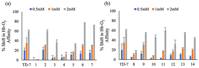
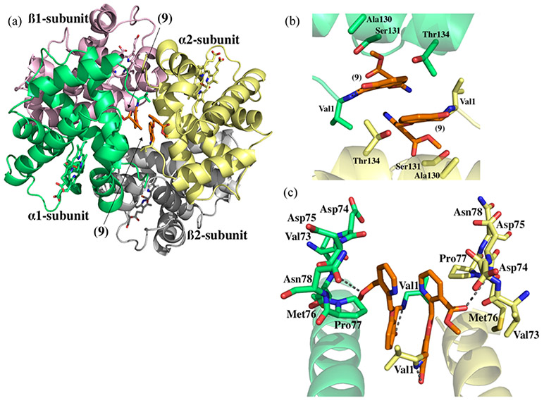
Aromatic aldehydes elicit their antisickling effects primarily by increasing the affinity of hemoglobin (Hb) for oxygen (O2). However, challenges related to weak potency and poor pharmacokinetic properties have hampered their development to treat sickle cell disease (SCD). Herein, we report our efforts to enhance the pharmacological profile of our previously reported compounds. These compounds showed enhanced effects on Hb modification, Hb-O2 affinity, and sickling inhibition, with sustained pharmacological effects in vitro. Importantly, some compounds exhibited unusually high antisickling activity despite moderate effects on the Hb-O2 affinity, which we attribute to an O2-independent antisickling activity, in addition to the O2-dependent activity. Structural studies are consistent with our hypothesis, which revealed the compounds interacting strongly with the polymer-stabilizing αF-helix could potentially weaken the polymer. In vivo studies with wild-type mice demonstrated significant pharmacologic effects. Our structure-based efforts have identified promising leads to be developed as novel therapeutic agents for SCD.


Figures

Figure 1.
Figure 1.
Structures of 5-HMF, vanillin, INN-298, INN-312, SAJ-310, TD-7, and GBT-440.
Figure 2.
Figure 2.
Concentration-dependent shift in the Hb-O2 affinity of (a) m-methoxy-substituted and, (b) o-hydroxy-substituted compounds in normal whole blood after 1.5 h incubation.
Figure 3.
Figure 3.
Time-dependent shift in the Hb-O2 affinity of (a) m-methoxy-substituted and, (b) o-hydroxy-substituted compounds in normal whole blood at a concentration of 2 mM.
Figure 4.
Figure 4.
Inhibition of sickling by (a) m-methoxy-substituted and, (b) o-hydroxy-substituted compounds in SS RBCs.
Figure 5.
Figure 5.
Concentration-dependent Hb modification (adduct formation) by (a) m-methoxy-substituted and, (b) o-hydroxy-substituted compounds in SS RBCs.
Figure 6.
Figure 6.
Concentration-dependent shift in Hb-O2 affinity by (a) m-methoxy-substituted and, (b) o-hydroxy-substituted compounds in SS RBCs.
Figure 7.
Figure 7.
Linear correlation between increase in the Hb-O2 affinity, antisickling, and Hb-modification properties for 0.5 mM (blue), 1 mM (green), and 2 mM (gray) concentrations of compounds.
Figure 8.
Figure 8.
Time-dependent recovery of parent compounds 9 and 10 at 0.5 and 1 mM; (a) representative chromatograms of 1 mM compound 9 and (b) compound 10 at 0, 4, 8, and 24 h.
Figure 9.
Figure 9.
Crystal structure of Hb in the R2 conformation in complex with two molecules of compound 9 bound at the α-cleft (a) overall crystal structure of Hb in the R2 conformation in the complex with two molecules of compound 9 in the central water cavity. (b) Close-up view of compound 9 bound at the α-cleft of Hb. (c) Close-up view of compound 9 showing interactions with the residues of the αF-helix. (Hb is shown as cartoon with α1 subunit in green, α2 in yellow, β1 in pink, and β2 in gray; hemes are shown as sticks; compound 9 is shown in orange sticks).
Figure 10.
Figure 10.
Crystal structure of Hb in the R2 conformation in complex with two molecules of compound 6 bound at the α-cleft (a) overall crystal structure of Hb in the R2 conformation in complex with two molecules of compound 6 in the central water cavity. (b) Closeup view of compound 6 bound at the α-cleft of Hb. (c) Closeup view of compound 6 showing interactions with the residues of the αF-helix. (Hb is shown as cartoon with α1 subunit in green, α2 in yellow, β1 in pink, and β2 in gray; hemes are shown as sticks; compound 6 is shown in cyan sticks; water molecules are shown as red spheres).
Figure 11.
Figure 11.
Structural comparison of compounds 6 and 9 bound at the α-cleft of Hb. (a) superposition of compounds 6 and 9 molecules showing interactions with residues from αF-helix. (b) 90° rotated view of (a). (Hb is shown as cartoon with α1 subunit in green and α2 in yellow; compounds 6 and 9 are shown in cyan and orange sticks, respectively).
Figure 12.
Figure 12.
Time-dependent modification of Hb in wild-type mice (n = 6) after 150 mg/kg IP administration of test compounds.
Figure 13.
Figure 13.
Time-dependent Hb-O2 affinity shifts in the wild-type mice (n = 6) after 150 mg/kg IP administration of test compounds.
Scheme 1.
Scheme 1.
Synthetic Route for Target Compounds

References

    1. Pauling L; Itano HA; Singer SJ; Wells IC Sickle Cell Anemia, a Molecular Disease. Science 1949, 110, 543–548. - PubMed
    1. Cretegny I; Edelstein SJ Double Strand Packing in Hemoglobin S Fibers. J. Mol. Biol 1993, 230, 733–738. - PubMed
    1. Ferrone FA Polymerization and Sickle Cell Disease: A Molecular View. Microcirculation 2004, 11, 115–128. - PubMed
    1. Ghatge MS; Ahmed MH; Omar ASM; Pagare PP; Rosef S; Kellogg GE; Abdulmalik O; Safo MK Crystal Structure of Carbonmonoxy Sickle Hemoglobin in R-State Conformation. J. Struct. Biol 2016, 194, 446–450. - PMC - PubMed
    1. Rogers SC; Ross JGC; d’Avignon A; Gibbons LB; Gazit V; Hassan MN; McLaughlin D; Griffin S; Neumayr T; Debaun M; DeBaun MR; Doctor A Sickle Hemoglobin Disturbs Normal Coupling among Erythrocyte O2 Content, Glycolysis, and Antioxidant Capacity. Blood 2013, 121, 1651–1662. - PMC - PubMed

Publication types

MeSH terms