. 2021 Jan;192(1):200-211.
doi: 10.1111/bjh.17137. Epub 2020 Nov 18.

Severe congenital neutropenia-associated JAGN1 mutations unleash a calpain-dependent cell death programme in myeloid cells

Affiliations

Severe congenital neutropenia-associated JAGN1 mutations unleash a calpain-dependent cell death programme in myeloid cells

Avinash Khandagale et al. Br J Haematol. 2021 Jan.

Abstract

Severe congenital neutropenia (SCN) of autosomal recessive inheritance, also known as Kostmann disease, is characterised by a lack of neutrophils and a propensity for life-threatening infections. Using whole-exome sequencing, we identified homozygous JAGN1 mutations (p.Gly14Ser and p.Glu21Asp) in three patients with Kostmann-like SCN, thus confirming the recent attribution of JAGN1 mutations to SCN. Using the human promyelocytic cell line HL-60 as a model, we found that overexpression of patient-derived JAGN1 mutants, but not silencing of JAGN1, augmented cell death in response to the pro-apoptotic stimuli, etoposide, staurosporine, and thapsigargin. Furthermore, cells expressing mutant JAGN1 were remarkably susceptible to agonists that normally trigger degranulation and succumbed to a calcium-dependent cell death programme. This mode of cell death was completely prevented by pharmacological inhibition of calpain but unaffected by caspase inhibition. In conclusion, our results confirmed the association between JAGN1 mutations and SCN and showed that SCN-associated JAGN1 mutations unleash a calcium- and calpain-dependent cell death in myeloid cells.

Keywords: apoptosis; calcium; calpain; necroptosis; neutropenia.


Figures

Fig 1
Fig 1
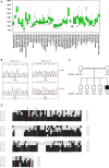
JAGN1 mutations in patients with severe congenital neutropenia. (A) Box plot depicting JAGN1 mRNA expression in normal human tissues. The data were derived from the in silico transcriptomics database, IST. 43 Each box represents the quartile distribution (25–75%) range with median indicated as a black horizontal line. The 95% range including individual outlier samples is also displayed. The y‐axis indicates the relative gene expression level. Note high expression of JAGN1 in haematopoietic stem cells, as well as in mesenchymal stem cells. (B) Sequence chromatograms illustrating the mutation c.40G>A, p.Gly14Ser (left panel) and c.63G>T; p.Glu21Asp (right panel) of JAGN1, NM_032492, in patients with SCN compared to the normal sequence in a healthy control. (C) Pedigree of the family of patient 1 illustrating the inheritance of the c.40G>A; p.Gly14Ser mutations in JAGN1. The filled symbol denotes the affected patient. (D) Sequence alignment of Jagunal homologues of C. elegans (NP_493559.1), D. melanogaster (NP_649585.1), D. rerio (NP_001005774.1), H. sapiens (NP_115881.3), and M. musculus (NP_080641.1). The human JAGN1 mutations identified in the present study are indicated by asterisks. The predicted transmembrane domains 12 are indicated by horizontal lines and the putative ER retention motif is boxed.
Fig 2
Fig 2
Expression of patient‐derived mutant JAGN1 in HL‐60 cells. (A) HL‐60 cells were transfected with either FLAG‐tagged wild‐type (wt) JAGN1 or patient‐derived JAGN1 mutation‐expressing plasmids. JAGN1 expression in HL‐60 cells was confirmed by Western blot using an anti‐FLAG antibody. The membrane was re‐probed for β‐actin to control for equal loading. (B) FLAG‐tagged JAGN1 co‐localises with the ER protein, calnexin. HL‐60 cells were transfected with FLAG‐tagged wt‐JAGN1 or E21D‐JAGN1 and cells were incubated with MitoTracker (red) or LysoTracker (red) before fixation or phycoerythrin (PE)‐labelled anti‐calnexin antibody (red) after fixation and subsequently stained with a FITC‐labelled anti‐FLAG antibody (green). Images were acquired with a confocal laser scanning microscope fitted with a ×63 objective. Scale bars, 20 μm. (C) Immunoprecipitation of FLAG‐tagged wt‐ and mutant‐JAGN1 proteins (G14S and E21D) with the endogenous ER protein, Grp78 in HL‐60 cells (see Table SI for yeast two‐hybrid screen results). (D) HL‐60 cells were transfected with either scrambled siRNA or JAGN1 specific siRNA and JAGN1 mRNA levels relative to glyceraldehyde‐3‐phosphate dehydrogenase (GAPDH) mRNA were quantified using RT‐PCR. Results are mean values ± SD (n = 3). ***P < 0·005, by Student’s t‐test.
Fig 3
Fig 3
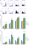
Increased apoptosis in mutant JAGN1‐expressing HL‐60 cells. HL‐60 cells expressing wt‐JAGN1, G14S‐JAGN1, or E21D‐JAGN1 were treated with vehicle alone or exposed to the indicated, pro‐apoptotic stimuli. (A) Representative flow cytometry results showing apoptosis as evidenced by the emergence of a sub‐G1 peak following treatment with staurosporine (STS) (2 µmol/l) for 3 h. (B) Quantification of apoptosis in cells stimulated with STS (2 µmol/l) for 3 h or thapsigargin (1 µmol/l) for 2 h, or stimulated for 6 h with etoposide (10 µmol/l). Results shown are mean values ± SD (n = 3). (C) Quantification of caspase‐3‐like activity as determined by DEVD‐AMC cleavage in HL‐60 cells expressing wt‐ or mutant‐JAGN1 stimulated as detailed above. Results shown are mean values ± SD (n = 5). *< 0·05, **< 0·01; ***P < 0·005; ****P < 0·001.
Fig 4
Fig 4
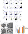
Impact of degranulation agonists in cells expressing mutant JAGN1. HL‐60 cells expressing wt‐JAGN1, G14S‐JAGN1, or E21D‐JAGN1 were exposed to vehicle alone or stimulated with cytochalasin D (10 ng/ml) and fMLP (10 µmol/l). (A) Cytosolic calcium was determined after 20 min by flow cytometry using the calcium‐sensitive probe, Fluo4‐AM. (B) Dissipation of the mitochondrial membrane potential (Δψm) was determined at 2 h using the fluorescent probe, tetramethylrhodamine ethyl ester (TMRE). The experiments were performed three times, and representative results are shown. (C) TEM images of peripheral blood neutrophils from a human donor (HD) and from patient 1, respectively. Note polarisation of granules to the periphery of the cell in patient neutrophils giving the impression of granule ‘congestion’, along with varying contents of dark, dense material, crystal‐like in appearance, but without discernible crystal structure, as well as several instances of disrupted granule membranes. Scale bars, 2 µm (left) and 500 nm (right). (D) HL‐60 cells were either mock‐transfected or transfected with wt‐ or mutant‐JAGN1 constructs. Exposure to vehicle alone increased mitochondrial calcium levels in cells expressing mutant JAGN1 and calcium levels were further increased by CytD/fMLP as shown by flow cytometric analysis after rhodamine‐2‐acetoxymethyl ester (Rhod2‐AM) staining. Data are mean values ± SD (n = 3). *< 0·05, ***P < 0·005. DMSO, dimethyl sulphoxide. [Colour figure can be viewed at wileyonlinelibrary.com]
Fig 5
Fig 5
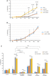
Degranulation agonist‐induced cell death is calpain‐dependent. (A) HL‐60 cells were mock‐transfected or transfected with wt‐ or mutant‐JAGN1 (G14S and E21D). Cells were then stimulated with cytochalasin D (10 ng/ml) + fMLP (10 µmol/l) for the indicated time‐points and cell death was determined using the LDH release assay. (B) HL‐60 cells were transiently transfected with JAGN1 specific siRNA versus control siRNA and stimulated as indicated in (A). Data shown in panels (A) and (B) are mean values ± SD (n = 3–4). (C) Cells were pretreated for 1 h with various inhibitors [i.e. the calpain inhibitor, PD150606 (60 µmol/l), the pan‐caspase inhibitor, zVAD‐fmk (10 µmol/l), the MPT inhibitor, bongkrekic acid (BA) (50 µmol/l), or the RIP1 kinase inhibitor, necrostatin‐1 (40 µmol/l)] prior to stimulation with cytochalasin D (10 ng/ml) + fMLP (10 µmol/l) for 12 h. Data are mean values ± SD (n = 3). *P < 0·05, **P < 0·01; ***P < 0·005; ****P < 0·001. [Colour figure can be viewed at wileyonlinelibrary.com]
Fig 6
Fig 6
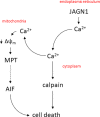
Schematic figure illustrating the putative role of JAGN1 in the calcium‐ and calpain‐dependent cell death pathway described in the present study. JAGN1 was found to interact with Grp78, a key regulator of calcium homeostasis in the ER (not shown). Cell death was completely prevented by PD150606, a selective calpain inhibitor. Bongkrekic acid, a specific inhibitor of ANT, also blocked cell death, pointing to a role for MPT in the present model, while the involvement of AIF remains to be demonstrated. Furthermore, a role for RIPK1, a key player in necroptosis, seems likely, as evidenced by the protective effect of nec‐1. However, caspase involvement was excluded in the present model. JAGN1, jagunal homolog 1; MPT, mitochondrial permeability transition; AIF, apoptosis‐inducing factor (note that AIF also participates in non‐apoptotic cell death); ANT, adenine nucleotide translocator; Δψm, mitochondrial membrane potential; RIPK1, receptor interacting serine/threonine kinase 1. [Colour figure can be viewed at wileyonlinelibrary.com]

Comment in

References

    1. Donadieu J, Beaupain B, Fenneteau O, Bellanné‐Chantelot C. Congenital neutropenia in the era of genomics: classification, diagnosis, and natural history. Br J Haematol. 2017;179:557–4. - PubMed
    1. Rosenberg PS, Zeidler C, Bolyard AA, Alter BP, Bonilla MA, Boxer LA, et al. Stable long‐term risk of leukaemia in patients with severe congenital neutropenia maintained on G‐CSF therapy. Br J Haematol. 2010;150:196–9. - PMC - PubMed
    1. Carlsson G, Fasth A, Berglöf E, Lagerstedt‐Robinson K, Nordenskjöld M, Palmblad J, et al. Incidence of severe congenital neutropenia in Sweden and risk of evolution to myelodysplastic syndrome/leukaemia. Br J Haematol. 2012;158:363–9. - PubMed
    1. Horwitz M, Benson KF, Person RE, Aprikyan AG, Dale DC. Mutations in ELA2, encoding neutrophil elastase, define a 21‐day biological clock in cyclic haematopoiesis. Nat Genet. 1999;23:433–6. - PubMed
    1. Dale DC, Person RE, Bolyard AA, Aprikyan AG, Bos C, Bonilla MA, et al. Mutations in the gene encoding neutrophil elastase in congenital and cyclic neutropenia. Blood. 2000;96:2317–22. - PubMed

Publication types

MeSH terms

Supplementary concepts