. 2020 Nov 17;53(5):1050-1062.e5.
doi: 10.1016/j.immuni.2020.10.013.

Production of the Cytokine VEGF-A by CD4+ T and Myeloid Cells Disrupts the Corneal Nerve Landscape and Promotes Herpes Stromal Keratitis

Affiliations

Production of the Cytokine VEGF-A by CD4+ T and Myeloid Cells Disrupts the Corneal Nerve Landscape and Promotes Herpes Stromal Keratitis

Hongmin Yun et al. Immunity. .

Abstract

Herpes simplex virus type 1 (HSV-1)-infected corneas can develop a blinding immunoinflammatory condition called herpes stromal keratitis (HSK), which involves the loss of corneal sensitivity due to retraction of sensory nerves and subsequent hyperinnervation with sympathetic nerves. Increased concentrations of the cytokine VEGF-A in the cornea are associated with HSK severity. Here, we examined the impact of VEGF-A on neurologic changes that underly HSK using a mouse model of HSV-1 corneal infection. Both CD4+ T cells and myeloid cells produced pathogenic levels of VEGF-A within HSV-1-infected corneas, and CD4+ cell depletion promoted reinnervation of HSK corneas with sensory nerves. In vitro, VEGF-A from infected corneas repressed sensory nerve growth and promoted sympathetic nerve growth. Neutralizing VEGF-A in vivo using bevacizumab inhibited sympathetic innervation, promoted sensory nerve regeneration, and alleviated disease. Thus, VEGF-A can shape the sensory and sympathetic nerve landscape within the cornea, with implications for the treatment of blinding corneal disease.

Keywords: CD4+ T cells; HSV-1; VEGF-A; cornea; myeloid cells; neuro-immune axis; sensory nerves; sympathetic nerves.


Conflict of interest statement

Declaration of Interests The authors declare no competing interests.

Figures
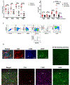
Figure 1.
Figure 1.. VEGF is produced by infiltrating leukocytes in HSV-1 infected corneas and VEGF levels correlate with HSK severity.
Mice were infected with 1 × 10⁵ PFU of HSV-1 (strain RE) and either received tarsorrhaphy at 4 dpi or were left untreated. (A) Mice were examined at 7, 10, 14 and 28 dpi for the severity of HSK and the corneal opacity was monitored in a blinded manner and recorded. (B) At each time point, 5 mice from each group were sacrificed, and corneas were excised and dispersed into single-cell suspensions and cultured in Neural basal A medium (with NGF and GDNF) for 24 hours. Cultures were then harvested and analyzed by sandwich ELISA for VEGF. (C) At each time point, corneas were digested, dispersed into single-cell suspensions and were stained with fluorescently conjugated antibodies to detect corneal leukocytes (CD45+) by flow cytometry. Immune cell populations that produced VEGF-A were quantified as CD11b+F4/80+Ly6G myeloid cells, CD11b+F4/80Ly6G+ myeloid cells CD11b+F4/80+Ly6G+ myeloid cells, and CD4+ T cells (CD4+). Flow plots are from 14 dpi. (D) At 14 dpi, HSK corneas were excised, flat mounted, and labeled with antibodies against CD4, F4/80, VEGF, and anti βIII Tubulin antibody. (Top row—high magnification) Arrows point to CD4+ cells in close proximity with VEGF producing F4/80+ cells. (Bottom row—low magnification) Arrows point to CD4+ cells and F4/80+ myeloid cells that were positive for anti-VEGF antibody staining. Data are pooled from 2–3 independent experiments and symbols represent individual mice. Bars represent the mean ± SEM. Significance was calculated using Student’s t test, *p < 0.05, **p < 0.01, ***p < 0.001,****p < 0.0001.
Figure 2.
Figure 2.. Protecting the corneal surface by tarsorrhaphy reduces VEGF-A production by leukocytes and inhibits sympathetic innervation of infected corneas.
Mice were infected with 1 × 10⁵ PFU of HSV-1 (strain RE) and either received tarsorrhaphy at 4 dpi or were left untreated. Five mice from each group were sacrificed at 7, 14 and 28 dpi, and the corneas were excised and dispersed into single-cell suspensions that were stained with fluorescently conjugated antibodies to detect corneal immune cells. Numbers represent (A) CD11b+ cells, (B) VEGF-A+ CD11b+ Cells, (C) VEGF-A+CD11b+F4/80+Ly6G myeloid cells,VEGF-A+CD11b+F4/80+Ly6G+ myeloid cells, VEGF-A+CD11b+F4/80 Ly6G+ myeloid cells, (D) CD4+ T cells, and (E) VEGF-A+ CD4+ T cells. (F) 28 dpi corneas with (Closed eye) or without (Open eye) tarsorrhaphy were excised, flat mounted and labeled with anti βIII Tubulin antibody which labels all the nerves, anti-Tyrosine Hydroxylase (TH) antibody which recognizes sympathetic nerves, anti-substance P (SP) antibody which recognizes sensory nerves, the arrows point to βIII +TH+ sympathetic nerves in the corneas. Results in all panels are represented as mean number of cells ± SEM from 2–3 independent experiments. Symbols represent individual mice. Significance was calculated using Student’s t test, *p < 0.05, **p < 0.01, ***p < 0.001,****p < 0.0001.
Figure 3.
Figure 3.. Depletion of CD4+ T cells combined with tarsorrhaphy reduces local VEGF-A production and promotes reinnervation of HSK corneas with sensory nerves.
Starting two days prior to infection, mice were given a subconjunctival injection of either anti-CD4 antibody or an isotype control antibody every other day until 28 dpi. Mice were then infected with HSV-1 (RE) and eyes from both groups were protected from desiccation by tarsorrhaphy at 4 dpi. Mice were sacrificed at 7, 14, and 28 dpi and corneas from each group were excised, digested with collagenase, dispersed and analyzed by flow cytometry. Leukocyte (CD45+) subpopulations were quantified as (A) CD4+ T cells, (B) VEGF-A+CD4+ T cells, and (C) CD11b+ cells. (D) CD11b+ cells which were further characterized as VEGF-A+F4/80+Ly6G myeloid cells, VEGF-A+F4/80+Ly6G+ myeloid cells, and VEGF-A+F4/80Ly6G+ myeloid cells. (E) In parallel, corneas from each group were excised and dispersed into single-cell suspensions and were then cultured in Neurobasal A medium (with NGF, GDNF) for 24 hours. Cells and supernatants were harvested and analyzed by sandwich ELISA for VEGF-A. For the apparent outlier, we performed a statistical outlier test offered by GraphPad; however, this point was not determined to be a statistical outlier. Omitting this point still achieves a p value of < 0.0001 for the cell number analysis and < 0.05 for the ELISA data analysis. (F) At 28 dpi, corneas from both groups were excised, flat mounted and labeled with anti βIII Tubulin antibody, anti-Tyrosine Hydroxylase (TH) antibody, and anti-substance P (SP) antibody. Arrows point to βIII +TH+ sympathetic nerves or βIII +SP+ sensory nerves. (G) Sympathetic nerve length in the corneas with mock or CD4 depletion at 28 dpi were quantified as the cumulative length of nerve fibers in the cornea. (H) The severity of HSK was monitored through 28dpi and the opacity scores (28dpi) were analyzed. Results in all panels are represented as mean ± SEM pooled from 2 independent experiments. Symbols represent individual mice. Significance was calculated using Student’s t test, *p < 0.05, **p < 0.01, ***p < 0.001,****p < 0.0001.
Figure 4.
Figure 4.. VEGF-A production by cells from corneas with HSK promotes growth of sympathetic neurites and retraction of sensory neurites.
Sensory (TG-derived) or sympathetic (SCG-derived) neurons were cultured in a microfluidic chamber (soma chamber) and allowed to extend neurites to grow through the microtubes to the axon chamber. (A) Images represent neuronal growth prior to addition of stimuli. (B-D) Single-cell suspensions of corneas from 14 dpi were added into the axon chamber and incubated for three days in the presence or absence of anti-VEGF antibody or bevacizumab. Each chamber was used to measure the ability of a single cornea to promote or inhibit the growth of sensory or sympathetic nerves. Images in section (B) represents sympathetic nerve growth three days after corneal cells addition. Images in section (C) represent sensory nerve growth three days after corneal cells addition. (D) Data represent the pooled imaging results from two independent experiments. Retraction was determined by the fragmentation of the neurites (arrows) in the axonal chamber. Non-retractions was determined by the elongation of the neurites (arrows) in the axonal chamber. Due to the clear growth or absence of growth of axons in the microfluidic chambers, a “yes” (axonal growth) or “no” (absence of axonal growth) was given for each chamber (cornea). Each experiment consisted of assessing four or five separate corneas resulting in an n value of four or five. To generate the pie charts, we pooled yes/no results and calculated the defined frequency in the pie charts. Significance was calculated using Fisher’s exact test of the yes/no results, and n values were biological replicates of wells that had or had not retracted.
Figure 5.
Figure 5.. Neutralizing VEGF in vivo during HSK inhibits sympathetic innervation, promotes reinnervation by sensory nerves, and reduces HSK severity.
Four groups of mice were infected with HSV-1 and were given subconjunctival injections of 500μg bevacizumab or isotype every other day starting from 9 dpi through 28dpi. Two groups of mice (bevacizumab and isotype) were protected by tarsorrhaphy. The severity of HSK was monitored and (A) corneal neovascularization and (B) corneal opacity at 28 dpi were recorded and analyzed. (C) At 28dpi, mice were sacrificed, and corneas were excised, flat mounted and labeled with antibodies to βIII Tubulin, Tyrosine Hydroxylase (TH), and substance P (SP). Arrows point to βIII +TH+ sympathetic nerves or βIII +SP+ sensory nerves. (D & E) Sympathetic nerves in the corneas protected by tarsorrhaphy were traced using the Simple Neurite Tracer (Longair et al., 2011) in the segmentation package in FIJI and was followed by analysis using the 3D Skeletonize (Arganda-Carreras et al., 2010) FIJI plugin. The z projection of the traced neurons from one cornea is shown. (F) The mice without tarsorrhaphy were assessed for corneal reflex at 28dpi. Results in all panels are represented as mean ± SEM pooled from 2 to 3 independent experiments. Significance was calculated using Student’s t test, *p < 0.05, **p < 0.01, ****p < 0.0001.
Figure 6.
Figure 6.. Soluble VEGF receptor 1 treatment prevents sympathetic nerve innervation and regenerates corneal sensitivity.
Mice were infected with HSV-1 (RE) and beginning at 9 dpi were given subconjunctival injections of soluble VEGF receptor 1 or isotype antibody every other day through 23dpi. (A-D) HSK severity was monitored as corneal opacity, neovascularization, and corneal reflex. Arrows point to the blood vessels in the corneas. (E) The corneas were excised, flat mounted and labeled with antibodies to βIII Tubulin, Tyrosine Hydroxylase (TH), and substance P (SP). Arrows point to βIII +TH+ sympathetic nerves or βIII +SP+ sensory nerves. Images in all panels represented the nerve staining from 2 independent experiments. Results in all panels are represented as mean ± SEM pooled from 2 independent experiments. Significance was calculated using Student’s t test,**p < 0.01.

References

    1. Acosta MC, Tan ME, Belmonte C, and Gallar J (2001). Sensations evoked by selective mechanical, chemical, and thermal stimulation of the conjunctiva and cornea. Invest Ophthalmol Vis Sci 42, 2063–2067. - PubMed
    1. Reeves Alexander G., R.S.S., Cohen Jeffrey, Fadul Camilo, Jenkyn Lawrence, Ward Thomas Facial sensations & movements In Disorders of the nervous system, Swenson RS, ed. (Dartmouth Medical School; ).
    1. Ambati BK, Nozaki M, Singh N, Takeda A, Jani PD, Suthar T, Albuquerque RJ, Richter E, Sakurai E, Newcomb MT, et al. (2006). Corneal avascularity is due to soluble VEGF receptor-1. Nature 443, 993–997. - PMC - PubMed
    1. Arbore G, West EE, Spolski R, Robertson AAB, Klos A, Rheinheimer C, Dutow P, Woodruff TM, Yu ZX, O’Neill LA, et al. (2016). T helper 1 immunity requires complement-driven NLRP3 inflammasome activity in CD4(+) T cells. Science 352, aad1210. - PMC - PubMed
    1. Arganda-Carreras I, Fernandez-Gonzalez R, Munoz-Barrutia A, and Ortiz-De-Solorzano C (2010). 3D reconstruction of histological sections: Application to mammary gland tissue. Microsc Res Tech 73, 1019–1029. - PubMed

Publication types

MeSH terms

Substances