. 2020 Nov 16;12(11):3388.
doi: 10.3390/cancers12113388.

Induction of Lysosomal Membrane Permeabilization Is a Major Event of FTY720-Mediated Non-Apoptotic Cell Death in Human Glioma Cells

Affiliations

Induction of Lysosomal Membrane Permeabilization Is a Major Event of FTY720-Mediated Non-Apoptotic Cell Death in Human Glioma Cells

Kyoung-Jin Min et al. Cancers (Basel). .

Abstract

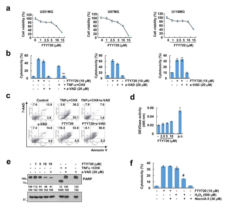
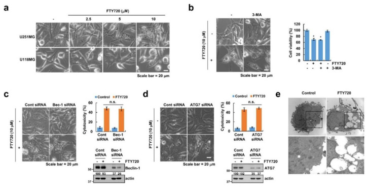
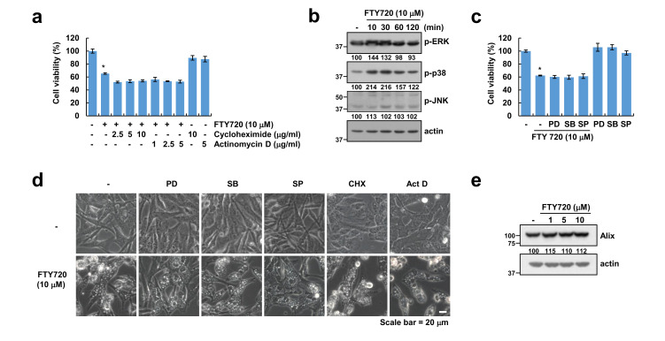
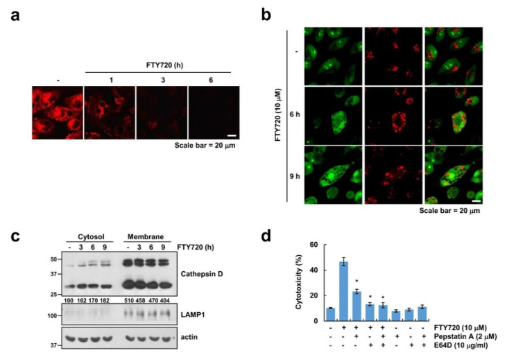
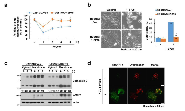
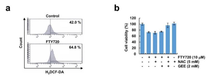
FTY720, a sphingosine-1-phosphate (S1P) receptor modulator, is a synthetic compound produced by the modification of a metabolite from I. sinclairii. Here, we found that FTY720 induced non-apoptotic cell death in human glioma cells (U251MG, U87MG, and U118MG). FTY720 (10 µM) dramatically induced cytoplasmic vacuolation in glioma cells. However, FTY720-mediated vacuolation and cell death are not associated with autophagy. Genetic or pharmacological inhibition of autophagy did not inhibit FTY720-induced cell death. Herein, we detected that FTY720-induced cytoplasmic vacuoles were stained with lysotracker red, and FTY720 induced lysosomal membrane permeabilization (LMP). Interestingly, cathepsin inhibitors (E64D and pepstatin A) and ectopic expression of heat shock protein 70 (HSP70), which is an endogenous inhibitor of LMP, markedly inhibited FTY720-induced cell death. Our results demonstrated that FTY720 induced non-apoptotic cell death via the induction of LMP in human glioma cells.

Keywords: FTY720; LMP; cathepsins; glioma; non-apoptotic cell death.


Conflict of interest statement

The authors declare that they have no conflict of interest.

Figures

Figure 1
Figure 1
FTY720 induces cell death in human glioma cells. (a) Cells (U251MG, U87MG, and U118MG) were treated with the indicated concentrations of FTY720 for 24 h. The cell viability was determined by XTT assay. (b) Cells (U251MG, U87MG, and U118MG) were treated with 10 μM FTY720 in the presence or absence of 20 μM z-VAD for 24 h. Cell cytotoxicity was detected by LDH assay. (ce) U251MG cells were treated with 10 μM FTY720 or 5 ng/mL TNF-α plus 2.5 μg/mL cycloheximide (CHX) (positive control; p.c.) in the presence or absence of 20 μM z-VAD for 24 h. Cell death was determined by staining with 7-AAD and Annexin V (c). Caspase activities were calculated using caspase-3 (DEVDase) assay kits (d). Protein expression was detected by Western blotting (e). (f) U251MG cells were treated with 10 μM FTY720 (18 h) and 250 μM H2O2 (2 h) in the presence or absence of 20 μM NecroX-5. Cell cytotoxicity was detected by LDH assay. The values in the graphs (a,b,d,f) represent the mean ± SD of three independent experiments. ** p < 0.01 compared to the TNF-α plus CHX. * p < 0.01 compared to the control. # p < 0.01 compared to the H2O2.
Figure 2
Figure 2
Effect of FTY720 on autophagy in human glioma U251MG cells. (a) Cells were treated with the indicated concentrations of FTY720 for 6 h. (b) U251MG cells were pretreated with 1 mM 3-MA for 30 min, and then treated with 10 μM FTY720 for 24 h. (c,d) U251MG cells were transfected with Beclin-1 (Bec-1) siRNA (c), ATG7 siRNA, (d) and/or control siRNA (Cont siRNA). Twenty-four hours after transfection, cells were treated with 10 μM FTY720 for 24 h. (e) U251MG cells were treated with 10 μM FTY720 for 12 h, and cell morphology was detected by electron microscopy. Cell morphology was detected by interference light microscopy. The mean number of microscopic images (ae) per sample is 10, and these images were taken randomly. Cell viability was determined by XTT assay (b). Protein expression was detected by Western blotting and cell death was analyzed by LDH assay (c,d). The values in the graphs (bd) represent the mean ± SD of three independent experiments. * p < 0.01 compared to the control. n.s. = no significance.
Figure 3
Figure 3
FTY720-induced cell death is independent of paraptosis in human glioma cells. (a) U251MG cells were pretreated with the indicated concentrations of cycloheximide or actinomycin D for 30 min, and then treated with 10 μM FTY720 for 24 h. (b) U251MG cells were treated with 10 μM FTY720 for the indicated time periods. (c,d) U251MG cells were pretreated with MAPK inhibitors [ERK inhibitor: 50 μM PD98059 (PD); p38 MAPK inhibitor: 10 μM SB203580 (SB); and JNK inhibitor: 10 μM SP600125 (SP)] for 30 min, and then treated with 10 μM FTY720 for 24 h. (e) U251MG cells were treated with the indicated concentrations of FTY720 for 24 h. Cell viability was analyzed by XTT assay (a,c), and protein expression was detected by Western blotting (b,e). Cell morphology was detected by interference light microscopy. The mean number of microscopic images per sample is 10, and these images were taken randomly (d). The values in the graphs (a,c) represent the mean ± SD of three independent experiments. * p < 0.01 compared to the control.
Figure 4
Figure 4
FTY720 induces lysosomal membrane permeabilization (LMP). (ac) U251MG cells were treated with 10 μM FTY720 for the indicated time periods. Cells were stained with lysotracker red (a) and acridine orange (b). Cytosol and membrane fractions (lysosome-rich fraction) were prepared, and the protein levels were determined by Western blotting. LAMP1 is shown as cell compartment controls (lysosome-rich membrane fraction) (c). (d) U251 cells were pretreated with 2 μM pepstatin A (Pep A) and/or 10 μg/mL E64D for 30 min, and then added along with 10 μM FTY720 for 24 h. Cell toxicity was determined using the LDH assay. The mean number of microscopic images (a,b) per sample is 10, and these images were taken randomly. The values in the graphs (d) represent the mean ± SD of three independent experiments. * p < 0.01 compared to the FTY720.
Figure 5
Figure 5
Ectopic expression of HSP70 inhibits FTY720-induced LMP. (ac) U251MG/vector and U251MG/HSP70 cells were treated with 10 μM FTY720 for the indicated time periods. Cells were stained with lysotracker red (a). Cell morphology was examined using interference light microscopy. Cell cytotoxicity was detected by LDH assay (b). Cytosol and membrane fractions (lysosome-rich fraction) were prepared, and the protein levels were determined by Western blotting. LAMP1 is shown as cell compartment controls (lysosome-rich membrane fraction) (c). (d) U251MG cells were treated with 10 μM NBD-FTY720 for 6 h, and then cell morphology was detected by confocal microscopy. The mean number of microscopic images (b,d) per sample is 10, and these images were taken randomly. The values in the graphs (a,b) represent the mean ± SD of three independent experiments. * p < 0.01 compared to the FTY720-treated U251MG/vector.
Figure 6
Figure 6
Reactive oxygen species (ROS) had no effect on FTY720-induced cell death in human glioma U251MG cells. (a) U251MG cells were treated with 10 μM FTY720 for 3 h, and then cells were stained with H2DCF-DA dye. Fluorescence was detected using flow cytometry. (b) U251MG cells pretreated with ROS scavengers [5 mM N-acetyl-cysteine (NAC) and 2 mM glutathione ethyl ester (GEE)] for 30 min, and then added along with 10 μM FTY720 for 24 h. Cell viability was determined by XTT assay. The values in the graphs (b) represent the mean ± SD of three independent experiments.

Similar articles

Cited by

References

    1. Permpongkosol S., Wang J.D., Takahara S., Matsumiya K., Nonomura N., Nishimura K., Tsujimura A., Kongkanand A., Okuyama A. Anticarcinogenic effect of FTY720 in human prostate carcinoma DU145 cells: Modulation of mitogenic signaling, FAK, cell-cycle entry and apoptosis. Int. J. Cancer. 2002;98:167–172. doi: 10.1002/ijc.10178. - DOI - PubMed
    1. Shen Y., Cai M., Xia W., Liu J., Zhang Q., Xie H., Wang C., Wang X., Zheng S. FTY720, a synthetic compound from Isaria sinclairii, inhibits proliferation and induces apoptosis in pancreatic cancer cells. Cancer Lett. 2007;254:288–297. doi: 10.1016/j.canlet.2007.03.013. - DOI - PubMed
    1. Perla A.S., Fratini L., Cardoso P.S., de Farias C.B., da Cunha Jaeger M., Roesler R. Fingolimod (FTY720) reduces viability and survival and increases histone H3 acetylation in medulloblastoma cells. Pediatr. Hematol. Oncol. 2020;37:170–175. doi: 10.1080/08880018.2019.1699213. - DOI - PubMed
    1. Young M.M., Bui V., Chen C., Wang H.G. FTY720 induces non-canonical phosphatidylserine externalization and cell death in acute myeloid leukemia. Cell Death Dis. 2019;10:847–895. doi: 10.1038/s41419-019-2080-5. - DOI - PMC - PubMed
    1. Ota K., Okuma T., Lorenzo A., Yokota A., Hino H., Kazama H., Moriya S., Takano N., Hiramoto M., Miyazawa K. Fingolimod sensitizes EGFR wildtype nonsmall cell lung cancer cells to lapatinib or sorafenib and induces cell cycle arrest. Oncol. Rep. 2019;42:231–242. - PubMed

LinkOut - more resources