. 2020 Dec;9(23):e017195.
doi: 10.1161/JAHA.120.017195. Epub 2020 Nov 19.

Dysfunctional Mitochondrial Dynamic and Oxidative Phosphorylation Precedes Cardiac Dysfunction in R120G-αB-Crystallin-Induced Desmin-Related Cardiomyopathy

Affiliations

Dysfunctional Mitochondrial Dynamic and Oxidative Phosphorylation Precedes Cardiac Dysfunction in R120G-αB-Crystallin-Induced Desmin-Related Cardiomyopathy

Shafiul Alam et al. J Am Heart Assoc. 2020 Dec.

Abstract

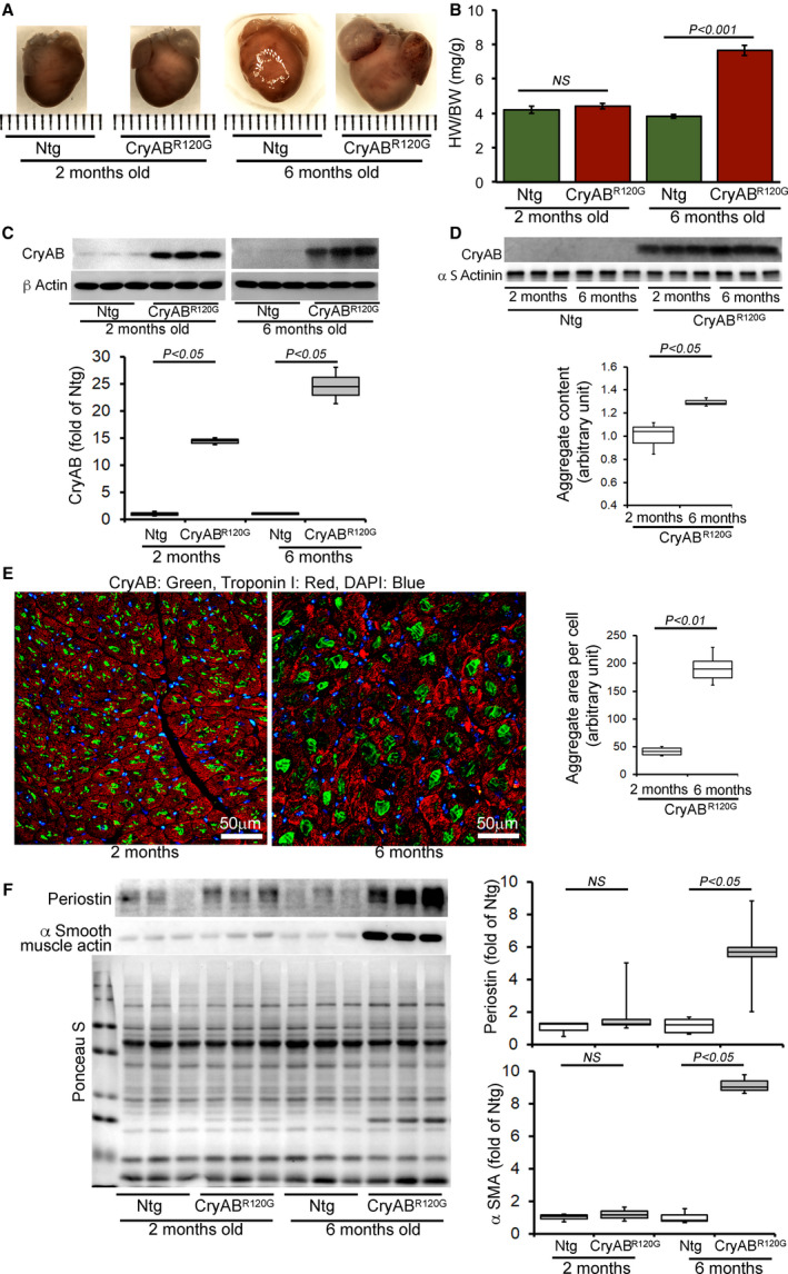
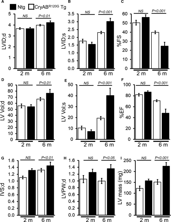
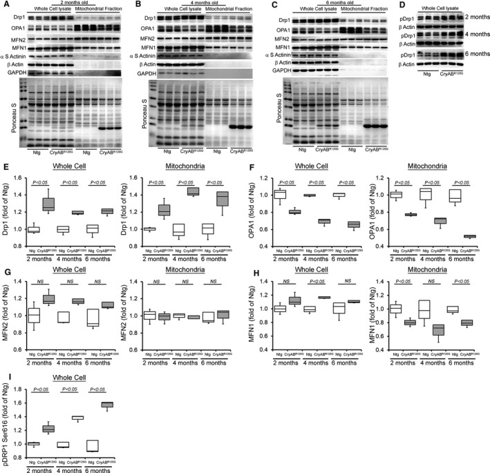
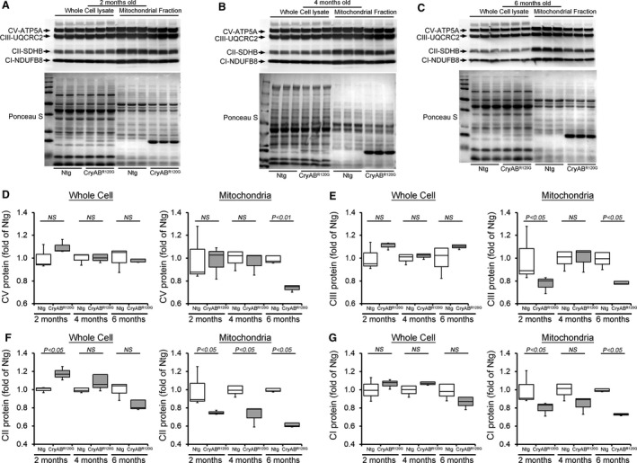
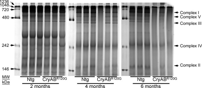
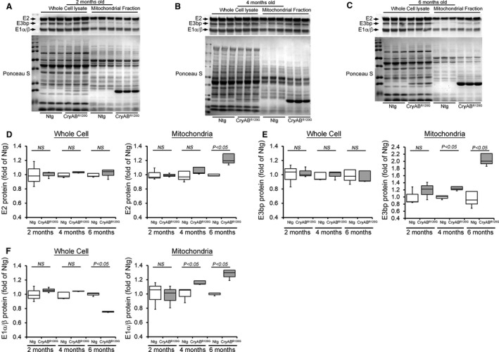
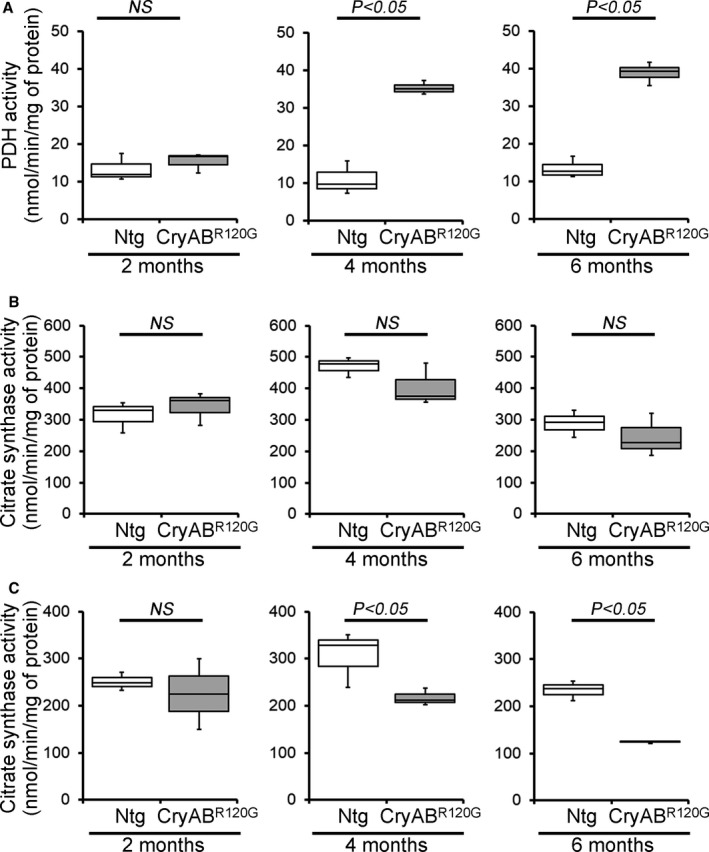
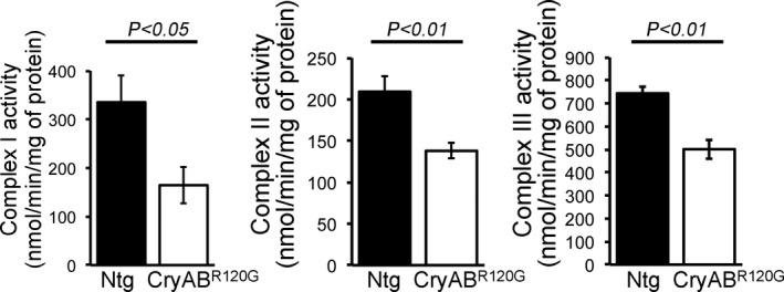
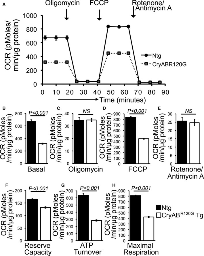
Background The mutated α-B-Crystallin (CryABR120G) mouse model of desmin-related myopathy (DRM) shows an age-dependent onset of pathologic cardiac remodeling and progression of heart failure. CryABR120G expression in cardiomyocytes affects the mitochondrial spatial organization within the myofibrils, but the molecular perturbation within the mitochondria in the relation of the overall course of the proteotoxic disease remains unclear. Methods and Results CryABR120G mice show an accumulation of electron-dense aggregates and myofibrillar degeneration associated with the development of cardiac dysfunction. Though extensive studies demonstrated that these altered ultrastructural changes cause cardiac contractility impairment, the molecular mechanism of cardiomyocyte death remains elusive. Here, we explore early pathological processes within the mitochondria contributing to the contractile dysfunction and determine the pathogenic basis for the heart failure observed in the CryABR120G mice. In the present study, we report that the CryABR120G mice transgenic hearts undergo altered mitochondrial dynamics associated with increased level of dynamin-related protein 1 and decreased level of optic atrophy type 1 as well as mitofusin 1 over the disease process. In association with these changes, an altered level of the components of mitochondrial oxidative phosphorylation and pyruvate dehydrogenase complex regulatory proteins occurs before the manifestation of pathologic adverse remodeling in the CryABR120G hearts. Mitochondria isolated from CryABR120G transgenic hearts without visible pathology show decreased electron transport chain complex activities and mitochondrial respiration. Taken together, we demonstrated the involvement of mitochondria in the pathologic remodeling and progression of DRM-associated cellular dysfunction. Conclusions Mitochondrial dysfunction in the form of altered mitochondrial dynamics, oxidative phosphorylation and pyruvate dehydrogenase complex proteins level, abnormal electron transport chain complex activities, and mitochondrial respiration are evident on the CryABR120G hearts before the onset of detectable pathologies and development of cardiac contractile dysfunction.

Keywords: R120G‐αB‐crystallin; desmin‐related myopathy; mitochondrial dynamics; mitochondrial respiration; oxidative phosphorylation.


Conflict of interest statement

None.

Figures

Figure 1
Figure 1. Pathological cardiac remodeling in CryABR120G Tg hearts.
A, Images of hearts and B, quantification of the heart‐weight‐to‐body‐weight ratios (HW/BW) showing cardiac hypertrophy in the CryABR120G transgenic hearts (n ≥ 6 or more mice per group) at 6 months of age. C, Representative Western blot and densitometric quantification of αB‐crystallin (CryAB) protein level in the whole cell lysate derived from 2‐ and 6‐month‐old CryABR120G transgenic and nontransgenic hearts (n=3 mice per group). β actin was used as a loading control. D, Representative Western blot and densitometric quantification of aggregated CryAB protein level present in the insoluble fractions derived from 2‐ and 6‐month‐old CryABR120G transgenic and nontransgenic hearts (n=3 mice per group). α‐Sarcomeric actinin was used as a loading control. E, Immunofluorescence staining for CryAB (green) with troponin I counterstaining (red) showed aggregate levels in 2‐ and 6‐month‐old CryABR120G transgenic hearts. Quantification of CryAB aggregates showed age‐dependent increased aggregate size in the heart. F, Representative Western blot and densitometric quantification of periostin and (αSMA) proteins level in the whole cell lysate derived from 2‐ and 6‐month‐old CryABR120G transgenic and nontransgenic hearts (n=3 mice per group). Boxes represent interquartile ranges, lines represent medians, whiskers represent ranges, and P values were determined by Kruskal–Wallis test. CryABR120G Tg indicates mutant αB‐crystallin transgenic mouse; DAPI, 4′,6‐diamidino‐2‐phenylindole; NS, not significant; Ntg, nontransgenic; and αSMA, α‐smooth muscle actin. Scale bars: 50 μm.
Figure 2
Figure 2. Echocardiography indices of cardiac structure and function in CryABR120G Tg and Ntg hearts.
M‐mode echocardiography indices measured in 2‐ and 6‐month‐old CryABR120G transgenic mice with age‐matched littermate nontransgenic mice. A, Left ventricular (LV) diastolic internal dimension (LVID;d). B, LV systolic internal dimension (LVID;s). C, Percent fractional shortening (%FS). D, LV diastolic volume (LV Vol;d). E, LV systolic volume (LV Vol;s). F, Percent ejection fraction (%EF). G, LV diastolic interventricular septum thickness (IVS;d). H, LV diastolic posterior wall thickness (LVPW;d). I, LV mass (n=5–6 mice per group). Bars represent mean±SEM. P values were determined by Student t test. CryABR120G Tg indicates mutant αB‐crystallin transgenic mouse; NS, not significant; and Ntg, nontransgenic.
Figure 3
Figure 3. Mitochondrial dynamics regulatory proteins levels in CryABR120G Tg and Ntg hearts.
Representative Western blot showing the mitochondrial dynamic regulatory proteins level in the whole cell lysate and the mitochondrial fraction of CryABR120G transgenic and nontransgenic hearts at (A) 2 months, (B) 4 months, and (C) 6 months of age: Drp1, OPA1, MFN2, and MFN1. (D) pDrp1 Ser616 protein level in the whole cell lysate of CryABR120G transgenic and nontransgenic hearts at 2, 4, and 6 months of age. GAPDH and β actin were used as a control to show the purity of the mitochondrial fraction. α‐Sarcomeric actinin was used to show the level of myofilament co‐enrichment in the mitochondrial fraction. Ponceau S protein staining of the transfer membrane confirmed approximately equal loading across the gel (n=3 mice per group). Densitometric quantification of the protein levels of (E) Drp1, (F) OPA1, (G) MFN2, (H) MFN1, and (I) pDrp1 Ser616 in the CryABR120G transgenic and nontransgenic hearts at 2, 4, and 6 months of age. Boxes represent interquartile ranges, lines represent medians, whiskers represent ranges, and P values were determined by Kruskal–Wallis test. CryABR120G Tg indicates mutant αB‐crystallin transgenic mouse; Drp1, dynamin‐related protein 1; MFN1, mitofusin 1; MFN1, mitofusin 2; Ntg, nontransgenic; OPA1, optic atrophy type 1; and NS, not significant.
Figure 4
Figure 4. OXPHOS complex protein levels in CryABR120G Tg and Ntg hearts.
Representative Western blot showing the OXPHOS complex regulatory proteins level in the whole cell lysate and the mitochondrial fraction of CryABR120G transgenic and nontransgenic hearts at (A) 2 months, (B) 4 months, and (C) 6 months of age: Complex I, Complex II, Complex III, and Complex V protein. Ponceau S protein stain of the transfer membrane was used to confirm approximately equal loading (n=3 mice per group). Densitometric quantification of the protein levels of (D) Complex V (CV), (E) Complex III (CIII), (F) Complex II (CII), and (G) Complex I (CI) in the whole cell lysate and the mitochondrial fraction of CryABR120G transgenic and nontransgenic hearts at 2, 4, and 6 months of age. Boxes represent interquartile ranges, lines represent medians, whiskers represent ranges, and P values were determined by Kruskal–Wallis test. CryABR120G Tg indicates mutant αB‐crystallin transgenic mouse; Ntg, nontransgenic; NS, not significant; and OXPHOS, oxidative phosphorylation system.
Figure 5
Figure 5. Representative gel image showing mitochondrial protein complexes resolved by nongradient BN‐PAGE in CryABR120G Tg and Ntg hearts.
Mitochondria isolated from CryABR120G transgenic and nontransgenic hearts at 2, 4, and 6 months of age were analyzed, with each lane containing mitochondria prepared from independent mice. An 8% separating gel was used and the gel was stained by Coomassie blue after gel electrophoresis. Arrows indicate mitochondrial electron transport chain complexes visualized following Coomassie blue staining. BN‐PAGE indicates blue native polyacrylamide gel electrophoresis; and CryABR120G, mutant αB‐crystallin transgenic mouse.
Figure 6
Figure 6. PDH complex protein levels in CryABR120G Tg and Ntg hearts.
Representative Western blot showing the PDH complex regulatory proteins level in the whole cell lysate and the mitochondrial fraction of CryABR120G transgenic and nontransgenic hearts at (A) 2 months, (B) 4 months, and (C) 6 months of age: E2, E3bp, and E1α/β protein. Ponceau S protein stain of the transfer membrane was used to confirm approximately equal loading (n=3 mice per group). Densitometric quantification of the protein levels of (D) E2, (E) E3bp, and (F) E1α/β component of the PDH complex in the whole cell lysate and the mitochondrial fraction of CryABR120G transgenic and nontransgenic hearts at 2, 4, and 6 months of age. Boxes represent interquartile ranges, lines represent medians, whiskers represent ranges, and P values were determined by Kruskal–Wallis test. CryABR120G Tg indicates mutant αB‐crystallin transgenic mouse; E1, pyruvate dehydrogenase; E2, dihydrolipoamide transacetylase; E3, dihydrolipoamide dehydrogenase; Ntg, nontransgenic; NS, not significant; and PDH, pyruvate dehydrogenase complex.
Figure 7
Figure 7. Mitochondrial PDH and CS activities in CryABR120G Tg and Ntg hearts.
A, PDH enzymatic activity in 2‐, 4‐, and 6‐month‐old CryABR120G transgenic and nontransgenic hearts (n=3 mice per group). B and C, CS activity in the mitochondrial fraction (B) and the whole heart homogenates (C) isolated from 2‐, 4‐, and 6‐month‐old CryABR120G transgenic and nontransgenic hearts (n=3 mice per group). Boxes represent interquartile ranges, lines represent medians, whiskers represent ranges, and P values were determined by Kruskal–Wallis test. CryABR120G Tg indicates mutant αB‐crystallin transgenic mouse; CS, citrate synthase; NS, not significant; Ntg, nontransgenic; and Tg, transgenic.
Figure 8
Figure 8. Mitochondrial electron transport chain complex activities in CryABR120G Tg and Ntg hearts.
Mitochondrial Complex I, II, and III activities in 2‐month‐old CryABR120G transgenic and nontransgenic hearts (n=6 mice per group). Bars represent mean±SEM. P values were determined by Student t test. CryABR120G Tg indicates mutant αB‐crystallin transgenic mouse; NS, not significant; and Ntg, nontransgenic.
Figure 9
Figure 9. Mitochondrial respiration in isolated mitochondria from CryABR120G Tg and Ntg mice hearts.
A, Mitochondrial oxygen consumption rate (OCR) profiles in isolated mitochondria from 2 months of age littermate CryABR120G Tg and Ntg hearts. Arrow indicates the sequential addition of oligomycin (1 µM), FCCP (4 µM), and rotenone (0.5 µM) plus antimycin A (0.5 µM). OCR profile is expressed as pmol O2/min per µg of protein, and each point represents average OCR values for 5 mice. Graphs showing OCR under (B) baseline as well as with the addition of (C) oligomycin, (D) FCCP, and (E) rotenone plus antimycin A. Key parameters of mitochondrial respiration, including (F) reserve capacity, (G) ATP turnover, and (H) maximal respiration were significantly decreased in CryABR120G Tg mitochondria. Bars represent mean±SEM. P values were determined by Student t test. CryABR120G Tg indicates mutant αB‐crystallin transgenic mouse; FCCP, carbonyl cyanide‐p‐trifluoromethoxy‐phenylhydrazone; NS, not significant; and Ntg, nontransgenic.

Similar articles

Cited by

References

    1. Wexler RK, Elton T, Pleister A, Feldman D. Cardiomyopathy: an overview. Am Fam Physician. 2009;79:778–784. - PMC - PubMed
    1. Knoll R, Hoshijima M, Hoffman HM, Person V, Lorenzen‐Schmidt I, Bang ML, Hayashi T, Shiga N, Yasukawa H, Schaper W, et al. The cardiac mechanical stretch sensor machinery involves a Z disc complex that is defective in a subset of human dilated cardiomyopathy. Cell. 2002;111:943–955. - PubMed
    1. Goldfarb LG, Dalakas MC. Tragedy in a heartbeat: malfunctioning desmin causes skeletal and cardiac muscle disease. J Clin Invest. 2009;119:1806–1813. - PMC - PubMed
    1. McLendon PM, Robbins J. Desmin‐related cardiomyopathy: an unfolding story. Am J Physiol Heart Circ Physiol. 2011;301:H1220–H1228. - PMC - PubMed
    1. Bennardini F, Wrzosek A, Chiesi M. Alpha B‐crystallin in cardiac tissue. Association with actin and desmin filaments. Circ Res. 1992;71:288–294. - PubMed

Publication types