. 2020 Nov 18;8(1):161.
doi: 10.1186/s40168-020-00917-7.

Spatial heterogeneity of bacterial colonization across different gut segments following inter-species microbiota transplantation

Affiliations

Spatial heterogeneity of bacterial colonization across different gut segments following inter-species microbiota transplantation

Na Li et al. Microbiome. .

Abstract

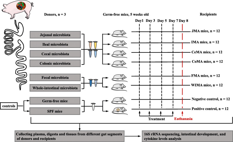
Background: The microbiota presents a compartmentalized distribution across different gut segments. Hence, the exogenous microbiota from a particular gut segment might only invade its homologous gut location during microbiota transplantation. Feces as the excreted residue contain most of the large-intestinal microbes but lack small-intestinal microbes. We speculated that whole-intestinal microbiota transplantation (WIMT), comprising jejunal, ileal, cecal, and colonic microbiota, would be more effective for reshaping the entire intestinal microbiota than conventional fecal microbiota transplantation fecal microbiota transplantation (FMT).

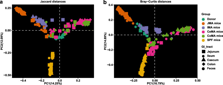
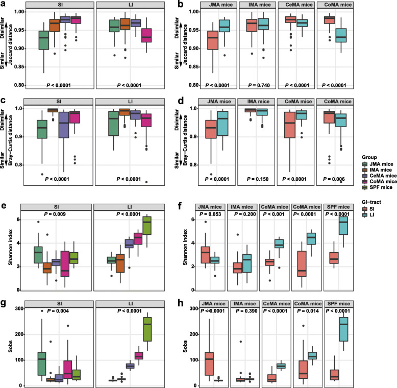
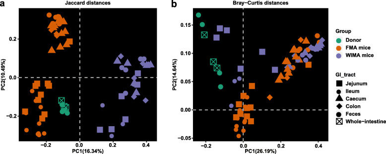
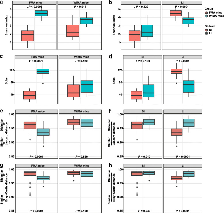
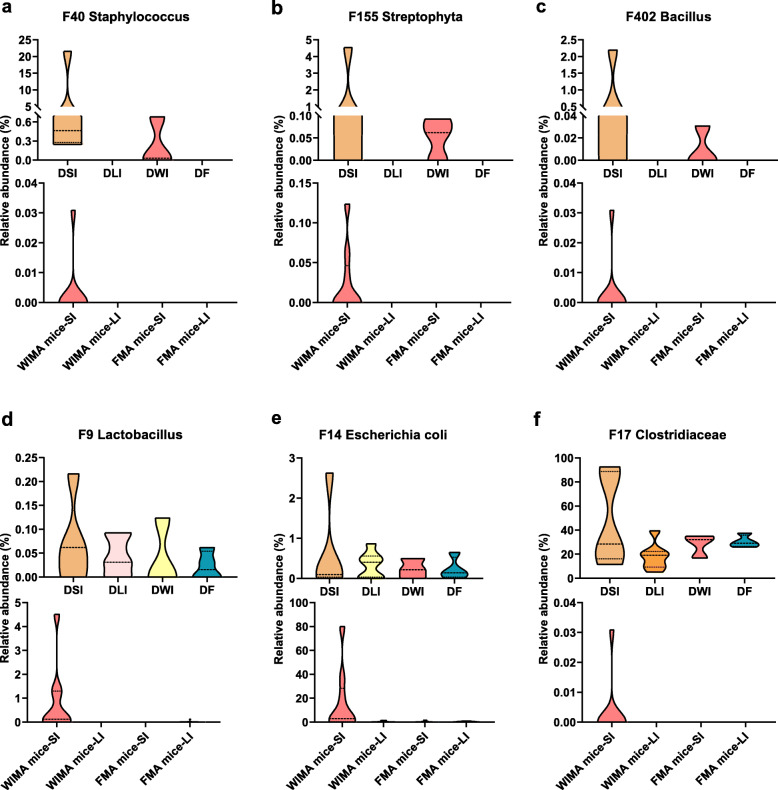
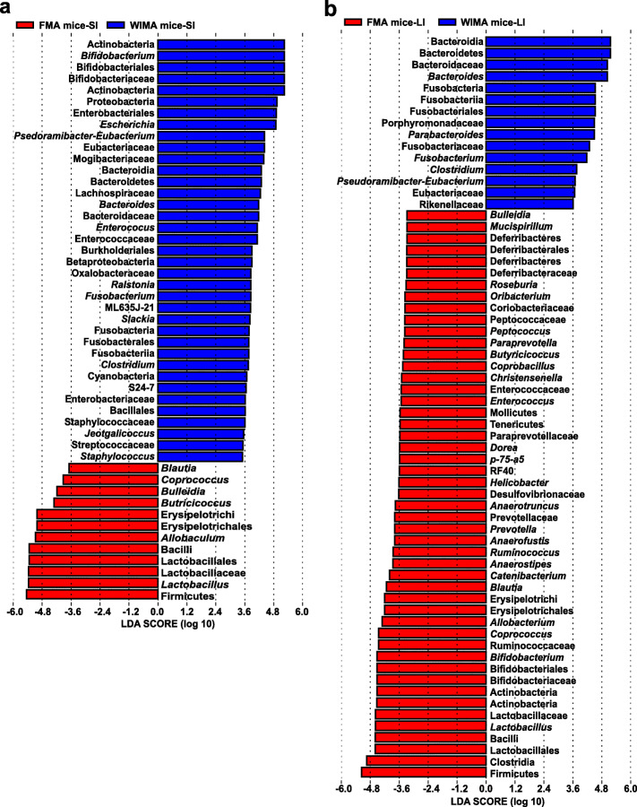
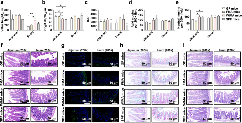
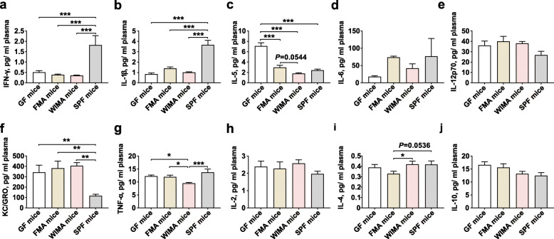
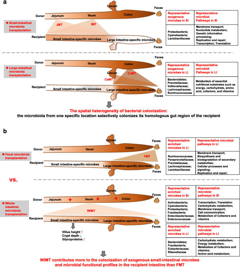
Results: We modeled the compartmentalized colonization of the gut microbiota via transplanting the microbiota from jejunum, ileum, cecum, and colon, respectively, into the germ-free mice. Transplanting jejunal or ileal microbiota induced more exogenous microbes' colonization in the small intestine (SI) of germ-free mice rather than the large intestine (LI), primarily containing Proteobacteria, Lactobacillaceae, and Cyanobacteria. Conversely, more saccharolytic anaerobes from exogenous cecal or colonic microbiota, such as Bacteroidetes, Prevotellaceae, Lachnospiraceae, and Ruminococcaceae, established in the LI of germ-free mice that received corresponding intestinal segmented microbiota transplantation. Consistent compartmentalized colonization patterns of microbial functions in the intestine of germ-free mice were also observed. Genes related to nucleotide metabolism, genetic information processing, and replication and repair were primarily enriched in small-intestinal communities, whereas genes associated with the metabolism of essential nutrients such as carbohydrates, amino acids, cofactors, and vitamins were mainly enriched in large-intestinal communities of germ-free mice. Subsequently, we compared the difference in reshaping the community structure of germ-free mice between FMT and WIMT. FMT mainly transferred LI-derived microorganisms and gene functions into the recipient intestine with sparse SI-derived microbes successfully transplanted. However, WIMT introduced more SI-derived microbes and associated microbial functions to the recipient intestine than FMT. Besides, WIMT also improved intestinal morphological development as well as reduced systematic inflammation responses of recipients compared with FMT.

Conclusions: Segmented exogenous microbiota transplantation proved the spatial heterogeneity of bacterial colonization along the gastrointestinal tract, i.e., the microbiota from one specific location selectively colonizes its homologous gut region. Given the lack of exogenous small-intestinal microbes during FMT, WIMT may be a promising alternative for conventional FMT to reconstitute the microbiota across the entire intestinal tract. Video Abstract.

Keywords: Different gut segments; Fecal microbiota transplantation; Gut microbiota; Spatial heterogeneity; Whole-intestinal microbiota transplantation.


Conflict of interest statement

The authors declare that they have no competing interests.

Figures

Fig. 1
Fig. 1
The timeline of treatments and sample collection. JMA mice: jejunal microbiota-associated mice; IMA: ileal microbiota-associated mice; CeMA: cecal microbiota-associated mice; CoMA: colonic microbiota-associated mice; FMA: fecal microbiota-associated mice; WIMA: whole-intestinal microbiota-associated mice; SPF: specific-pathogen-free mice
Fig. 2
Fig. 2
Gut microbiota structure of recipient mice, SPF mice, and donor pigs. Principal coordinate analysis (PCoA) plots based on the Jaccard distance (a) and Bray-Curtis distance (b) showed distinct clusters in donor and mouse samples. JMA mice: jejunal microbiota-associated mice; IMA: ileal microbiota-associated mice; CeMA: cecal microbiota-associated mice; CoMA: colonic microbiota-associated mice; SPF: specific-pathogen-free mice
Fig. 3
Fig. 3
Differences in beta and alpha diversities of gut microbiota in recipient mice and SPF mice. Differences in the Jaccard distance from the recipient to the donor among different groups (a) and between SI and LI of recipients (b). Differences in the Bray-Curtis distance from the recipient to the donor among different groups (c) and between SI and LI of recipients (d). Differences in the community diversity (Shannon index) among different groups (e) and between SI and LI of recipients (f). Differences in the community richness (sobs) among different groups (g) and between SI and LI of recipients (h). Jejunal and ileal samples of recipients were pooled into small-intestinal samples. Cecal, colonic, and fecal samples of recipients were pooled into large-intestinal samples. JMA mice: jejunal microbiota-associated mice; IMA: ileal microbiota-associated mice; CeMA: cecal microbiota-associated mice; CoMA: colonic microbiota-associated mice; SPF mice: specific-pathogen-free mice; SI: small intestine; LI: large intestine
Fig. 4
Fig. 4
Gut microbiota structure of FMA mice, WIMA mice, and donor pigs. Principal coordinate analysis (PCoA) plots based on the Jaccard distance (a) and Bray-Curtis distance (b) showed distinct clusters in donor and mouse samples. FMA mice: fecal microbiota-associated mice; WIMA mice: whole-intestinal microbiota-associated mice
Fig. 5
Fig. 5
Differences in alpha and beta diversities of gut microbiota in FMA and WIMA mice. Differences in the community diversity (Shannon index) between SI and LI of recipients (a) and between FMA mice and WIMA mice (b). Differences in the community richness (sobs) between SI and LI of recipients (c) and between FMA mice and WIMA mice (d). Differences in the Jaccard distance from the recipient to the donor between SI and LI of recipients (e) and between FMA mice and WIMA mice (f). Differences in the Bray-Curtis distance from the recipient to the donor between SI and LI of recipients (g) and between FMA mice and WIMA mice (h). Jejunal and ileal samples of mice were pooled into small-intestinal samples. Cecal, colonic, and fecal samples of mice were pooled into large-intestinal samples. FMA mice: fecal microbiota-associated mice; WIMA mice: whole-intestinal microbiota-associated mice; SI: small intestine; LI: large intestine
Fig. 6
Fig. 6
Small intestinal-specific microbes in donors that successfully colonized the SI of WIMA mice. Bacterial features that were either absent (a, b, c) or less abundant in the donor feces than in the donor whole intestine (d, e, f) only colonizing the SI of WIMA mice, were referred to as “small intestine-specific microbes” during WIMT. Jejunal and ileal samples of mice were pooled into small-intestinal samples. Cecal, colonic, and fecal samples of mice were pooled into large-intestinal samples. DSI: donor small-intestine; DLI: donor large-intestine; DWI: donor whole-intestine; DF: donor feces; SI: small intestine; LI: large intestine; FMA mice: fecal microbiota-associated mice; WIMA mice: whole-intestinal microbiota-associated mice
Fig. 7
Fig. 7
Differentially abundant taxa between FMA and WIMA mice. Histograms of a linear discriminant analysis (LDA) score (threshold ≥ 2) in small-intestinal samples (a) and large-intestinal samples (b) are plotted. Jejunal and ileal samples of recipients were pooled into small-intestinal samples. Cecal, colonic, and fecal samples of recipients were pooled into large-intestinal samples. FMA mice: fecal microbiota-associated mice; WIMA mice: whole-intestinal microbiota-associated mice; SI: small intestine; LI: large intestine
Fig. 8
Fig. 8
The development of small-intestinal epithelial morphology of germ-free, FMA, WIMA, and SPF mice. Differences in the villus height (a), crypt depth (b), the number of apoptotic positive cells (c), and the number of acid mucins (d) and neutral mucins (e) in the jejunum and ileum among different groups are presented. The hematoxylin and eosin staining of the jejunum and ileum of different groups (f). The TUNEL staining of the jejunum and ileum of different groups (g), the green fluorescent cell nuclei were selected as the apoptotic positive cells. The Alcian Blue staining of the jejunum and ileum of different groups (h), the acidic mucins were stained in blue. The Periodic Acid-Schiff staining of the jejunum and ileum of different groups (i), the neutral mucins were stained in magenta red. Data are shown as mean±SEM. *P < 0.05, **P < 0.01. GF mice: germ-free mice, FMA mice: fecal microbiota-associated mice; WIMA mice: whole-intestinal microbiota-associated mice; SPF mice: specific-pathogen-free mice; IOD: integrated optical density
Fig. 9
Fig. 9
Plasma inflammatory profiles of germ-free, FMA, WIMA, and SPF mice. Differences in concentrations of IFN-γ (a), IL-1β (b), IL-5 (c), IL-6 (d), IL-12p70 (e), KC/GRO (f), TNF-α (g), IL-2 (h), IL-4 (i), and IL-10 (j) among different groups are presented. Data are shown as mean±SEM. *P < 0.05, **P < 0.01, ***P < 0.001. GF mice: germ-free mice, FMA mice: fecal microbiota-associated mice; WIMA mice: whole-intestinal microbiota-associated mice; SPF mice: specific-pathogen-free mice
Fig. 10
Fig. 10
Integrative diagram showing the main results obtained from the present work. JMT: jejunal microbiota transplantation; IMT: ileal microbiota transplantation; CeMT: cecal microbiota transplantation; CoMT: colonic microbiota transplantation; FMT: fecal microbiota transplantation; WIMT: whole-intestinal microbiota transplantation; SI: small intestine; LI: large intestine

Similar articles

Cited by

References

    1. Rooks MG, Garrett WS. Gut microbiota, metabolites and host immunity. Nat Rev Immunol. 2016;16(6):341–352. doi: 10.1038/nri.2016.42. - DOI - PMC - PubMed
    1. Lemon KP, Armitage GC, Relman DA, Fischbach MA, et al. Sci Transl Med. 2012;4(137):137rv5. doi: 10.1126/scitranslmed.3004183. - DOI - PMC - PubMed
    1. McIlroy J, Ianiro G, Mukhopadhya I, Hansen R, Hold GL. Review article: the gut microbiome in inflammatory bowel disease-avenues for microbial management. Aliment Pharmacol Ther. 2018;47(1):26–42. doi: 10.1111/apt.14384. - DOI - PubMed
    1. Gareau MG, Sherman PM, Walker WA. Probiotics and the gut microbiota in intestinal health and disease. Nat Rev Gastroenterol Hepatol. 2010;7(9):503–514. doi: 10.1038/nrgastro.2010.117. - DOI - PMC - PubMed
    1. Suez J, Zmora N, Zilberman-Schapira G, Mor U, Dori-Bachash M, Bashiardes S, Zur M, Regev-Lehavi D, Ben-Zeev Brik R, Federici S, et al. Post-antibiotic gut mucosal microbiome reconstitution is impaired by probiotics and improved by autologous FMT. Cell. 2018;174(6):1406-23.e16. - PubMed

Publication types

LinkOut - more resources