. 2020 Nov 18;13(1):582.
doi: 10.1186/s13071-020-04299-5.

Identification keys to the Anopheles mosquitoes of South America (Diptera: Culicidae). II. Fourth-instar larvae

Affiliations

Identification keys to the Anopheles mosquitoes of South America (Diptera: Culicidae). II. Fourth-instar larvae

Maria Anice Mureb Sallum et al. Parasit Vectors. .

Abstract

Background: Accurate species identification of South American anophelines using morphological characters of the fourth-instar larva is problematic, because of the lack of up-to-date identification keys. In addition, taxonomic studies, employing scanning electron microscopy of the eggs and DNA sequence data, have uncovered multiple complexes of morphologically similar species, and resulted in the resurrection of other species from synonymy, mainly in the subgenus Nyssorhynchus. Consequently, the identification keys urgently need to be updated to provide accurate morphological tools to identify fourth-instar larvae of all valid species and species complexes.

Methods: Morphological characters of the fourth-instar larvae of South American species of the genus Anopheles were examined and employed to elaborate a fully illustrated identification key. For species for which no specimens were available, illustrations were based on published literature records.

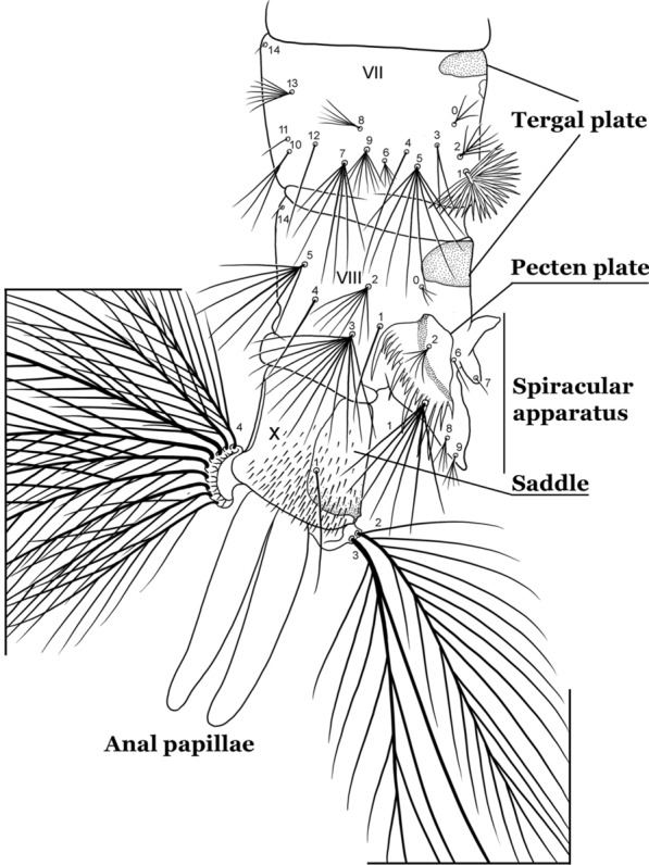
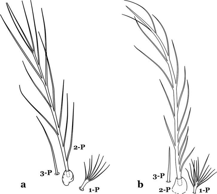
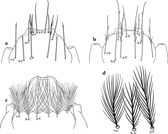
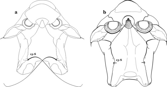
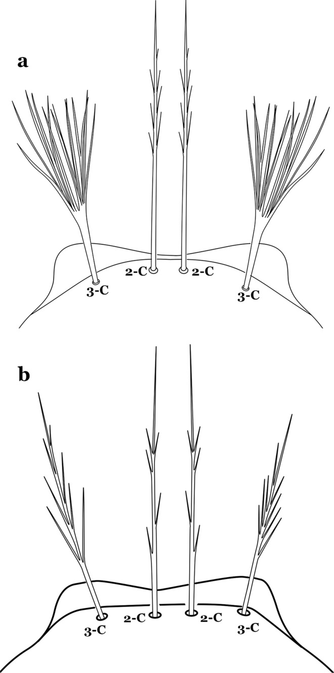
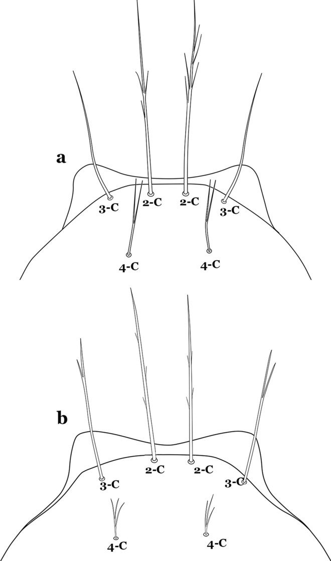
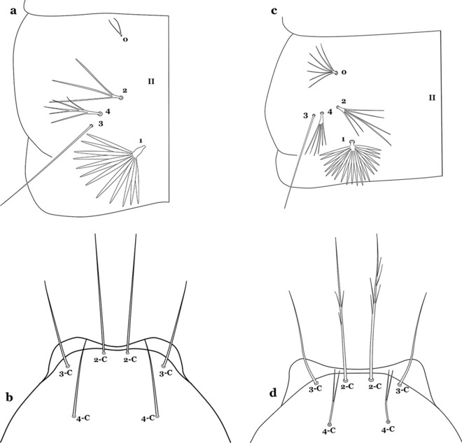
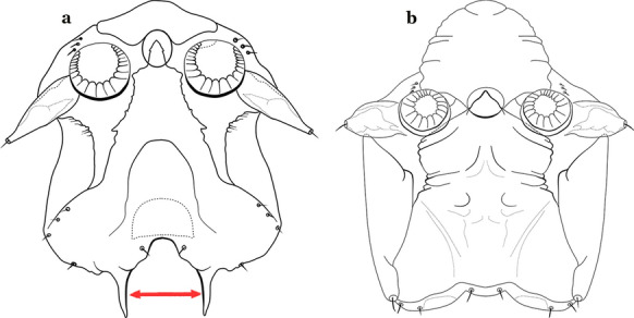
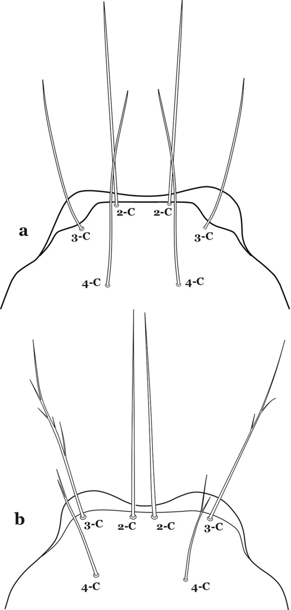
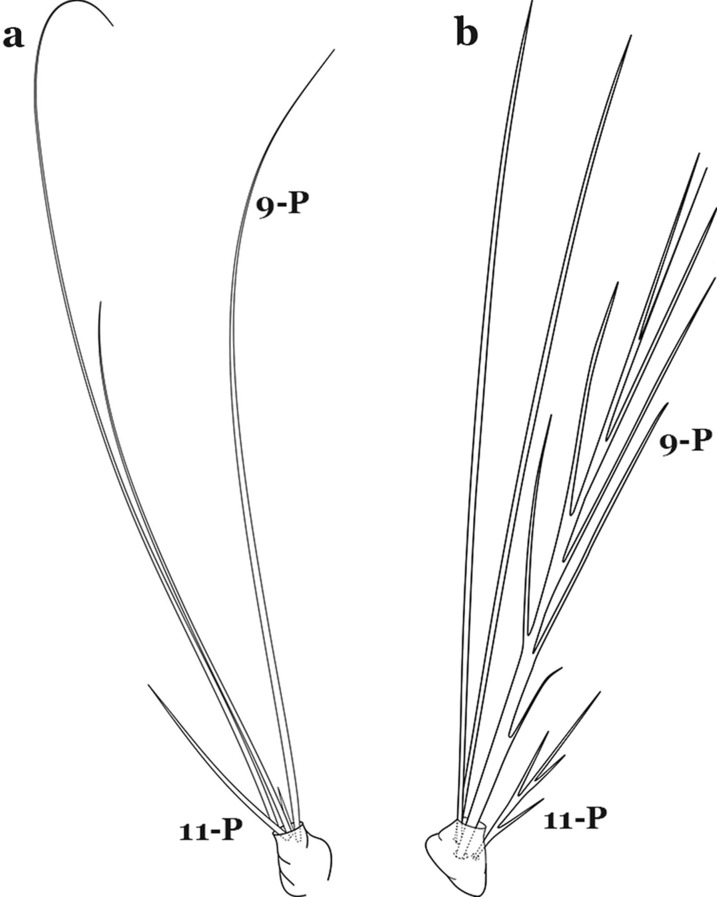
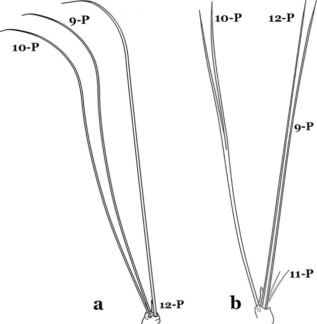
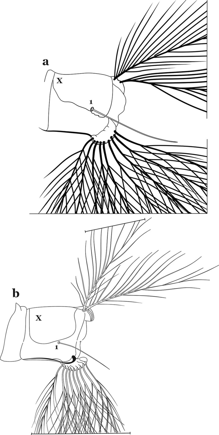
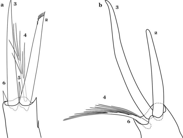
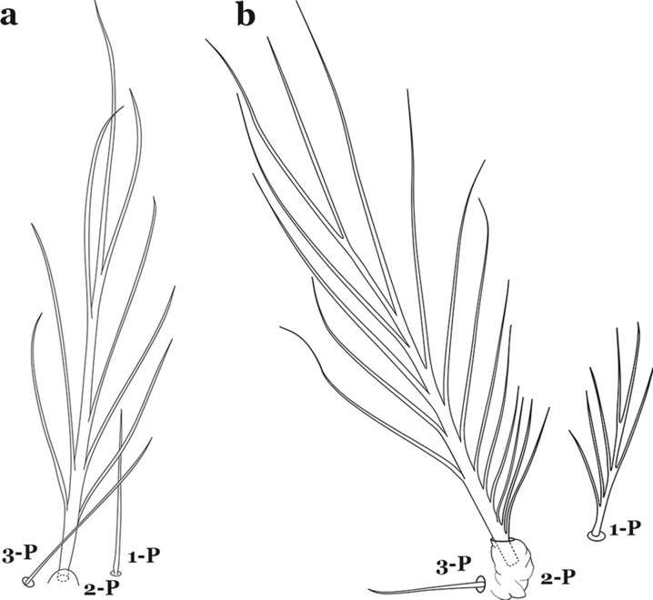
Results: A fully illustrated key to the fourth-instar larvae of South American species of the genus Anopheles (Diptera: Culicidae) is presented. Definitions of the morphological terms used in the key are provided and illustrated.

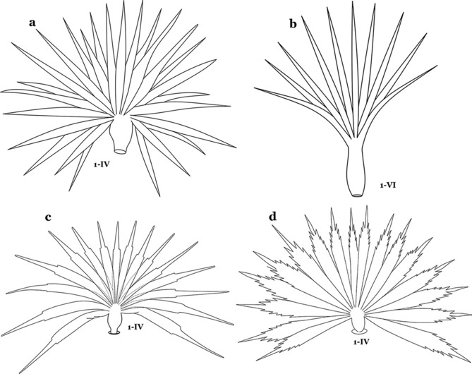
Conclusions: Morphological identification of South American Anopheles species based on the fourth-instar larvae has been updated. Characters of the spiracular apparatus were determined useful for the identification of morphologically similar species, in the Strodei Group and some taxa in the Myzorhynchella Section. The single versus branched abdominal seta 6-IV used to differentiate Myzorhynchella species from other Nyssorhynchus species was shown to be variable in Myzorhynchella species. Also, the abdominal setae 1-IV,V of Anopheles atacamensis and Anopheles pictipennis were shown to be slightly serrate at the edges. Recognition of this character is important to avoid inaccurate identification of these species as members of the subgenus Anopheles.

Keywords: Anopheles; Fourth-instar larvae; Identification key; Illustrations; Morphology.


Conflict of interest statement

The authors declare that they have no competing interests.

Figures

Fig. 1
Fig. 1
Fourth-instar larva of An. goeldii Rozeboom & Gabaldon, 1941. a Head, dorsal aspect, left, ventral aspect, right. b Thorax, dorsal aspect, left, ventral aspect, right. c Abdominal segments I–VI, dorsal aspect, left, ventral aspect, right. d VII–X, lateral aspect
Fig. 2
Fig. 2
Types of common setae on larval Anopheles. a Palmate with serrate margins. b Dendritic. c Palmate with smooth margins. d Branched seta with filamentous branches. e Simple. f Aciculate. g Bifurcate. h Plumose
Fig. 3
Fig. 3
Head of fourth-instar larva of An. goeldii. a Antenna. b Dorsal apotome
Fig. 4
Fig. 4
Thorax of An. goeldii, dorsal aspect, left; ventral aspect, right. P, prothorax; M, mesothorax; T, metathorax
Fig. 5
Fig. 5
Larval abdomen of An. goeldii. Abdominal segments I–VI, dorsal aspect, left, ventral aspect, right
Fig. 6
Fig. 6
Spiracular apparatus of fourth-instar larva of Anopheles species, dorsal aspect
Fig. 7
Fig. 7
Segments VII–X of abdomen of fourth-instar larva of An. goeldii, lateral aspect
Fig. 8
Fig. 8
a An. nimbus (Theobald, 1902), fourth-instar larva, spiracular apparatus. b An. albimanus Wiedemann, 1820, abdominal segment, dorsal aspect, showing palmate seta 1
Fig. 9
Fig. 9
Prothoracic setae 1–3-P. a An. thomasi Shannon, 1933. b An. nimbus
Fig. 10
Fig. 10
Prothoracic setae 1–3-P. a An. kompi Edwards, 1930. b An. nimbus
Fig. 11
Fig. 11
Abdominal seta 1. a, b An. bellator Dyar & Knab, 1906 seta 1-IV, VI. c An. pseudopunctipennis Theobald, 1901 seta 1-IV. d An. mattogrossensis Lutz & Neiva, 1911 seta 1-IV
Fig. 12
Fig. 12
Larval head, setae 2–7-C. a An. cruzii Dyar & Knab, 1906. b An. laneanus Corrêa & Cerqueira, 1944. c An. darlingi Root, 1926, setae 5–7-C. d An. goeldii, setae 5–7-C
Fig. 13
Fig. 13
Abdominal seta 1-I. a An. cruzii. b An. bellator
Fig. 14
Fig. 14
Larval head, setae 5–7-C. a An. rollai Cova Garcia, Pulido F. & Escalante de Ugueto, 1977. b An. boliviensis (Theobald, 1905)
Fig. 15
Fig. 15
a, b An. gonzalezrinconesi Cova Garcia, Pulido F. & Escalante de Ugueto, 1977. a Abdominal seta 9-IV. b Head seta 11-C. c An. boliviensis Head seta 11-C
Fig. 16
Fig. 16
Abdominal seta 1-II–VI. An. neivai Howard, Dyar & Knab, 1913
Fig. 17
Fig. 17
a An. neivai abdominal seta 6-VI. An. pholidotus Zavortink, 1973. b Abdominal seta 1-VII. c Abdominal seta 6-VI
Fig. 18
Fig. 18
Abdominal seta 13-III. a An. neivai. b An. auyantepuiensis Harbach & Navarro, 1996
Fig. 19
Fig. 19
a, b An. pholidotus. a Head seta 3-C. b Prothoracic seta 11-P. c, d An. lepidotus. c Head seta 3-C. d Prothoracic seta 11-P
Fig. 20
Fig. 20
a, b An. bambusicolus Komp, 1937. a Abdominal seta 1-I, II. b Abdominal seta 6-IV. c An. bellator, abdominal seta 1-II–IV. d An. cruzii, abdominal seta 6-VI
Fig. 21
Fig. 21
Pecten, siphonal seta 1-S. a An. bellator. b An. laneanus
Fig. 22
Fig. 22
Abdominal segment X. a An. homunculus Komp, 1937. b An. cruzii. c An. laneanus
Fig. 23
Fig. 23
Larva head, dorsal view, setae 2–8-C. a An. laneanus. b An. cruzii
Fig. 24
Fig. 24
Abdominal seta 6-IV, V. a An. darlingi. b An. antunesi Galvão & Franco do Amaral, 1940. c An. guarani Shannon, 1928
Fig. 25
Fig. 25
Prothoracic setae 1–3-P. a An. darlingi. b An. albimanus. c An. braziliensis (Chagas, 1907). d An. marajoara Galvão & Damasceno, 1942
Fig. 26
Fig. 26
Spiracular apparatus, seta 13-S. a An. darlingi. b An. albimanus
Fig. 27
Fig. 27
Larva head, dorsal view, seta 3-C. a An. lanei Galvão & Franco do Amaral, 1938. b An. albimanus
Fig. 28
Fig. 28
Prothoracic setae 1–3-P. a An. albimanus. b An. argyritarsis Robineau-Desvoidy, 1827
Fig. 29
Fig. 29
a, b An. sawyeri Causey, Deane, Deane & Sampaio, 1943. a Prothoracic seta 3-T. b Abdominal seta 1-I. c, d An. argyritarsis. c Prothoracic seta 3-T. d Abdominal seta 1-I
Fig. 30
Fig. 30
Larva head, dorsal view, setae 2, 3-C. a An. braziliensis. b An. marajoara
Fig. 31
Fig. 31
a, b An. braziliensis. a Protoracic setae 1–3-P. b Larva head, dorsal view, setae 2–4-C. c, d An. strodei Root, 1926. c Protoracic setae 1–3-P. d Larva head, dorsal view, setae 2–4-C
Fig. 32
Fig. 32
Protoracic setae 1–3-P. a An. marajoara. b An. benarrochi Gabaldon, Cova-Garcia & Lopez, 1941
Fig. 33
Fig. 33
Larva head, dorsal view, setae 2, 3-C. a An. albitarsis Lynch Arribálzaga, 1878. b An. deaneorum Rosa-Freitas, 1989
Fig. 34
Fig. 34
Larva head, dorsal view, setae 2, 3-C. a An. benarrochi. b An. triannulatus (Neiva & Pinto, 1922)
Fig. 35
Fig. 35
a, b An. benarrochi. a Antennal seta 1-A. b Abdominal seta 1-X. c, d An. konderi Galvão & Damasceno, 1942. c Antennal seta 1-A. d Larva head, dorsal view, setae 2, 3-C
Fig. 36
Fig. 36
ac An. aquasalis Curry, 1932. a Larva head, dorsal view, setae 2, 3-C. b Abdominal seta 1-X. c Spiracular apparatus. d-f An. konderi. d Larva head, dorsal view, setae 2, 3-C. e Abdominal seta 1-X. f Spiracular apparatus
Fig. 37
Fig. 37
Spiracular apparatus. a An. triannulatus. b An. nuneztovari Gabaldon, 1940
Fig. 38
Fig. 38
ac An. ininii Senevet & Abonnenc, 1938. a Abdominal setae 11–13-I. b Prothoracic setae 1–3-P. c Spiracular apparatus. df An. triannulatus. d Abdominal setae 11–13-I. e Prothoracic setae 1–3-P. f Spiracular apparatus
Fig. 39
Fig. 39
a, b An. triannulatus. a Prothoracic setae 1–3-P. b Spiracular apparatus. c, d An. halophylus Silva do Nascimento & Lourenço-de-Oliveira, 2002. c Prothoracic setae 1–3-P. d Spiracular apparatus
Fig. 40
Fig. 40
a, b An. halophylus. a Abdominal setae 11,13-I. b Prothoracic setae 1–3-P. c, d An. nuneztovari. c Abdominal setae 11,13-I. d Prothoracic setae 1–3-P
Fig. 41
Fig. 41
Larva head, dorsal view, setae 2–4-C. a An. nuneztovari. b An. rangeli Gabaldon, Cova-Garcia & Lopez, 1940
Fig. 42
Fig. 42
a, b An. trinkae Faran, 1979. a Abdominal setae 0–4-II. b Larva head, dorsal view, setae 2–4-C. c, d An. nuneztovari. c Abdominal setae 0–4-II. d Larva head, dorsal view, setae 2–4-C
Fig. 43
Fig. 43
Larva head, dorsal view, setae 2–4-C. a An. rangeli. b An. evansae (Brèthes, 1926)
Fig. 44
Fig. 44
ac An. galvaoi Causey, Deane & Deane, 1943. a Prothoracic setae 1-3-P. b Lateral arm of median plate of spiracular apparatus. c Abdominal segment X. df An. evansae. d Prothoracic setae 1-3-P. e Spiracular apparatus. f Abdominal segment X
Fig. 45
Fig. 45
Abdominal seta 6-IV-VI. a An. antunesi. b An. guarani Shannon, 1928
Fig. 46
Fig. 46
ac An. antunesi. a Prothoracic seta 14-P. b Larva head, dorsal view, setae 2,4-C. c Seta 8-C. df An. lutzii Cruz, 1901. d Prothoracic seta 14-P. e Larva head, dorsal view, setae 2,4-C. f Seta 8-C
Fig. 47
Fig. 47
Spiracular apparatus. a An. guarani. b An. parvus (Chagas, 1907)
Fig. 48
Fig. 48
ac An. parvus. a Spiracular apparatus. b Abdominal seta 1-IV. c Antennal seta 1-A. df An. pristinus Nagaki & Sallum, 2010. d Spiracular apparatus. e Abdominal seta 1-IV. f Antennal seta 1-A
Fig. 49
Fig. 49
Spiracular apparatus. a An. pseudopunctipennis. b An. pictipennis (Philippi, 1865)
Fig. 50
Fig. 50
Larval abdominal seta 1. a An. atacamensis González & Sallum, 2010. b An. mattogrossensis
Fig. 51
Fig. 51
Larva head, dorsal view, setae 2–4-C. a An. atacamensis. b An. oiketorakras Osorno-Mesa, 1947
Fig. 52
Fig. 52
a, b An. atacamensis. a Larva head, dorsal view, setae 2–7-C. b Prothoracic setae 1–3-P. c, d An. pictipennis. c Larva head, dorsal view, setae 2–7-C. d Prothoracic setae 1–3-P
Fig. 53
Fig. 53
Antennal setae 2–4-A. a An. atacamensis. b An. calderoni Wilkerson, 1991
Fig. 54
Fig. 54
Larval prothoracic setae 9,11-P. a An. mattogrossensis. b An. squamifemur Antunes, 1937
Fig. 55
Fig. 55
Larva head, dorsal view, setae 2–4-C. a An. mattogrossensis. b An. tibiamaculatus (Neiva, 1906)
Fig. 56
Fig. 56
Larval prothoracic setae 1–3-P. a An. mattogrossensis. b An. peryassui Dyar & Knab, 1908
Fig. 57
Fig. 57
Larva head, dorsal view, setae 2–4-C. a An. peryassui. b An. shannoni Davis, 1931
Fig. 58
Fig. 58
Larva head, dorsal view, setae 2–4-C. a An. eiseni Coquillett, 1902. b An. tibiamaculatus (Neiva, 1906)
Fig. 59
Fig. 59
Larva head, dorsal view, setae 2–4-C. a An. gilesi Neiva, 1908. b An. vargasi Gabaldon, Cova-Garcia & Lopez, 1941
Fig. 60
Fig. 60
Larva head, dorsal view, setae 2,3-C. a An. neomaculipalpus Curry, 1931. b An. calderoni
Fig. 61
Fig. 61
An. neomaculipalpus, larval prothoracic setae 1–3-P
Fig. 62
Fig. 62
Larval prothoracic setae 1–3-P. a An. mattogrossensis. b An. calderoni
Fig. 63
Fig. 63
Larval prothoracic setae 9–12-P. a An. anchietai Corrêa & Ramalho, 1968. b An. calderoni
Fig. 64
Fig. 64
Abdominal segment X. a An. malefactor Dyar & Knab, 1907. b An. mediopunctatus (Lutz, 1903)
Fig. 65
Fig. 65
Antennal setae 2–6-A. a An. calderoni. b An. mediopunctatus
Fig. 66
Fig. 66
Larva head, dorsal view, setae 2,3-C. a An. medialis Harbach, 2018. b An. fluminensis Root, 1927
Fig. 67
Fig. 67
Larval prothoracic setae 1–3-P. a An. anchietai. b An. fluminensis

References

    1. Sallum MAM, Gonzalez Obando R, Carrejo N, Wilkerson RC. Identification keys to the Anopheles mosquitoes of South America (Diptera: Culicidae). I. Introduction. Parasit Vectors. 2020. 10.1186/s13071-020-04298-6. - PMC - PubMed
    1. Cohen JM, Le Menach A, Pothin E, et al. Mapping multiple components of malaria risk for improved targeting of elimination interventions. Malar J. 2017;16:459. doi: 10.1186/s12936-017-2106-3. - DOI - PMC - PubMed
    1. WHO. Guidelines for malaria vector control. Geneva: World Health Organization; 2019. https://www.who.int/publications/i/item/guidelines-for-malaria-vector-co.... - PubMed
    1. Baird JK. Malaria control by commodities without practical malariology. BMC Public Health. 2017;17:590. doi: 10.1186/s12889-017-4454-x. - DOI - PMC - PubMed
    1. Harbach RE, Knight KL. Taxonomistsʼ glossary of mosquito anatomy. Marlton: Plexus Publishing, Inc.; 1980.

LinkOut - more resources