. 2021 Jul;20(4):664-672.
doi: 10.1016/j.jcf.2020.10.006. Epub 2020 Nov 15.

Defective immunometabolism pathways in cystic fibrosis macrophages

Affiliations

Defective immunometabolism pathways in cystic fibrosis macrophages

Kaitlin Hamilton et al. J Cyst Fibros. 2021 Jul.

Abstract

Background: Mitochondria play a key role in immune defense pathways, particularly for macrophages. We and others have previously demonstrated that cystic fibrosis (CF) macrophages exhibit weak autophagy activity and exacerbated inflammatory responses. Previous studies have revealed that mitochondria are defective in CF epithelial cells, but to date, the connection between defective mitochondrial function and CF macrophage immune dysregulation has not been fully elucidated. Here, we present a characterization of mitochondrial dysfunction in CF macrophages.

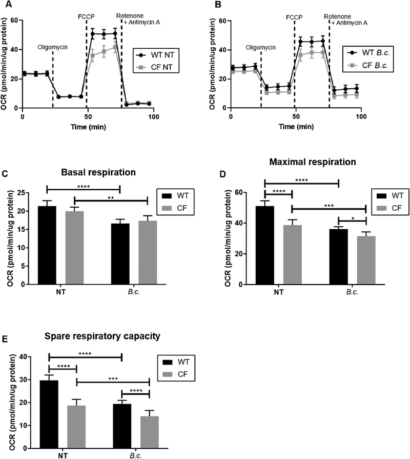
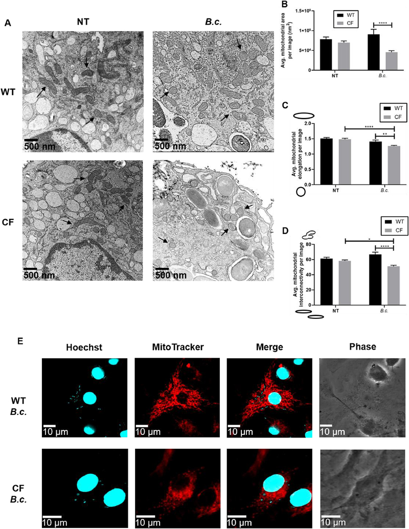
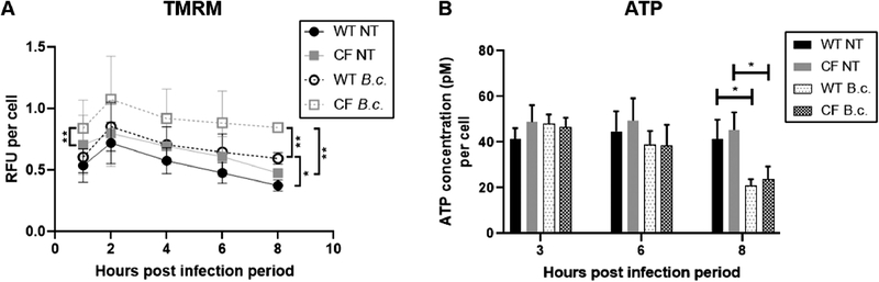
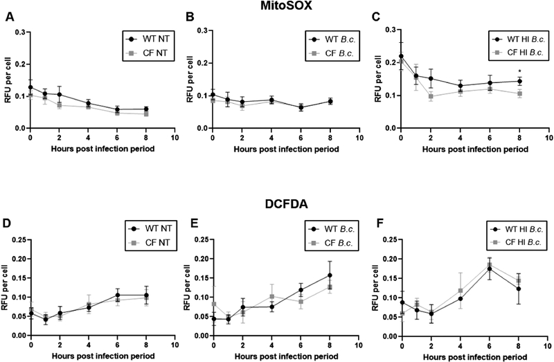
Methods: Mitochondrial function in wild-type (WT) and CF F508del/F508del murine macrophages was measured using the Seahorse Extracellular Flux analyzer. Mitochondrial morphology was investigated using transmission electron and confocal microscopy. Mitochondrial membrane potential (MMP) as well as mitochondrial reactive oxygen species (mROS) were measured using TMRM and MitoSOX Red fluorescent dyes, respectively. All assays were performed at baseline and following infection by Burkholderia cenocepacia, a multi-drug resistant bacterium that causes detrimental infections in CF patients.

Results: We have identified impaired oxygen consumption in CF macrophages without and with B. cenocepacia infection. We also observed increased mitochondrial fragmentation in CF macrophages following infection. Lastly, we observed increased MMP and impaired mROS production in CF macrophages following infection with B. cenocepacia.

Conclusions: The mitochondrial defects identified are key components of the macrophage response to infection. Their presence suggests that mitochondrial dysfunction contributes to impaired bacterial killing in CF macrophages. Our current study will enhance our understanding of the pathobiology of CF and lead to the identification of novel mitochondrial therapeutic targets for CF.

Keywords: Bacterial infection; Immunometabolism; Macrophage; Mitochondria; Reactive oxygen species.


Figures

Fig. 1:
Fig. 1:. Mitochondria in CF macrophages are less capable of responding to an increased energy demand at baseline and following B. cenocepacia infection
Seahorse Cell Mito Stress Test performed in WT and CF macrophages at baseline (NT = no treatment) or following 5 h B. cenocepacia (B.c.) infection. (A-B) Oxygen consumption rate (OCR) was measured following the addition of ETC inhibitors to investigate mitochondrial function. (C-E) Mitochondrial function parameters calculated from (A-B). Graphs show mean and SEM. N=7 WT and CF mice. Statistics: Linear mixed effects model with Holm’s post-test. *, p≤0.05; **, p≤0.01; ***, p≤0.001; ****, p≤0.0001.
Fig. 2:
Fig. 2:. Decreased oxygen consumption in CF macrophages is not due to a difference in mitochondrial mass
Immunoblot for mitochondrial proteins in WT and CF macrophages at baseline (NT) and following 6 h B. cenocepacia (B.c.) infection. (A) Representative immunoblots. (B-D) Densitometry analysis. Graphs show mean and SEM. N=5 WT and CF mice. Statistics: Linear mixed effects model with Holm’s post-test.
Fig. 3:
Fig. 3:. B. cenocepacia infection induces mitochondrial fragmentation in CF macrophages
Microscopy analysis of mitochondrial morphology in WT and CF macrophages at baseline (NT) and following 6 h B. cenocepacia (B.c.) infection. (A) Representative electron microscopy images taken at 22,500x magnification. Several mitochondria are indicated with arrows. (B-D) Mitochondrial morphology parameters calculated from electron microscopy images. Parameters were calculated for individual mitochondria then averaged per image. For the elongation and interconnectivity graphs, sketches next to the y-axis indicate the morphology represented by low vs. high scores. Graphs show mean and SEM. N=1 WT and CF mouse; 14–16 images analyzed per condition. Statistics: Two-way ANOVA with Holm’s post-test. *, p≤0.05; **, p≤0.01; ***, p≤0.001; ****, p≤0.0001. (E) Representative fluorescence microscopy images. MitoTracker Deep Red was used to stain the mitochondria. Hoechst 33342 was used to stain the macrophage nuclei and B. cenocepacia.
Fig. 4:
Fig. 4:. Mitochondria in CF macrophages have a higher mitochondrial membrane potential following B. cenocepacia infection
TMRM (A) and ATP (B) assays performed in WT and CF macrophages at baseline (NT) and following B. cenocepacia (B.c.) infection. RFU = Relative fluorescence units. Graphs show mean and SEM. N=3 WT and CF mice for (A) and N=4 WT and CF mice for (B). Statistics: Linear mixed effects model with Holm’s post-test for each time point. *, p≤0.05; **, p≤0.01.
Fig. 5:
Fig. 5:. Mitochondria in CF macrophages produce less mitochondrial ROS following B. cenocepacia infection
MitoSOX (A-C) and DCFDA (D-E) assays performed in WT and CF macrophages at baseline (NT) and following infection with live or heat-inactivated (HI) B. cenocepacia (B.c.). Graphs show mean and SEM. N=10–14 WT and CF mice for (A), N=5–7 WT and CF mice for (B), N=5–7 WT and CF mice for (C), N=8–10 WT and CF mice for (D), N=5–7 WT and CF mice for (E), and N=3 WT and CF mice for (F). Statistics: Two-tailed paired t-test for each time point. *, p≤0.05.

References

    1. Abdulrahman BA, Khweek AA, Akhter A, Caution K, Kotrange S, Abdelaziz DHA, et al.Autophagy stimulation by rapamycin suppresses lung inflammation and infection by Burkholderia cenocepacia in a model of cystic fibrosis. Autophagy 2011;7:1359–70. doi:10.4161/auto.7.11.17660. - DOI - PMC - PubMed
    1. Abdulrahman BA, Khweek AA, Akhter A, Caution K, Tazi M, Hassan H, et al.Depletion of the ubiquitin-binding adaptor molecule SQSTM1/P62 from macrophages harboring cftr Δf508 Mutation Improves the Delivery of Burkholderia cenocepacia to the Autophagic Machinery. J Biol Chem 2013;288:2049–58. doi:10.1074/jbc.M112.411728. - DOI - PMC - PubMed
    1. Elborn JS, Balfour-Lynn IM, Bilton D. Respiratory disease: Infectious complications. In: Bush A, Hodson ME, Bilton D, editors. Hodson Geddes’ Cyst. Fibros. 4th ed., CRC Press; 2015, p. 205–20.
    1. Mehta MM, Weinberg SE, Chandel NS. Mitochondrial control of immunity: beyond ATP. Nat Rev Immunol 2017. doi:10.1038/nri.2017.66. - DOI - PubMed
    1. Mills EL, Kelly B, Logan A, Costa ASH, Varma M, Bryant CE, et al.Succinate Dehydrogenase Supports Metabolic Repurposing of Mitochondria to Drive Inflammatory Macrophages. Cell 2016;167:457–470.e13. doi:10.1016/j.cell.2016.08.064. - DOI - PMC - PubMed

Publication types