. 2021 Oct;70(10):1954-1964.
doi: 10.1136/gutjnl-2020-322509. Epub 2020 Nov 18.

Sphingomyelin synthase 1 mediates hepatocyte pyroptosis to trigger non-alcoholic steatohepatitis

Affiliations

Sphingomyelin synthase 1 mediates hepatocyte pyroptosis to trigger non-alcoholic steatohepatitis

Eun Hee Koh et al. Gut. 2021 Oct.

Abstract

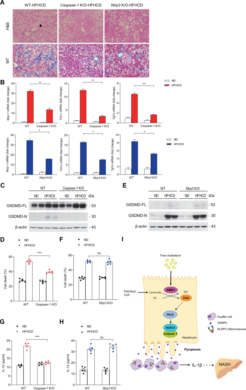
Objective: Lipotoxic hepatocyte injury is a primary event in non-alcoholic steatohepatitis (NASH), but the mechanisms of lipotoxicity are not fully defined. Sphingolipids and free cholesterol (FC) mediate hepatocyte injury, but their link in NASH has not been explored. We examined the role of free cholesterol and sphingomyelin synthases (SMSs) that generate sphingomyelin (SM) and diacylglycerol (DAG) in hepatocyte pyroptosis, a specific form of programmed cell death associated with inflammasome activation, and NASH.

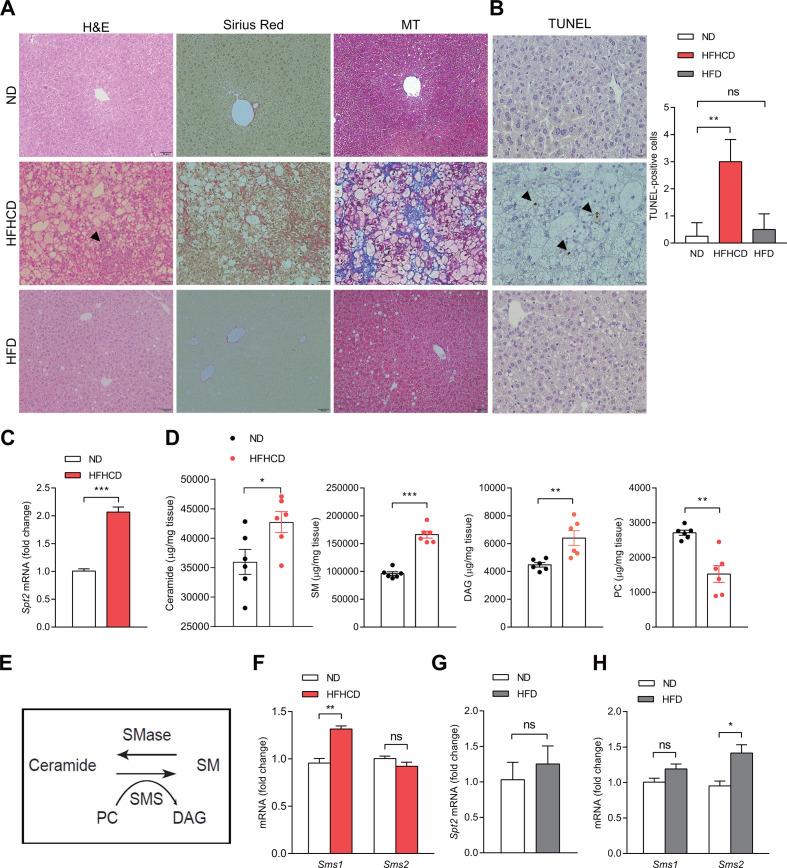
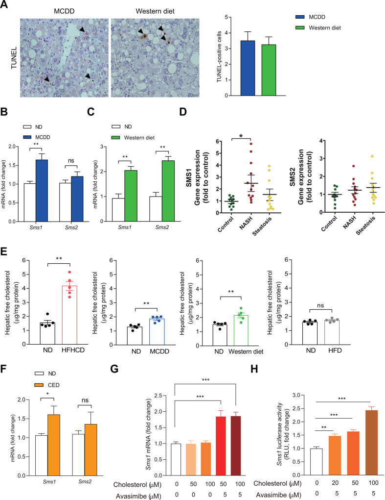
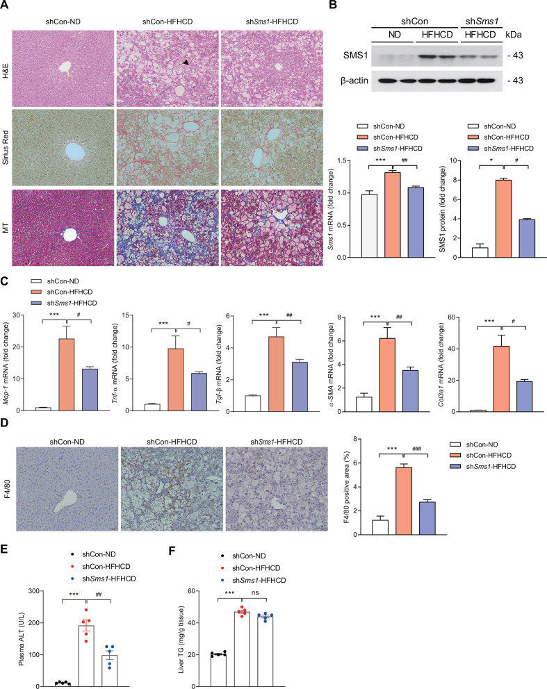
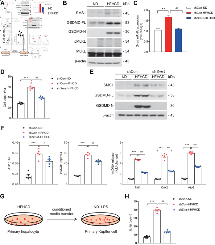
Design: Wild-type C57BL/6J mice were fed a high fat and high cholesterol diet (HFHCD) to induce NASH. Hepatic SMS1 and SMS2 expressions were examined in various mouse models including HFHCD-fed mice and patients with NASH. Pyroptosis was estimated by the generation of the gasdermin-D N-terminal fragment. NASH susceptibility and pyroptosis were examined following knockdown of SMS1, protein kinase Cδ (PKCδ), or the NLR family CARD domain-containing protein 4 (NLRC4).

Results: HFHCD increased the hepatic levels of SM and DAG while decreasing the level of phosphatidylcholine. Hepatic expression of Sms1 but not Sms2 was higher in mouse models and patients with NASH. FC in hepatocytes induced Sms1 expression, and Sms1 knockdown prevented HFHCD-induced NASH. DAG produced by SMS1 activated PKCδ and NLRC4 inflammasome to induce hepatocyte pyroptosis. Depletion of Nlrc4 prevented hepatocyte pyroptosis and the development of NASH. Conditioned media from pyroptotic hepatocytes activated the NOD-like receptor family pyrin domain containing 3 inflammasome (NLRP3) in Kupffer cells, but Nlrp3 knockout mice were not protected against HFHCD-induced hepatocyte pyroptosis.

Conclusion: SMS1 mediates hepatocyte pyroptosis through a novel DAG-PKCδ-NLRC4 axis and holds promise as a therapeutic target for NASH.

Keywords: hepatocyte; immunology in hepatology; lipid mediators; nonalcoholic steatohepatitis.


Conflict of interest statement

Competing interests: None declared.

Figures

Figure 1
Figure 1
Hepatic Sms1 expression in murine NASH models. Liver tissues from mice fed control diet (ND), HFHCD or HFD for 12 weeks showing representative H&E, Sirius Red and MT staining. Scale bar, 50 µM. Arrowhead indicates inflammatory foci (A) and TUNEL staining. TUNEL staining positivity was observed in hepatocytes (arrowhead). Scale bar, 20 µm. The graph represents the percentage of TUNEL-positive cells in each group (B). (C) Relative mRNA expression of Spt2 in ND-fed or HFHCD-fed mice. (D) Levels of hepatic ceramide, SM, DAG and PC. (E) Schematic figure depicting the action of the SMSs. (F) Relative mRNA expression of Sms1 and Sms2. (G, H) Relative mRNA expression of Spt2, Sms1 and Sms2 in mice fed ND or HFD. Data are presented as mean±SEM (n=6). *p<0.05, **p<0.01 and ***p<0.001 vs control mice. DAG, diacylglycerol; HFD, high fat diet; HFHCD, high fat, high cholesterol diet; ns, not significant; PC, phosphatidylcholine; SM, sphingomyelin; TUNEL, terminal deoxynucleotidyl transferase-mediated dUTP nick‐end labelling.
Figure 2
Figure 2
FC induces Sms1 expression in hepatocytes. (A) TUNEL staining in the liver sections following MCDD (8 weeks) or Western diet (16 weeks). Arrowheads show TUNEL positive hepatocytes, which are quantitated in the graph. Scale bar, 20 µm. (B, C) mRNA expression of Sms1 and Sms2 in the livers of mice fed MCDD (B) or Western diet (C) (n=7). (D) Hepatic mRNA expression of SMS1 and SMS2 in liver samples of patients with NASH/cirrhosis and subjects with steatosis (n=12). Surgical specimens of donor livers were used as controls (n=11). (E) Hepatic FC levels of mice fed ND, HFHCD, MCDD, Western diet or HFD (n=5). (F) mRNA expression of Sms1 and Sms2 in the livers of mice fed ND or CED for 2 days (n=7). (G) AML12 cells were treated with cholesterol with Avasimibe for 2 hours at the indicated dose to determine Sms1 mRNA expression (n=4). (H) AML12 cells were transfected with mouse Sms1 promoter-Luc to determine Sms1 transcriptional activity. After 48 hours of transfection, cells were treated with vehicle or cholesterol with Avasimibe for 3 hours (n=6). Data are presented as mean±SEM *p< 0.05, **p<0.01 and ***p<0.001 vs control. CED, cholesterol-enriched diet; FC, free cholesterol; HFD, high fat diet; HFHCD, high fat, high cholesterol diet; MCDD, methionine and choline-deficient diet; ns, not significant; TUNEL, terminal deoxynucleotidyl transferase-mediated dUTP nick‐end labelling.
Figure 3
Figure 3
Depletion of SMS1 prevents NASH development. Mice were injected with AAV carrying control shRNA (shCon) or Sms1-specific shRNA (shSms1) and then fed ND or HFHCD for 12 weeks. (A) Representative H&E, Sirius Red and MT liver staining. Scale bar, 50 µM. Arrowhead indicates inflammatory foci. (B) Relative mRNA expression, and Western blot of SMS1. (C) mRNA expression of Mcp-1, Tnf-α, Tgf-β1, α-Sma and Col3a1. (D) Immunohistochemical staining and quantitative analysis of F4/80-positive area in the liver. (E) Plasma ALT level. (F) Liver TG contents. Data are presented as mean±SEM of mice (n=5). *p<0.05 and ***p<0.001 vs control mice. #p<0.05, ##p<0.01, ###p<0.001 vs shCon-HFHCD mice.HFHCD, high fat, high cholesterol diet; ns, not significant; SMS, sphingomyelin synthase; TG, triglyceride.
Figure 4
Figure 4
SMS1 causes hepatocyte pyroptosis. (A) Cell death measured by LDH level in the media of primary hepatocytes isolated from mice fed HFHCD (4 weeks). (B) Representative Western blots of SMS1, GSDMD-FL, GSDMD-N and MLKL. (C–E) Mice were infected with AAV carrying control shRNA (shCon) or Sms1-specific shRNA (shSms1) and then fed with ND or HFHCD (4 weeks) to determine mRNA expression of Sms1 in the hepatocyte (C), cell death by LDH release (D) and Western blots of SMS1, GSDMD-FL and -N (E). (F–H) DAMPs released from pyroptotic hepatocytes activate NLRP3 inflammasome in Kupffer cells. (F) Levels of ATP, HMGB1 and mtDNA in the supernatant. (G, H) Primary hepatocyte-conditioned media was transferred to primary Kupffer cells primed with 10 ng/ml of LPS for 4 hours (G) to determine IL-1β in the culture supernatants of primary hepatocytes (H). Data are presented as mean±SEM (n=5). **p< 0.01 and ***p<0.001 vs control mice. #p<0.05, ##p<0.01 and ###p<0.001 vs shCon-HFHCD mice. DAMPs, damage-associated molecular pattern molecules; GSDMD-FL, full-length gasdermin D; GSDMD-N, N-terminal fragment of gasdermin-D; HFHCD, high fat, high cholesterol diet; HMGB1, high mobility group protein box 1; LDH, lactate dehydrogenase; LPS, lipopolysaccharides; MLKL, mixed linkage kinase domain-like protein; NLRC4, NLR family CARD domain-containing protein 4; NLRP3, NOD-like receptor family pyrin domain containing 3; SMS, sphingomyelin synthase.
Figure 5
Figure 5
NLRC4-dependent pyroptosis in hepatocytes from HFHCD-fed mice through PKCδ. (A–F) Hepatocytes from mice infected with AAV carrying control shRNA (shCon), Sms1-specific shRNA (shSms1) (A) or Nlrc4-specific shRNA (shNlrc4) (B–F) and fed ND or HFHCD for 12 weeks (B, C) or 4 weeks (A, D–G) to determine Western blots for NLRC4 and NLRP3 (A). (B) Representative H&E and MT staining. Scale bar, 50 µM. Arrowhead indicates inflammatory foci. (C) Liver TG. (D) Representative Western blots of GSDMD-FL and GSDMD-N. (E) Cell death by LDH level. (F) IL-1β from hepatocyte-conditioned media transferred to Kupffer cells primed with 10 ng/ml of LPS for 4 hours. (G, H) Representative Western blots of PKCδ (G) and NLRC4 (H) of hepatocytes from mice infected with AAV carrying control shRNA (shCon), Sms1-specific shRNA (shSms1) (G) or Pkcδ-specific shRNA (shPkcδ) (H). (I) mRNA expression and protein levels of PKCδ. Data are presented as mean±SEM (n=5). *p<0.05, **p<0.01 and ***p<0.001 vs control mice. #p<0.05, ##p<0.01 vs shCon-HFHCD mice. GSDMD-FL, full-length gasdermin D; GSDMD-N, N-terminal fragment of gasdermin-D; HFHCD, high fat, high cholesterol diet; LDH, lactate dehydrogenase; NLRC4, NLR family CARD domain-containing protein 4; NLRP3, NOD-like receptor family pyrin domain containing 3; ns, not significant; PKC, protein kinase C; TG, triglyceride.
Figure 6
Figure 6
Genetic ablation of caspase-1 but not NLRP3 protects against hepatocyte pyroptosis. Caspase-1 K/O mice and Nlrp3 K/O mice were fed HFHCD for 12 weeks (A, B) or 4 weeks (C–H) to determine (A) H&E and MT staining. Scale bar, 50 µm. Arrowhead indicates inflammatory foci. (B) mRNA expression of Mcp-1, Tnf-α and Tgf-β1. Representative Western blots of GSDMD (C) and cell death (D) of hepatocytes from WT and Caspase-1 K/O mice. Representative Western blots of GSDMD-FL and GSDMD-N (E) and cell death (F) in hepatocytes of WT and Nlrp3 K/O mice. IL-1β from Caspase-1 K/O (G) or Nlrp3 K/O (H) hepatocyte-conditioned media transferred to primary Kupffer cells primed with 10 ng/ml of LPS for 4 hours. (I) Conceptual model depicting the role of SMS1, PKCδ and NLRC4 inflammasome in causing hepatocyte pyroptosis and NLRP3 inflammasome activation in Kupffer cells in the pathogenesis of NASH. Data are presented as mean±SEM (n=4-5). *p<0.05, **p<0.01 and ***p<0.001 vs WT-HFHCD mice. GSDMD-FL, full-length gasdermin D; GSDMD-N, N-terminal fragment of gasdermin-D; HFHCD, high fat, high cholesterol diet; LPS, lipopolysaccharides; NLRC4, NLR family CARD domain-containing protein 4; NLRP3, NOD-like receptor family pyrin domain containing 3; ns, not significant; SMSs, sphingomyelin synthases; WT, wild type.

References

    1. Friedman SL, Neuschwander-Tetri BA, Rinella M, et al. . Mechanisms of NAFLD development and therapeutic strategies. Nat Med 2018;24:908–22. 10.1038/s41591-018-0104-9 - DOI - PMC - PubMed
    1. Tilg H, Moschen AR. Evolution of inflammation in nonalcoholic fatty liver disease: the multiple parallel hits hypothesis. Hepatology 2010;52:1836–46. 10.1002/hep.24001 - DOI - PubMed
    1. Ratziu V, Goodman Z, Sanyal A. Current efforts and trends in the treatment of NASH. J Hepatol 2015;62:S65–75. 10.1016/j.jhep.2015.02.041 - DOI - PubMed
    1. Machado MV, Diehl AM. Pathogenesis of nonalcoholic steatohepatitis. Gastroenterology 2016;150:1769–77. 10.1053/j.gastro.2016.02.066 - DOI - PMC - PubMed
    1. Wallach D, Kang T-B, Dillon CP, et al. . Programmed necrosis in inflammation: toward identification of the effector molecules. Science 2016;352:aaf2154. 10.1126/science.aaf2154 - DOI - PubMed

Publication types

Substances