. 2021 Jan 13;41(2):215-233.
doi: 10.1523/JNEUROSCI.1284-20.2020. Epub 2020 Nov 18.

Golgi-Dependent Copper Homeostasis Sustains Synaptic Development and Mitochondrial Content

Affiliations

Golgi-Dependent Copper Homeostasis Sustains Synaptic Development and Mitochondrial Content

Cortnie Hartwig et al. J Neurosci. .

Abstract

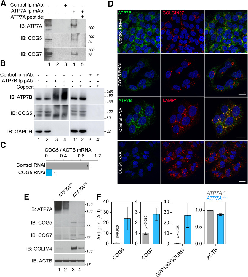
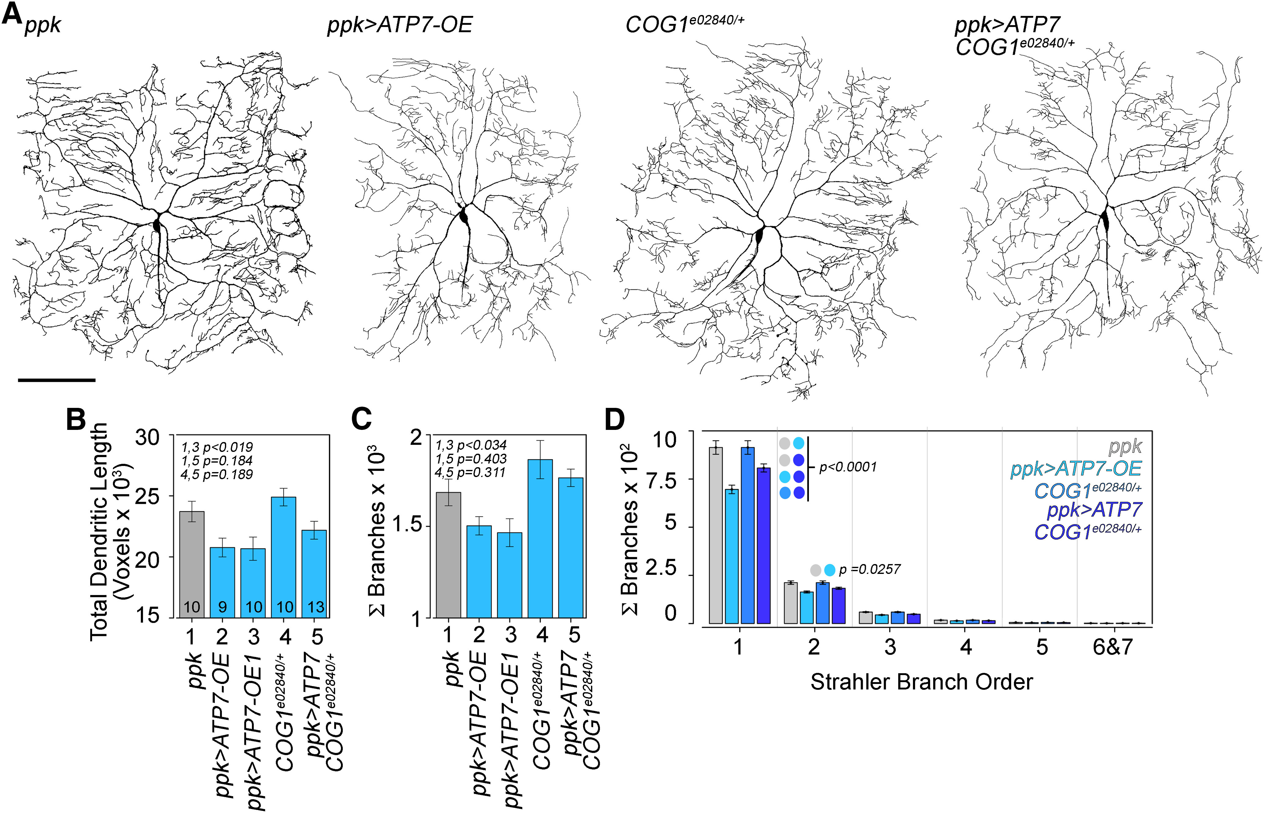
Rare genetic diseases preponderantly affect the nervous system causing neurodegeneration to neurodevelopmental disorders. This is the case for both Menkes and Wilson disease, arising from mutations in ATP7A and ATP7B, respectively. The ATP7A and ATP7B proteins localize to the Golgi and regulate copper homeostasis. We demonstrate genetic and biochemical interactions between ATP7 paralogs with the conserved oligomeric Golgi (COG) complex, a Golgi apparatus vesicular tether. Disruption of Drosophila copper homeostasis by ATP7 tissue-specific transgenic expression caused alterations in epidermis, aminergic, sensory, and motor neurons. Prominent among neuronal phenotypes was a decreased mitochondrial content at synapses, a phenotype that paralleled with alterations of synaptic morphology, transmission, and plasticity. These neuronal and synaptic phenotypes caused by transgenic expression of ATP7 were rescued by downregulation of COG complex subunits. We conclude that the integrity of Golgi-dependent copper homeostasis mechanisms, requiring ATP7 and COG, are necessary to maintain mitochondria functional integrity and localization to synapses.SIGNIFICANCE STATEMENT Menkes and Wilson disease affect copper homeostasis and characteristically afflict the nervous system. However, their molecular neuropathology mechanisms remain mostly unexplored. We demonstrate that copper homeostasis in neurons is maintained by two factors that localize to the Golgi apparatus, ATP7 and the conserved oligomeric Golgi (COG) complex. Disruption of these mechanisms affect mitochondrial function and localization to synapses as well as neurotransmission and synaptic plasticity. These findings suggest communication between the Golgi apparatus and mitochondria through homeostatically controlled cellular copper levels and copper-dependent enzymatic activities in both organelles.

Keywords: ATP7A; Golgi; Menkes; Wilson; copper; mitochondria.


Figures

Figure 1.
Figure 1.
Biochemical and genetic interactions between COG complex subunits and ATP7 paralogs. SH-SY5Y (A, E, F) and HepG2 (B) cells were incubated in normal media or media supplemented with a 200 μm copper excess (B, even lanes). Detergent soluble extracts from (A) SH-SY5Y and (B) HepG2 cells were immunoprecipitated with antibodies against ATP7A or ATP7B, respectively. The presence of COG complex in the immunoprecipitates was detected with antibodies against COG5 and COG7. Controls were performed with unrelated antibodies (A, lane 3 or B, lanes 3′−4′) or with the ATP7A antigenic peptide (A, lane 5). Asterisk marks non-specific band. A, Lane 1 corresponds to input. B, Lanes 1,2,1′ and 2′ correspond to inputs. C, siRNA knock-down of COG5 in HepG2 cells. Relative expression corresponds to the ratio between COG5 and ACTB (n = 3). D, Immunomicroscopy analysis of the expression and localization of ATP7B in Golgi (untreated cells) and lysosomes (CuSO4-treated cells). These compartments were detected with antibodies against GOLGIN97 and LAMP1, respectively. Scale bar: 10 µm. E, ATP7A control (lanes 1–2) and null clone (lanes 3–4) cell extracts were immunoblotted with the indicated antibodies. F, Quantification of blots presented in E; n = 4 Wilcoxon–Mann–Whitney rank-sum test.
Figure 2.
Figure 2.
Model of copper homeostasis by the COG complex and copper transporters and copper content in Drosophila. A, Model of predicted larval and brain copper status in different genotypes. Brain and gut are depicted in systemic genetic models, while only brain is depicted in the neuronal cell autonomous models. Color intensities of organs show predicted copper content (color code in the left of the figure). Magnitude of brain copper influx and efflux are depicted in the size of blue arrows in the bottom row. Note that changes in copper content in brain and whole larvae can be independently modulated by neuronal cell autonomous (C155>) or systemic (actin driver, Act>) modifications of ATP7 gene expression. B, ICP-MS determinations of total larval copper content in UAS-ATP7 or UAS-ATP7 RNAi transgenes expressed from a ubiquitous Actin-GAL4 driver. The mean difference for two comparisons against the shared control Act are shown in the above Cumming estimation plot. The raw data are plotted on the upper axes. On the lower axes, mean differences are plotted as bootstrap sampling distributions. Each mean difference is depicted as a dot. Each 95% confidence interval is indicated by the ends of the vertical error bars. The p value of the two-sided permutation t test is indicated. The number of larvae analyzed is depicted by italic figures in parentheses.
Figure 3.
Figure 3.
Copper homeostasis by the COG complex and copper transporters modulate glutamatergic synapse morphology. Muscle 6–7 neuromuscular junctions from wild-type animals and animals expressing ATP7, COG, and CTR1 UAS-transgenes stained with anti-HRP antibodies (A, C, E). C, Synapses from animals carrying the UAS-ATP7 transgene expressed under the systemic driver Actin-GAL4, the COG1e02840/+ genotype, each in isolation or their combination. Scale bar: 50 µm. B, Total branch length quantifications of animals in A. C, Synapses from animals carrying the UAS-ATP7 transgene, UAS-COG1 RNAi expressed under the C155-GAL4 driver, or the COG1e02840/+ genotype, each in isolation or their combinations. D, Quantifications of animals in C. E, Synapses from animals carrying the UAS-ATP7 RNAi, UAS-CTR1 RNAi expressed with the C155-GAL4 driver, or their combinations. F, G, Total branch length and bouton size area quantifications, respectively, of animals in E. Number of animals per genotype (n) are presented by the number at the base each column. One-way ANOVA followed by two-tailed Fisher's least significant difference comparison.
Figure 4.
Figure 4.
The COG complex and ATP7 modulate sensory C-IV da neuron dendrite architecture. A, Representative live confocal images of C-IV da neurons of the specified genotypes labeled by GFP driven by CIV-GAL4 (477;ppk). B–D, Quantitative analysis of total dendritic length and total number of branches in the specified genotypes. OE and OE1 represent different transgenic animal strains overexpressing ATP7 (Norgate et al., 2006). Scale bar: 200 µm. D, Reversed Strahler branch order analysis. Terminal branches correspond to Strahler numbers 1 and 2. Average ± SEM n of animals per genotype are presented by the number at the base each column. For B, C, comparisons were made with one-way ANOVA followed by two-tailed Fisher's least significant difference comparison. D, Analysis was made with two-way ANOVA followed by Tukey's multiple comparisons test.
Figure 5.
Figure 5.
Interactions between ATP7 and the COG complex are conserved in Drosophila adult dopaminergic neurons. Male and female adult animals expressing UAS-ATP7A (A), UAS-ATP7 RNAi (B), or RNAi against COG1, COG5, or COG8 alone or in combination in dopaminergic neurons using the ddc-GAL4 driver (A, B). Mortality was induced by copper feeding for 2–3 d. For experiments assessing UAS-ATP7 in both sexes, we used 10 experiments with 86–96 animals in total per genotype and sex, except for UAS-COG8 RNAi experiments were n = 7–8 with 46–77 animals used in total. For experiments assessing UAS-ATP7 RNAi in both sexes, we used 12–38 experiments with 80–336 animals in total per genotype and sex; p values calculated with Kruskal–Wallis test followed by pairwise comparisons with two-tailed Mann–Whitney rank-sum test. See Extended Data Figure 5-1.
Figure 6.
Figure 6.
The COG complex activity is required for mitochondrial respiration. A–C, Evolutionary rate covariation (ERC) between gene groups constituted by human ATP7A and ATP7B or the COG complex subunits with subunits of the five mitochondrial respiratory complexes defined by the CORUM database. A, Group probability of evolutionary covariation. B, ERC for pairs of genes in a gene group that includes ATP7A and ATP7B, the eight COG complex subunits, and the 38 nuclear encoded subunits of the mitochondrial respiratory Complex I (CORUM complex ID:388). ERC threshold >0.3 identifies covariated gene pairs. C, p values for genes pairs presented in B; α <0.05 Wilcoxon rank-sum test against 100,000 permutations. ERC and p values are represented by circle color and size. D–H, COG null cells have defective respiration that can be ameliorated with a copper ionophore. Extracellular Seahorse flow oxymetry was used to measure oxygen consumption rate (OCR) in wild-type, COG1Δ/Δ, and COG8Δ/Δ HEK293 cells with or without the addition of 100 nm disulfiram in the presence of 180 nm copper. (D) OCR each time points over 300 min. E, Average basal OCR. F, Acute response. Mitochondrial respiration was isolated by the addition of 0.1 μm oligomycin (G), and non-mitochondrial respiration was measured by the addition of 0.5 μm rotenone and antimycin A (H). N = 4, non-parametric Kruskal–Wallis test followed by pairwise Mann–Whitney U test comparisons. See Extended Data Figure 6-1.
Figure 7.
Figure 7.
The COG complex and ATP7 are required to maintain mitochondrial content in glutamatergic synapses. A, B, Muscle 6–7 neuromuscular junctions from third-instar larvae from wild-type animals and animals expressing ATP7 transgene, carrying the COG1e02840/+ allele or the combination were crossed to the mitochondrial UAS-mitochondrial-GFP reporter. Reporter genes were expressed with the neuronal C155-GAL4 driver. Dissected larvae were stained with anti-HRP antibodies and anti-GFP. Scale bar: 50 µm. B–D, Quantification of synaptic branches and mitochondria content expressed as HRP or GFP area and their ratios. Average ± SEM n of animals per genotype are presented by the number at the base each column. One-way ANOVA followed by two-tailed Fisher's least significant difference comparison. E, Third-instar larvae from wild-type animals and animals expressing ATP7 transgene, carrying the COG1e02840/+ allele, or their combination were crossed to the mitochondrial UAS-GFP reporter. Transgenes were expressed with the neuronal C155-GAL4 driver. Dissected ventral cords were stained with anti-GFP and imaged by confocal microscopy. Scale bar: 25 µm.
Figure 8.
Figure 8.
The COG complex and ATP7 are required to maintain mitochondria in sensory C-IV da neuron dendrites. A, Representative live confocal images of C-IV da neurons of the specified genotypes expressing a mitochondria-targeted GFP UAS-transgene and a UAS-plasma membrane marker (CD4-td-Tomato) under the control of the 477-GAL4 driver. B, C, Quantification of mitochondria content as organelles per branch length unit or as organelles at branching points. Scale bar: 100 µm. Average ± SEM n of animals per genotype are presented by the number at the base each column. One-way ANOVA followed by two-tailed Fisher's least significant difference comparison.
Figure 9.
Figure 9.
The COG complex and ATP7 modifies nerve-evoked synaptic transmission and fast plasticity. Representative nEJCs in each genotype at 0.2 mm extracellular Ca2+ (A) and the average amplitude (B). Overlapping of the average normalized nEJCs in each genotype magnified in a new time window (C) and the skew factor of kurtosis in each genotype (D). Facilitation in each genotype induced by two pulses spaced at 50 ms (E) and the average (F). Representative recording of nEJCs during short-term tetanic facilitation induced with nerve stimulation at 20 Hz (G) and the average tetanic facilitation (H); n of animals per genotype are presented by the number at the base each column. One-way ANOVA followed by Tukey's HSD post hoc test.
Figure 10.
Figure 10.
The COG complex and ATP7 alter short-term synaptic-plasticity in conditioning and postconditioning episodes. A, Average amplitude of nEJCs during the application of a short-term synaptic memory protocol in each genotype, and representative electrophysiological recordings of synaptic transmission (inset). The average instantaneous skewness in each condition is displayed at the top. Test and conditioning stimulation were conducted at 0.5 and 20 Hz, respectively. B, Average responses during the low-frequency test stimulation (0.5 Hz) before and after the tetanic episode normalized by the pretetanic value, and the average increase in synaptic efficacy during tetanic activity in each genotype. C, The average estimation for tetanic potentiation, PTP, and depression, with the time constant of transition. All graphs in C have the same n of animals per genotype. These are presented by the number at the base the tau columns. One-way ANOVA followed by Tukey's HSD post hoc test.

References

    1. Acharya U, Edwards MB, Jorquera RA, Silva H, Nagashima K, Labarca P, Acharya JK (2006) Drosophila melanogaster Scramblases modulate synaptic transmission. J Cell Biol 173:69–82. 10.1083/jcb.200506159 - DOI - PMC - PubMed
    1. Allensworth JL, Evans MK, Bertucci F, Aldrich AJ, Festa RA, Finetti P, Ueno NT, Safi R, McDonnell DP, Thiele DJ, Van Laere S, Devi GR (2015) Disulfiram (DSF) acts as a copper ionophore to induce copper-dependent oxidative stress and mediate anti-tumor efficacy in inflammatory breast cancer. Mol Oncol 9:1155–1168. 10.1016/j.molonc.2015.02.007 - DOI - PMC - PubMed
    1. Ashrafi G, Schlehe JS, LaVoie MJ, Schwarz TL (2014) Mitophagy of damaged mitochondria occurs locally in distal neuronal axons and requires PINK1 and Parkin. J Cell Biol 206:655–670. 10.1083/jcb.201401070 - DOI - PMC - PubMed
    1. Astorga C, Jorquera RA, Ramírez M, Kohler A, López E, Delgado R, Córdova A, Olguín P, Sierralta J (2016) Presynaptic DLG regulates synaptic function through the localization of voltage-activated Ca(2+) channels. Sci Rep 6:32132. 10.1038/srep32132 - DOI - PMC - PubMed
    1. Bahadorani S, Bahadorani P, Marcon E, Walker DW, Hilliker AJ (2010) A Drosophila model of Menkes disease reveals a role for DmATP7 in copper absorption and neurodevelopment. Dis Model Mech 3:84–91. 10.1242/dmm.002642 - DOI - PubMed

Publication types

LinkOut - more resources