. 2020 Nov 19;5(22):e135775.
doi: 10.1172/jci.insight.135775.

Hyaluronan control of the primary vascular barrier during early mouse pregnancy is mediated by uterine NK cells

Affiliations

Hyaluronan control of the primary vascular barrier during early mouse pregnancy is mediated by uterine NK cells

Ron Hadas et al. JCI Insight. .

Abstract

Successful implantation is associated with a unique spatial pattern of vascular remodeling, characterized by profound peripheral neovascularization surrounding a periembryo avascular niche. We hypothesized that hyaluronan controls the formation of this distinctive vascular pattern encompassing the embryo. This hypothesis was evaluated by genetic modification of hyaluronan metabolism, specifically targeted to embryonic trophoblast cells. The outcome of altered hyaluronan deposition on uterine vascular remodeling and postimplantation development were analyzed by MRI, detailed histological examinations, and RNA sequencing of uterine NK cells. Our experiments revealed that disruption of hyaluronan synthesis, as well as its increased cleavage at the embryonic niche, impaired implantation by induction of decidual vascular permeability, defective vascular sinus folds formation, breach of the maternal-embryo barrier, elevated MMP-9 expression, and interrupted uterine NK cell recruitment and function. Conversely, enhanced deposition of hyaluronan resulted in the expansion of the maternal-embryo barrier and increased diffusion distance, leading to compromised implantation. The deposition of hyaluronan at the embryonic niche is regulated by progesterone-progesterone receptor signaling. These results demonstrate a pivotal role for hyaluronan in successful pregnancy by fine-tuning the periembryo avascular niche and maternal vascular morphogenesis.

Keywords: Angiogenesis; Embryonic development; Mouse models; Reproductive Biology.


Conflict of interest statement

Conflict of interest: The authors have declared that no conflict of interest exists.

Figures

Figure 1
Figure 1. Hyaluronan deposition and vascular remodeling in the implantation site during early pregnancy.
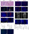
Female mice were mated with Venus+ males, and their uterine horns were harvested after implantation (E5.5–E6.5, n = 4 dams). (A) H&E staining of decidua. Black arrows designate embryos. (B) Newly formed CD34+ uterine blood vessels, reflecting decidual angiogenesis. (C) Hyaluronan localization indicated by IHC. White arrow designate embryo. (D) Glycosaminoglycans were separated from uteri of pregnant mice, at different time points, subjected to native PAGE next to hyaluronan standard and stained for hyaluronan (n = 2 dams; 3 implantation sites per dam). AMP, antimesometrial pole; MP, mesometrial pole.
Figure 2
Figure 2. Hyaluronan metabolism following embryo implantation.
Female mice were mated with Venus+ males, and their uterine horns were harvested after implantation (E5.5–E6.5) and subjected to histological analysis. Venus (embryo) and keratin detects ectoplacental trophoblasts and visceral endoderm cells (n = 4 dams, 10 implantation sites). Right panels are magnifications of left panels. (A and B) IHC analysis of hyaluronan synthesizing enzymes HAS-1 and HAS-2. (C) IHC analysis of Hyal-2, hyaluronan degrading enzyme. (D) IHC analysis of the most prominent hyaluronan receptor, CD44.
Figure 3
Figure 3. Pharmacological blockade of the progesterone receptor after attachment.
Female ICR mice were mated with ICR males and administered with RU-486 at E4.5. Their uterine horns were harvested at E5.5 (n = 3 dams). (A) Representative images of VEGF-A staining in E5.5 decidua. (B) Smaller decidua and abnormal embryonic morphology, detected by H&E staining. (C) Comparison of cytokeratin-7–expressing trophoblast cells and their distribution; white arrows indicate embryos. (D) Representative images of decreased Hyal-2 decidual distribution. (E) Western blot analysis of Hyal-2 following RU-486 treatment (n = 4 dams; pools of 3 decidua). (F) Representative images of Hyal-1 staining in E5.5 decidua.
Figure 4
Figure 4. Genetic modifications in embryonic trophoblast cells.
(A) Morulae were retrieved from pregnant mice (E2.5), grown to blastocysts, infected with lentiviral vectors, and transferred to pseudopregnant mice. Immediately thereafter, Hyal-2 was overexpressed in the trophectoderm. (B) eGFP expression prior to embryo transfer. (CE) Maximal intensity projections of whole-mount immunofluorescence for Hyal-2 in blastocysts following lentiviral transduction prior to embryo transfer, blastocysts over-expressing HAS-2 in their trophectoderm visualized for eGFP expression, and HAS-2 overexpression following viral infection. (F) Validation of Hyal-2 and HAS-2 overexpression by Western blot analysis. (G) Quantification of Western blot analysis following infection with lentiviral vectors. (H) Histological assessment of hyaluronan deposition as a result of Hyal-2 and HAS-2 overexpression. (n = 3 mice). (I) E6.5 trophoblast cells exclusively expressing eGFP alongside the WT epiblast and endoderm.
Figure 5
Figure 5. Trophectoderm overexpression of Hyal-2 resulted in enhanced implantation rate but early embryonic lethality.
Implantation sites were studied at E6.5, containing sham-infected embryos and embryos overexpressing Hyal-2 in their trophoblast. (A) H&E staining (n = 6 dams; white arrows indicate decidua). (B) TUNEL staining revealed cell death at the embryonic niche of Hyal-2 OEx (white arrow indicate embryonic cells; n = 4 dams). (C and D) Quantification of observed decidua, as well as their calculated area (2.9 ± 0.5; 4.75 ± 0.7; P = 0.02) (n = 12 dams in control, n = 10 dams in Hyal-2 OEx) divided by (3.42-fold change± 0.07; 0.03; P = 0.0006) (n = 8 dams, 27 implantation sites in control; n = 6 dams, 23 implantation sites for Hyal-2 OEx). (E and F) Detection of MAC-2+ macrophages and their quantification (5.9-fold change ± 0.35; P = 0.02) (n = 5 dams, 9 decidua in each group). (G) Presence of MAC-2+ macrophages was further demonstrated using immunostaining of macrophages in implantation sites harvested from surrogate mothers injected with rhodamine-labeled (ROX-labeled) lectin. Tissues were made transparent using modified tissue clearing procedure, thus enabling visualization of whole decidua by confocal microscopy. (n = 2 dams from each group). (H) Flow cytometry analysis of CD11b+F4/80+ cells in E6.5 decidua (12.33 ± 0.31 [control]; 17.02 ± 1.49 [overexpression]; P = 0.04) (n = 3 dams in control, n = 5 dams in Hyal-2 OEx). The statistical analysis applied was Student’s t test (C, D, F, and H).
Figure 6
Figure 6. Detrimental decidual hypervascularity and breach of the maternal-embryo barrier is induced by trophectoderm Hyal-2 overexpression.
(A) Ectopic presence of newly formed maternal blood vessels in the embryonic niche was observed in dams carrying Hyal-2 OEx embryos (n = 5 dams). (B) GE-T1–weighted MRI images of decidua, acquired from pregnant mice at E6.5, 3 minutes after administration of biotin-BSA-GdDTPA; white arrows point at implantation sites. (C–E) Linear regression plots from DCE MRI analysis. Decidual blood volume fraction (fBV; 0.023 ± 0.01 [control]; 0.072 ± 0.009 [overexpression], P = 0.0005) and permeability (PS; 0.00092 ± 0.0003; 0.0074 ± 0.0031, P = 0.09) were calculated from DCE MRI (n = 5 dams 13 implantation sites in control; 6 dams 24 implantation sites in Hyal-2 OEx). (F and G) Increased levels of VEGF-A and VEGFR-2 in decidua harvested at E6.5 (n = 3 dams, 2 implantation sites from each group). (H) Increased VEGFR-2 expression on decidual vessels in mesometrial orientation, as well as in cytotrophoblast cells, in Hyal-2 OEx foster dams (n = 4 dams in each group). (I) Declined VEGFR-3+ VSFs endothelial expression, demonstrated by immunofluorescence (n = 3 dams, 6 implantation sites from each group). (J) Hyperpermeable blood vessels in the embryonic niche were visualized by staining of biotin-BSA-GdDTPA, 40 minutes after intravenous injection (n = 5 dams). (K) Increased MMP-9 levels in E6.5 trophoblast cells OEx Hyal-2 (n = 5 dams in each group). (L) Quantification of MMP-9 expression in E6.5 decidua by immunofluorescence of histological sections (1.858-fold change ± 0.14 [control]; 0.25 [overexpression]; P = 0.01) (n = 5 dams in each group). (M) Increased levels of pro MMP-9 and mature MMP-9 in decidua harvested at E6.5 (n = 3 dams, 2 implantation sites in each group). The statistical analysis applied was Student’s t test (D, E, and L).
Figure 7
Figure 7. Trophoblast Hyal-2 overexpression impairs uterine NK cell recruitment and function.
(A) Representative images of DBA+ uterine NK cells staining in E6.5 decidua (n = 4 dams in each group). (B) Immunofluorescence of colocalized DBA+ uterine NK cells with hyaluronan receptor RHAMM at E6.5 decidua. (C) Impaired uterine NK cell recruitment demonstrated by decreased IL-15/IL-15R complexes detected by ELISA (n = 5 dams in each group 323.4 ± 23.49 pg/mL [control]; 213.65 ± 30.43 pg/mL [overexpression], P = 0.2). (D) Western blot analysis for NCR-1 in E6.5 decidual extracts demonstrated decreased accumulation of NCR-1–expressing NK cells in Hyal-2 OEx foster dams (n = 3 dams, 2 implantation sites in each group). (E) Flow cytometry analysis of E6.5 implantation sites harvested from foster dams of both groups. Note decreased ratio of CD45+NCR-1+ population in the Hyal-2 OEx group. (F) Immunofluorescence of TNFRSF9 expressed by uterine NK cells, in the mesometrial pole in E6.5 decidua. The statistical analysis applied was Student’s t test (C).
Figure 8
Figure 8. Uterine NK cells transcriptome is dysregulated in foster dams carrying Hyal-2 OEx embryos.
NCR1+ uterine NK cells were sorted from E6.5 decidua of surrogate mice (n = 3 dams; pools of 4 implantation sites each). (A) Volcano plot of all genes detected in RNA sequencing analysis. All points above the gray dotted horizontal line are statistically significant. Raw P values were adjusted for multiple testing using the procedure of Benjamini and Hochberg. Genes associated with antiviral and innate immune response are indicated in Hyal-2 OEx. Genes associated with uterine NK cells classification are indicated in Control. (B) Heatmap of differentially expressed (DE) genes in uterine NK cells sorted from control dams. Bolt DE genes in green were associated serine-type peptidase activity, characteristic of uterine NK cells by GO: molecular function. Bolt DE genes in orange were associated Cytokine Signaling in Immune System by GO: Pathways. (C) GSEA analysis of uterine NK cells, sorted from Hyal-2 OEx dams, tested for transcriptional enrichment in the above-indicated pathways. Genes clustered above the dotted line are significantly enriched. (D) GSEA analysis of uterine NK cells, sorted from control dams, tested for transcriptional enrichment in the above-indicated pathways. Genes clustered above the dotted line are significantly enriched.
Figure 9
Figure 9. Trophectoderm overexpression of HAS-2 resulted in early embryonic lethality and attenuated decidual angiogenesis.
(A) H&E staining of implantation sites revealed embryo resorption upon HAS-2 OEx (n = 4 dams). (B) Remnants of embryo overexpressing HAS-2 by trophoblast cells (CK-7) as opposed to decidual cells expressing HAS-2 in the embryonic niche in the control (n = 3 dams). (C) Profound embryonic cell death indicated by TUNEL staining was detected upon HAS-2 OEx, (n = 3 dams). (D) Similar pattern of MAC-2+ macrophages, confined to the mesometrial pole, away from the embryonic niche, observed in both groups (n = 3 dams). (E) Quantification MAC-2 staining by fluorescent microscopy (1.066-fold change ± 0.18 [control]; 0.41 [overexpression]; n = 5 dams, 7 implantation sites, P = 0.879). (F) Number of embryo implantation site per dam was assessed, at E6.5 by gross morphology inspection, as well as by examination of histological sections (3 ± 0.63 [control]; 2.88 ± 0.61 [overexpression], P = 0.88) (n = 6 dams in control; 8 dams in HAS-2 OEx). (G) Representative image of DBA+ NK cells in E6.5 decidua (n = 3 dams). (H) Representative image of MMP-9 in E6.5 decidua (n = 3 dams). (I) Impaired development of CD34+ newly formed blood vessels was observed in pregnant mice carrying HAS-2 OEx embryos, reflected by confinement of vessels away from the embryonic niche (n = 3 dams). (J) T1 weighted GE-MRI of embryo implantation sites, acquired from pregnant mice at E6.5 30 minutes after administration of biotin-BSA-GdDTPA. Little accumulation of biotin-BSA-GdDTPA was observed in the in dams carrying HAS-2 OEx embryos in comparison with control (white arrows indicate implantation sites; n = 6 dams). (K) Visualization of hyperpermeable blood vessels in the embryonic niche was achieved by staining of biotin-BSA-GdDTPA, 40 minutes after i.v. injection. Hyperpermeable vessels were not detected at HAS-2 OEx decidua in contrast to those detected in the control group (n = 3 dams). (L–N) These observations were consistent with DCE-MRI of biotin-BSA-GdDTPA (L), fBV (M) (0.041 ± 0.005; 0.023 ± 0.002; P = 0.004) and PS (N) (0.0017 ± 0.0003; 0.0009 ± 0.00031; P = 0.2) (control: 5 dams 9 implantation sites; HAS-2 OEx: 6 dams 19 implantation sites). The statistical analysis applied was Student’s t test (E, F, M, and N).

References

    1. Cross JC, Werb Z, Fisher SJ. Implantation and the placenta: key pieces of the development puzzle. Science. 1994;266(5190):1508–1518. puzzle. Science. doi: 10.1126/science.7985020. - DOI - PubMed
    1. Cha J, Sun X, Dey SK. Mechanisms of implantation: strategies for successful pregnancy. Nat Med. 2012;18(12):1754–1767. doi: 10.1038/nm.3012. - DOI - PMC - PubMed
    1. Morris SA, Zernicka-Goetz M. Formation of distinct cell types in the mouse blastocyst. Results Probl Cell Differ. 2012;55:203–217. doi: 10.1007/978-3-642-30406-4_11. - DOI - PubMed
    1. Dey SK, et al. Molecular cues to implantation. Endocr Rev. 2004;25(3):341–373. doi: 10.1210/er.2003-0020. - DOI - PubMed
    1. Kim M, et al. VEGF-A regulated by progesterone governs uterine angiogenesis and vascular remodelling during pregnancy. EMBO Mol Med. 2013;5(9):1415–1430. doi: 10.1002/emmm.201302618. - DOI - PMC - PubMed

Publication types

MeSH terms