Review
. 2020 Dec 15;60(12):581-593.
doi: 10.2176/nmc.ra.2020-0197. Epub 2020 Nov 18.

Recent Advancement of Technologies and the Transition to New Concepts in Epilepsy Surgery

Affiliations
Review

Recent Advancement of Technologies and the Transition to New Concepts in Epilepsy Surgery

Takamichi Yamamoto. Neurol Med Chir (Tokyo). .

Abstract

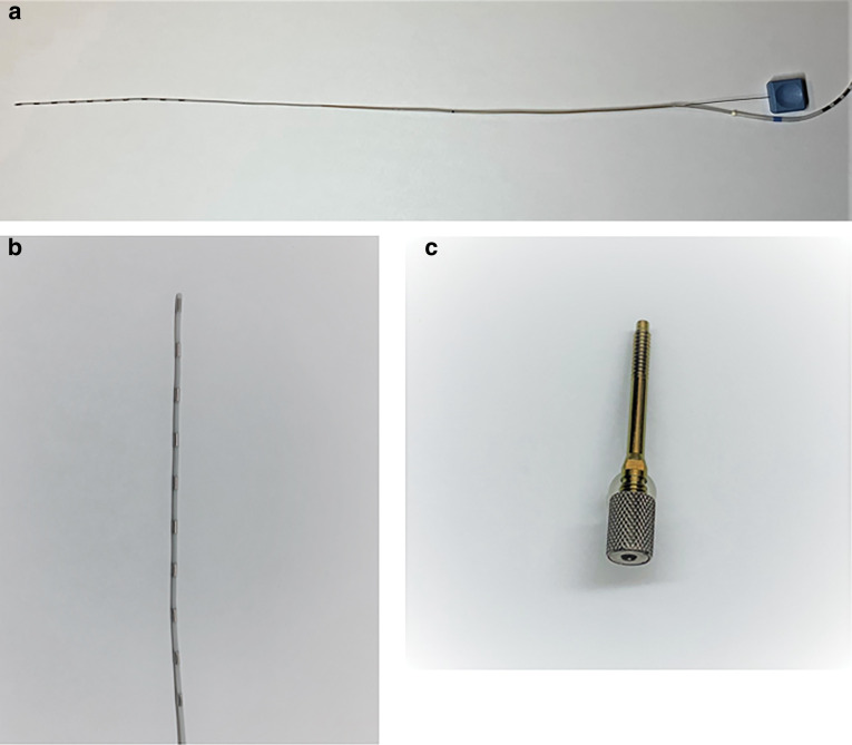
Fruitful progress and change have been accomplished in epilepsy surgery as science and technology advance. Stereotactic electroencephalography (SEEG) was originally developed by Talairach and Bancaud at Hôspital Sainte-Anne in the middle of the 20th century. SEEG has survived, and is now being recognized once again, especially with the development of neurosurgical robots. Many epilepsy centers have already replaced invasive monitoring with subdural electrodes (SDEs) by SEEG with depth electrodes worldwide. SEEG has advantages in terms of complication rates as shown in the previous reports. However, it would be more indispensable to demonstrate how much SEEG has contributed to improving seizure outcomes in epilepsy surgery. Vagus nerve stimulation (VNS) has been an only implantable device since 1990s, and has obtained the autostimulation mode which responds to ictal tachycardia. In addition to VNS, responsive neurostimulator (RNS) joined in the options of palliative treatment for medically refractory epilepsy. RNS is winning popularity in the United States because the device has abilities of both neurostimulation and recording of ambulatory electrocorticography (ECoG). Deep brain stimulation (DBS) has also attained approval as an adjunctive therapy in Europe and the United States. Ablative procedures such as SEEG-guided radiofrequency thermocoagulation (RF-TC) and laser interstitial thermal therapy (LITT) have been developed as less invasive options in epilepsy surgery. There will be more alternatives and tools in this field than ever before. Consequently, we will need to define benefits, indications, and limitations of these new technologies and concepts while adjusting ourselves to a period of fundamental transition in our foreseeable future.

Keywords: RNS; SEEG; VNS; ablative surgery; epilepsy surgery.


Conflict of interest statement

Conflicts of Interest Disclosure

The author who is a member of the Japan Neurosurgical Society (JNS) reports no conflict of interest (COI) regarding this article and has made declaration of COI with a self-reported COI disclosure statement form to the JNS Office in the preceding three years.

Figures

Fig. 1
Fig. 1
A subdural grid electrode was placed to cover the right frontal and temporal cortexes. Six subdural strips around the grid and two depth electrodes through the grid were additionally implanted. A photo from the author’s surgical experience. Consent was obtained from the patient.
Fig. 2
Fig. 2
(a) A whole view of a depth electrode with an inner wire stylet. (b) The tip of a depth electrode. (c) An anchor bolt to secure a depth electrode in the skull. Courtesy of Ad-Tech Medical Instrument Corporation, Oak Creek, WI, USA.
Fig. 3
Fig. 3
Three depth electrodes were implanted on each side for an exploratory evaluation of SEEG by conventional frame-based implantation. Courtesy of Dr. Yuichi Kubota, Department of Neurosurgery, Tokyo Women’s Medical University Medical Center East, Tokyo, Japan.
Fig. 4
Fig. 4
ROSA, a neurosurgical robot is useful for frameless implantation of depth electrodes for SEEG. In addition, this robot has been utilized for other neurosurgical procedures such as neuroendoscopy, stereotactic biopsy, pallidotomy, shunt placement, DBS procedures, and stereotactic cyst aspiration. Courtesy of Zimmer Biomet, Warsaw, IN, USA, 2020. DBS: deep brain stimulation, SEEG: stereotactic electroencephalography.
Fig. 5
Fig. 5
A screen of the dedicated software demonstrates trajectories of depth electrodes as preoperative planning. Pictures of neuroimaging such as an MRI and a CT scan can be imported. The gadolinium T1-weighted sequence is indispensable in locating an entry point to avoid hemorrhagic complications. Courtesy of Zimmer Biomet, Warsaw, IN, USA, 2020. CT: computed tomography; MRI: magnetic resonance imaging.
Fig. 6
Fig. 6
Thirteen depth electrodes were implanted with particular interest on the left temporal lobe by frameless navigation of the ROSA system. The neurosurgical robot reduces the required time and brings convenience for implantation of depth electrodes. Courtesy of Dr. Jorge González-Martínez, Department of Neurosurgery, University of Pittsburgh Medical Center, PA, USA.
Fig. 7
Fig. 7
AspireSR (Model 106) of the VNS Therapy System responds to ictal tachycardia. The previous systems had only Normal Mode and Magnet Mode functioning as an open-loop system. Ultimately, the system from Model 106 has gained AutoStim Mode as a closed-loop system. Courtesy of LivaNova USA, Inc.
Fig. 8
Fig. 8
The number of the VNS® Therapy Systems implanted each year in Japan since its approval in 2010. A total of 2731 implantation procedures were carried out by the end of 2019. A small decrement was observed in 2017 possibly because several newer AEDs came onto the Japanese market and showed an influence to a certain extent. Courtesy of LivaNova Japan KK. AEDs: anti-epileptic drugs.
Fig. 9
Fig. 9
An implantable part of the RNS System is comprised of a neurostimulator and depth and/or subdural cortical leads. The neurostimulator connects one or two leads placed surgically at the seizure focus. Courtesy of NeuroPace, Inc.
Fig. 10
Fig. 10
The cranially seated neurostimulator continually senses electrocorticographic activity, and then provides stimulation when it detects abnormal activity. Detection and stimulation are performed through the two leads, and these parameters are adjusted for optimizing control of seizures. Courtesy of NeuroPace, Inc.
Fig. 11
Fig. 11
Utilization of the RNS System in the United States. The line graph demonstrates the number of cumulative patients treated with the RNS System. Courtesy of NeuroPace, Inc.

Similar articles

Cited by

References

    1. Wheless JW, Gienapp AJ, Ryvlin P: Vagus nerve stimulation (VNS) therapy update. Epilepsy Behav 88: 2–10, 2018 - PubMed
    1. Kang JY, Sperling MR: Epileptologist’s view: laser interstitial thermal ablation for treatment of temporal lobe epilepsy. Epilepsy Res 142: 149–152, 2018 - PubMed
    1. Wennberg R, Ladino LD, Téllez-Zenteno JF: On the renaissance of stereotactic EEG and its interpretation. Can J Neurol Sci 45: 255–258, 2018 - PubMed
    1. Morrell MJ: RNS System in Epilepsy Study Group: Responsive cortical stimulation for the treatment of medically intractable partial epilepsy. Neurology 77: 1295–1304, 2011 - PubMed
    1. Salanova V: Deep brain stimulation for epilepsy. Epilepsy Behav 88: 21–24, 2018 - PubMed

MeSH terms