

Multispectral imaging detects gastritis consistently in mouse model and in humans
- PMID: 33208839
- PMCID: PMC7674504
- DOI: 10.1038/s41598-020-77145-4
Multispectral imaging detects gastritis consistently in mouse model and in humans
Erratum in
-
Author Correction: Multispectral imaging detects gastritis consistently in mouse model and in humans.Sci Rep. 2021 Aug 17;11(1):17045. doi: 10.1038/s41598-021-94899-7. Sci Rep. 2021. PMID: 34404827 Free PMC article. No abstract available.
Abstract
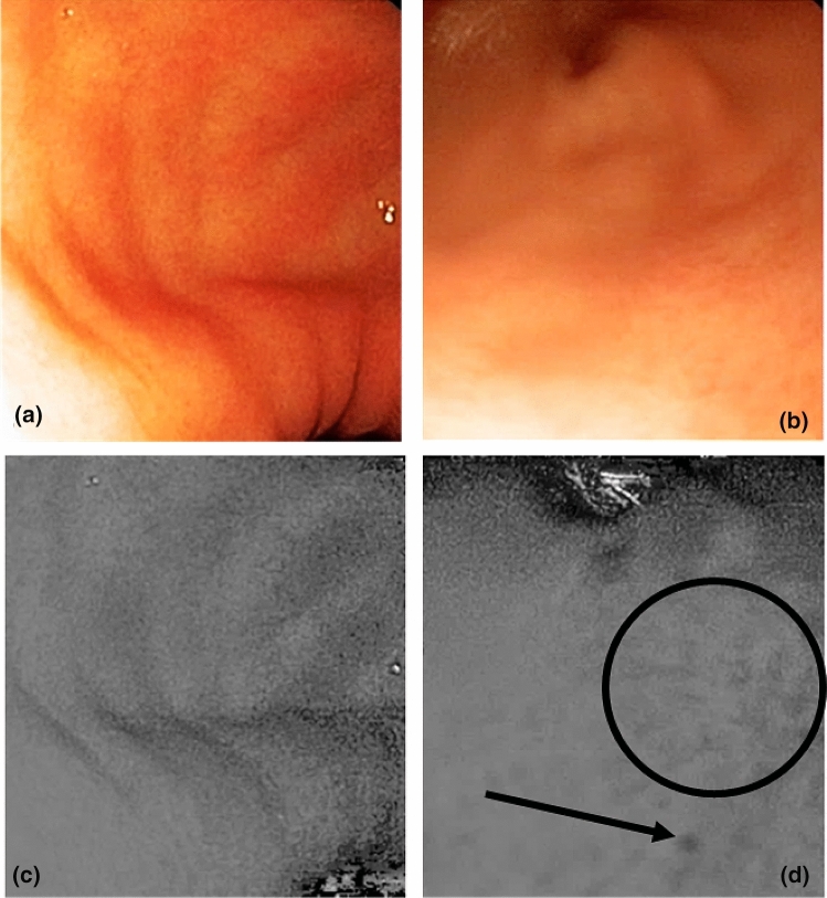
Gastritis constitutes the initial step of the gastric carcinogenesis process. Gastritis diagnosis is based on histological examination of biopsies. Non-invasive real-time methods to detect mucosal inflammation are needed. Tissue optical properties modify reemitted light, i.e. the proportion of light that is emitted by a tissue after stimulation by a light flux. Analysis of light reemitted by gastric tissue could predict the inflammatory state. The aim of our study was to investigate a potential association between reemitted light and gastric tissue inflammation. We used two models and three multispectral analysis methods available on the marketplace. We used a mouse model of Helicobacter pylori infection and included patients undergoing gastric endoscopy. In mice, the reemitted light was measured using a spectrometer and a multispectral camera. We also exposed patient's gastric mucosa to specific wavelengths and analyzed reemitted light. In both mouse model and humans, modifications of reemitted light were observed around 560 nm, 600 nm and 640 nm, associated with the presence of gastritis lesions. These results pave the way for the development of improved endoscopes in order to detect real-time gastritis without the need of biopsies. This would allow a better prevention of gastric cancer alongside with cost efficient endoscopies.
Conflict of interest statement
The authors declare no competing interests.
Figures


Similar articles
-
Multimodal imaging as optical biopsy system for gastritis diagnosis in humans, and input of the mouse model.EBioMedicine. 2021 Jul;69:103462. doi: 10.1016/j.ebiom.2021.103462. Epub 2021 Jul 3. EBioMedicine. 2021. PMID: 34229278 Free PMC article. Clinical Trial.
-
Accuracies of Endoscopic Diagnosis of Helicobacter pylori-Gastritis: Multicenter Prospective Study Using White Light Imaging and Linked Color Imaging.Digestion. 2020;101(5):624-630. doi: 10.1159/000501634. Epub 2019 Jul 23. Digestion. 2020. PMID: 31336366
-
Prevalence of Helicobacter pylori infection in patients with large gastric folds: evaluation and follow-up with endoscopic ultrasound before and after antimicrobial therapy.Am J Gastroenterol. 1995 Nov;90(11):1969-73. Am J Gastroenterol. 1995. PMID: 7485002
-
Helicobacter pylori-induced inflammation and epigenetic changes during gastric carcinogenesis.World J Gastroenterol. 2015 Dec 7;21(45):12742-56. doi: 10.3748/wjg.v21.i45.12742. World J Gastroenterol. 2015. PMID: 26668499 Free PMC article. Review.
-
Can image-enhanced endoscopy improve the diagnosis of Kyoto classification of gastritis in the clinical setting?Dig Endosc. 2020 Jan;32(2):191-203. doi: 10.1111/den.13540. Epub 2019 Oct 10. Dig Endosc. 2020. PMID: 31550395 Review.
Cited by
-
Multimodal imaging as optical biopsy system for gastritis diagnosis in humans, and input of the mouse model.EBioMedicine. 2021 Jul;69:103462. doi: 10.1016/j.ebiom.2021.103462. Epub 2021 Jul 3. EBioMedicine. 2021. PMID: 34229278 Free PMC article. Clinical Trial.
References
-
- Calabrese C, et al. Correlation between endoscopic features of gastric antrum, histology and Helicobacter pylori infection in adults. Ital. J. Gastroenterol. Hepatol. 1999;31:359–365. - PubMed
Publication types
MeSH terms
LinkOut - more resources
Full Text Sources
Medical
