. 2020 Nov;587(7835):638-643.
doi: 10.1038/s41586-020-2927-z. Epub 2020 Nov 18.

Host ANP32A mediates the assembly of the influenza virus replicase

Affiliations

Host ANP32A mediates the assembly of the influenza virus replicase

Loïc Carrique et al. Nature. 2020 Nov.

Abstract

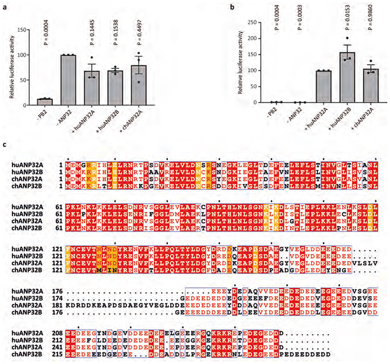
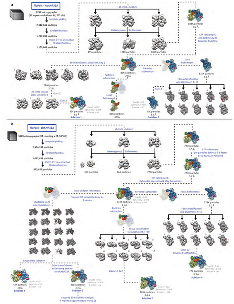
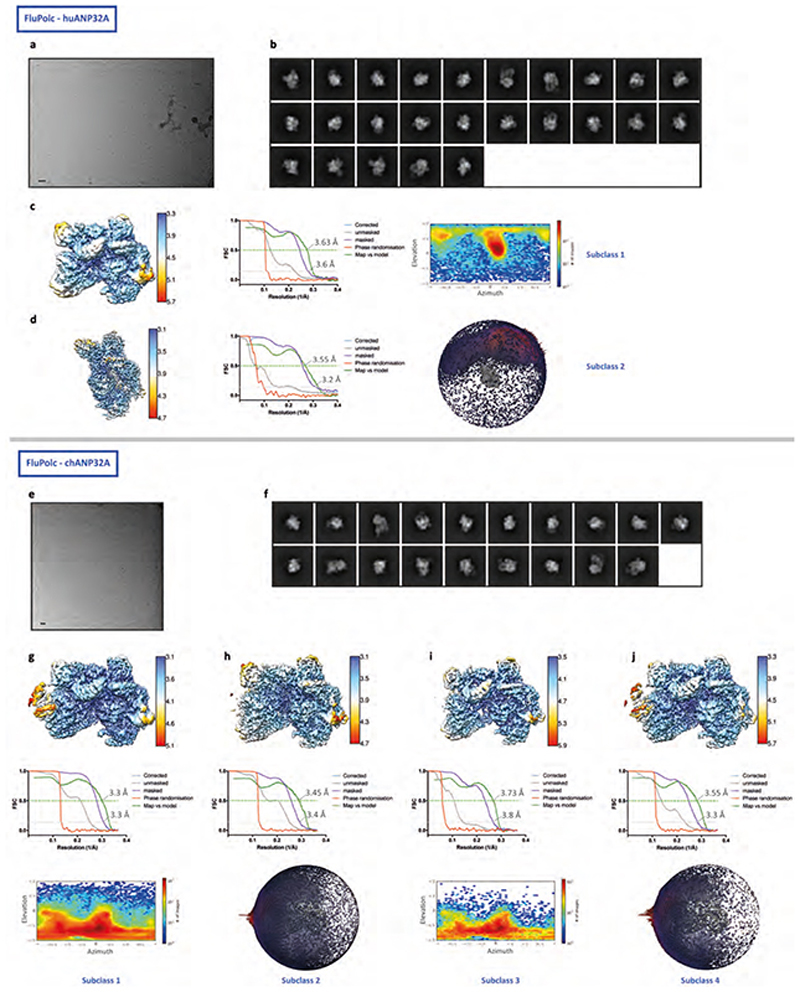
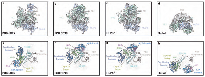
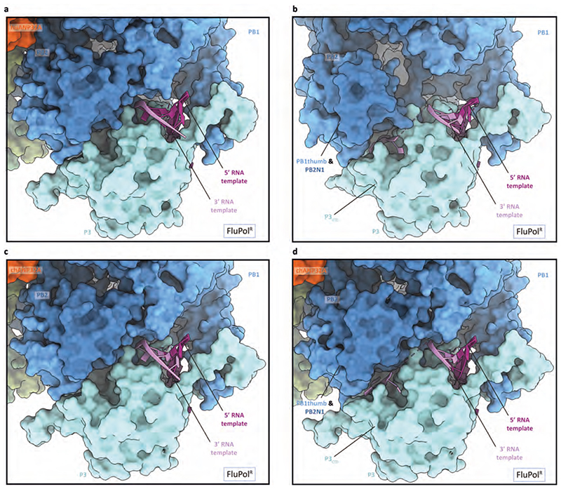
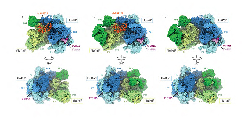
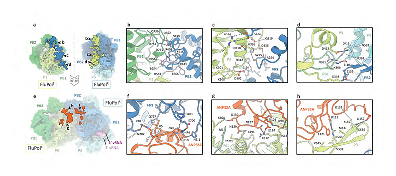
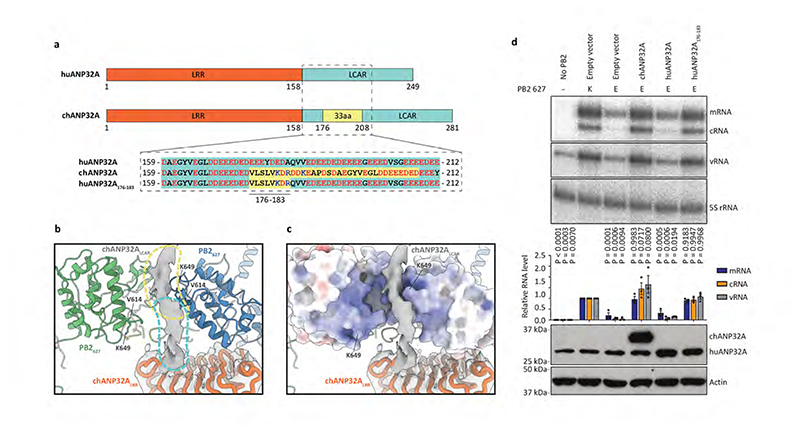
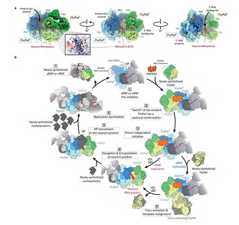
Aquatic birds represent a vast reservoir from which new pandemic influenza A viruses can emerge1. Influenza viruses contain a negative-sense segmented RNA genome that is transcribed and replicated by the viral heterotrimeric RNA polymerase (FluPol) in the context of viral ribonucleoprotein complexes2,3. RNA polymerases of avian influenza A viruses (FluPolA) replicate viral RNA inefficiently in human cells because of species-specific differences in acidic nuclear phosphoprotein 32 (ANP32), a family of essential host proteins for FluPol activity4. Host-adaptive mutations, particularly a glutamic-acid-to-lysine mutation at amino acid residue 627 (E627K) in the 627 domain of the PB2 subunit, enable avian FluPolA to overcome this restriction and efficiently replicate viral RNA in the presence of human ANP32 proteins. However, the molecular mechanisms of genome replication and the interplay with ANP32 proteins remain largely unknown. Here we report cryo-electron microscopy structures of influenza C virus polymerase (FluPolC) in complex with human and chicken ANP32A. In both structures, two FluPolC molecules form an asymmetric dimer bridged by the N-terminal leucine-rich repeat domain of ANP32A. The C-terminal low-complexity acidic region of ANP32A inserts between the two juxtaposed PB2 627 domains of the asymmetric FluPolA dimer, suggesting a mechanism for how the adaptive PB2(E627K) mutation enables the replication of viral RNA in mammalian hosts. We propose that this complex represents a replication platform for the viral RNA genome, in which one of the FluPol molecules acts as a replicase while the other initiates the assembly of the nascent replication product into a viral ribonucleoprotein complex.


Conflict of interest statement

Competing interests

The authors declare no competing interests.

Figures

Extended Data Fig. 1
Extended Data Fig. 1. FluPolC activity depends on ANP32A and alignment of ANP32 proteins.
a, b, Luciferase reporter gene activities reflecting FluPolC activity in control (a) and dKO (b) eHAP cells in the presence or absence of overexpressed huANP32A, huANP32B or chANP32A. Data are presented as mean values ± s.e.m. n = 3 biologically independent samples from n = 3 independent experiments. Ordinary one-way ANOVA with Dunnett’s post hoc test for multiple comparisons. P < 0.05 is considered significant to reject the null hypothesis. c, Sequence alignment of huANP32A, huANP32B, chANP32A, chANP32B. Residues involved in hydrogen bonding interactions with FluPolC are indicated in orange. The chANP32A avian-specific 33 amino acid insertion is highlighted in cyan. The SUMO interaction motif (SIM) sequence is indicated by black triangles. The figure was prepared with Espript 3.0.
Extended Data Fig. 2
Extended Data Fig. 2. Data collection, processing and analysis scheme.
a, b, Flowchart for the processing and the classification of the FluPolC-huANP32A complex (a) and FluPolC-chANP32A complex (b).
Extended Data Fig. 3
Extended Data Fig. 3. Single-particle cryo-EM analysis of FluPolC-huANP32A and FluPolC-chANP32A complexes.
a, e, Representative micrograph of FluPolC-huANP32A (a) and FluPolC-chANP32A (e) embedded in vitreous ice. Scale bar, 200 Å. b, f, Representative 2D class averages of FluPolC-huANP32A (b) and FluPolC-chANP32A (f). c, d, Data analysis for FluPolC-huANP32A Subclass1 (c) and Subclass2 (d). 3D reconstruction locally filtered and coloured according to RELION local resolution (left panel). FSC curve indicating overall map resolution and model-to-map FSC (middle panel). Curves are shown for phase randomisation, unmasked, masked and phase-randomisation-corrected masked maps. Angular distribution of particle projections with the cryo-EM map shown in grey (right panel). g-j, Data analysis for FluPolC-chANP32A Subclass1 (g), Subclass2 (h), Subclass3 (i) and Subclass4 (j). 3D reconstruction locally filtered and coloured according to RELION local resolution (top panel). FSC curve indicating overall map resolution and the model-to-map FSC (middle panel). Curves are shown for phase randomisation, unmasked, masked and phase-randomisation-corrected masked maps. Angular distribution of particle projections with the cryo-EM map shown in grey (bottom panel).
Extended Data Fig. 4
Extended Data Fig. 4. Comparison of FluPolR and FluPolE structures with the transcriptase and apo conformations of FluPol.
a-d, Comparison of structures of human influenza A/NT/60/68 (H3N2) bound to vRNA and capped RNA in the transcriptase conformation (PDB: 6RR7) (a) and human influenza C/Johannesburg/1/66 in the apo conformation (PDB: 5D98) (b) with structures of FluPolR (c) and FluPolE (d) in the FluPolC-chANP32A complex. e-h, Comparison of the PB2 domain arrangements in the complexes shown in a-d.
Extended Data Fig. 5
Extended Data Fig. 5. Close-up view of the interaction of 5’ and 3’ vRNA termini with FluPolR.
a, c, Close-up view of the 3’ vRNA pointing towards the active site in the FluPolC-huANP32A (a) and FluPolC-chANP32A (c) structures. b, d, Close-up view of the 3’ vRNA binding in a groove located between P3CTD and the PB1thumb and PB2N1 subdomains in the FluPolC-huANP32A (b) and FluPolC-chANP32A (d) structures.
Extended Data Fig. 6
Extended Data Fig. 6. Effect of FluPolR-FluPolE dimer interface mutations on FluPolA activity.
a, b, Effect of mutations at the FluPolR-FluPolE dimer interface on FluPolA activity in viral minigenome assays (a) and cRNA encapsidation by FluPolA (b). Data are presented as mean values ± s.e.m. n = 3 biologically independent samples from n = 3 independent experiments. Ordinary one-way ANOVA with Dunnett’s post hoc test for multiple comparisons. P < 0.05 is considered significant to reject the null hypothesis. Western blot analyses were repeated from n = 3 independent experiments with similar results. For gel source data, see Supplementary Fig. 2.
Extended Data Fig. 7
Extended Data Fig. 7. Effect of FluPolA mutations at the FluPolA-ANP32A interface on FluPolA activity and interaction with huANP32A.
a, b, Effect of FluPolA mutations at the FluPolA-ANP32A interface on FluPolA activity in viral minigenome assays on (a) and FluPolAANP32A interaction (b). Data are presented as mean values ± s.e.m. n = 3 biologically independent samples from n = 3 independent experiments. Ordinary one-way ANOVA with Dunnett’s post hoc test for multiple comparisons. P < 0.05 is considered significant to reject the null hypothesis. Western blot analyses were repeated from n = 3 independent experiments with similar results. For gel source data, see Supplementary Fig. 2.
Extended Data Fig. 8
Extended Data Fig. 8. Structural comparison of PB2627 domains of FluPolA and FluPolC.
Structures of the PB2627 domains from crystal structures of FluPol from influenza C/Johannesburg/1/1966 (a, PDB ID: 5D98) and A/NT/60/1968 (H3N2) (b, PDB ID: 6QNW) viruses are aligned and shown in cartoon mode. Residues discussed in this study are highlighted in stick mode and coloured in orange.
Fig. 1
Fig. 1. Structures of dimers of FluPolC heterotrimers with or without bound ANP32A.
a-c, Cryo-EM structures of dimers of FluPolC heterotrimers bound to huANP32A (a), chANP32A (b), and without ANP32A (c).
Fig. 2
Fig. 2. FluPolC-FluPolC and ANP32A-FluPolC interaction interfaces.
a, FluPolC-FluPolC dimer interface with interacting regions in FluPolR and FluPolE highlighted on the molecular surface, by peeling apart the two molecules each by 30°. The letters denote the close-up views shown in panels b-d, Close-up views of the FluPolC-FluPolC interaction interface. Dashed lines indicate hydrogen bonds. e, ANP32A-FluPolC surface interface with interacting regions in FluPolC highlighted. The letters denote the close-up views shown in panels f-h, Close-up views of the ANP32A-FluPolC interaction interface. Dashed lines indicate hydrogen bonds.
Fig. 3
Fig. 3. Interaction of ANP32LCAR with FluPolC and the effect of ANP32A on FluPolA activity.
a, Schematic of huANP32A and chANP32A highlighting the 33 amino acid (33 aa) insertion in chANP32A and sequence differences. b, Close-up view of the cryo-EM density attributed to chANP32A (grey, threshold 0.934) with the chANP32ALRR domain represented in cartoon (orange) and the positions of PB2627 of FluPolR (blue) and FluPolE (green). Regions potentially corresponding to the N-terminal part of ANP32ALCAR and part of the avian 33 aa insertion are highlighted in cyan and yellow, respectively. V614 and K649 in FluPolC corresponds to K/Q591 and K/E627 in FluPolA, respectively. c, Same view as shown in (b), with the PB2627 of FluPolR and FluPolE shown in surface representation. d, Effect of chANP32A and huANP32A, wild type or mutant (huANP32A176-183), on the activity of FluPolA with mammalian-adapted PB2 K627 or avian-signature PB2 E627 in a viral RNP reconstitution assay in mammalian HEK 293T cells. Data of primer extension analysis of viral RNA levels with quantitation (upper panels) and western blot analysis of ANP32A levels (lower panel) with molecular weight markers is shown. Data are presented as mean values ± s.e.m. n = 3 biologically independent samples from n = 3 independent experiments. Ordinary one-way ANOVA with Dunnett’s post hoc test for multiple comparisons. P < 0.05 is considered significant to reject the null hypothesis. Western blot analyses were repeated from n = 3 independent experiments with similar results. For gel source data, see Supplementary Fig. 2.
Fig. 4
Fig. 4. Functional implications of the FluPolC-ANP32A complex.
a, Relative positions of the RNA product exit channel in FluPolR and the 5’ RNA binding site in FluPolE in the chANP32A-FluPolC complex. The positions of the RNA product exit channel in FluPolR and the 5’ RNA binding site in FluPolE were determined by superposing FluPolR and FluPolE with the structure of FluPolA bound to capped RNA and vRNA template (PDB 6RR7). b, Model for the role of FluPol-ANP32A complex in the replication of the influenza virus RNA genome and its assembly into RNP. FluPol in the context of vRNP or cRNP is flexible (1) but is stabilised in a replicase FluPolR conformation upon binding of a newly synthesised FluPol in the presence of ANP32A (2). FluPolR initiates replication in a primer independent manner (3) with a trans-activating FluPol involved in cRNA to vRNA replication by promoting cRNA template realignment (3’). As the 5’ end of the nascent replication product is released from the polymerisation active site of FluPolR, it is captured in the 5’ RNA binding pocket of the encapsidating FluPolE bound to FluPolR (4), initiating the encapsidation of the nascent RNA with NP (5). Nascent vRNA or cRNA assemble into vRNP or cRNP, respectively (6), and are released upon FluPolR termination. FluPolE becomes the resident polymerase of the newly produced vRNP or cRNP.

References

    1. Krammer F, et al. Influenza. Nat Rev Dis Primers. 2018;4:3. - PMC - PubMed
    1. Fodor E, Te Velthuis AJW. Structure and Function of the Influenza Virus Transcription and Replication Machinery. Cold Spring Harb Perspect Med. 2019 - PMC - PubMed
    1. Wandzik JM, Kouba T, Cusack S. Structure and Function of Influenza Polymerase. Cold Spring Harb Perspect Med. 2020 - PMC - PubMed
    1. Long JS, et al. Species difference in ANP32A underlies influenza A virus polymerase host restriction. Nature. 2016;529:101–4. - PMC - PubMed
    1. Walker AP, Fodor E. Interplay between Influenza Virus and the Host RNA Polymerase II Transcriptional Machinery. Trends Microbiol. 2019;27:398–407. - PMC - PubMed

Publication types

MeSH terms