Case Reports
. 2020 Jul-Sep;13(3):233-236.
doi: 10.4103/JCAS.JCAS_148_19.

A Rare Case of Nasal Glial Heterotopia in an Infant

Affiliations
Case Reports

A Rare Case of Nasal Glial Heterotopia in an Infant

Karishma Tahlan et al. J Cutan Aesthet Surg. 2020 Jul-Sep.

Abstract

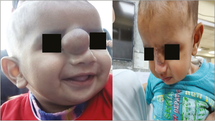
Glial heterotopias are the displacement of neuroglial tissue in extracranial sites. Nasal glial heterotopias can be of three types-extranasal, intranasal and mixed. Root of the nose is the most common location. These are rare anomalies with an incidence of 1 case in 20,000-40,000 live births. Here we report the case of a 6-month-old infant with a congenital mass located at the root of the nose. Non-contrast computed tomography studies showed no evidence of intracranial communication of the lesion. The mass was excised, and on histopathological examination, it showed glial tissue with astrocytes in a fibrillary background and fibroconnective tissue. Masson's trichrome stain showed the red staining of the glial tissue, whereas the background fibrosis was stained blue. On immunohistochemistry, glial fibrillary acidic protein was positive. Hence, the diagnosis of nasal glial heterotropia was made. The patient had an uneventful postoperative period.

Keywords: Nasal glial heterotopia; infant; nasal glioma.


Conflict of interest statement

There are no conflicts of interest.

Figures

Figure 1
Figure 1
(A) Preoperative photograph of the child with swelling at the root of nose. (B) Postoperative photograph of the child
Figure 2
Figure 2
NCCT showing no connection between the mass and the dura or brain
Figure 3
Figure 3
(A) Cut section of the mass showing homogenous gray-white solid soft tissue lesion. No hemorrhage or necrosis is seen. (B) Glial tissue with intervening fibrosis (hematoxylin and eosin [H and E], ×100). (C) Astrocytes in a fibrillary background (H and E, ×100). Few gemistocytes seen (arrow)
Figure 4
Figure 4
(A) The glial tissue is immunopositive for GFAP (immunohistochemistry, ×100). (B) The glial tissue stained dusky pink to red in color on Masson’s trichrome stain (×100), whereas the fibrous tissue stained blue

References

    1. Sharma JK, Pippal SK, Sethi Y, Arora S, Raghuwanshi SK. Nasal glioma: A case report. Indian J Otolaryngol Head Neck Surg. 2006;58:413–15. - PMC - PubMed
    1. Friel MT, Flores RL. Resection of nasal glial heterotopia using a nasal subunit approach. Ochsner J. 2018;18:176–9. - PMC - PubMed
    1. Gan W, Xiang Y, Tang Y, He X, Hu J, Yang F, et al. A CARE-compliant article: Extranasal glial heterotopia in a female infant: A case report. Medicine (Baltimore) 2018;97:e12000. - PMC - PubMed
    1. Hagiwara A, Nagai N, Ogawa Y, Suzuki M. A case of nasal glial heterotopia in an adult. Case Rep Otolaryngol. 2014;2014:354672. - PMC - PubMed
    1. Altissimi G, Ascani S, Falcetti S, Cazzato C, Bravi I. Central nervous system tissue heterotopia of the nose: Case report and review of the literature. Acta Otorhinolaryngol Ital. 2009;29:218–21. - PMC - PubMed

Publication types