. 2020 Sep 1;24(5):260-266.
doi: 10.1080/19768354.2020.1813805.

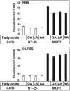
Arachidonic acid induces ER stress and apoptosis in HT-29 human colon cancer cells

Affiliations

Arachidonic acid induces ER stress and apoptosis in HT-29 human colon cancer cells

Sijeong Bae et al. Anim Cells Syst (Seoul). .

Abstract

Polyunsaturated fatty acids (PUFAs) have important functions in biological systems. The beneficial effects of dietary PUFAs against inflammatory diseases, cardiovascular diseases, and metabolic disorders have been shown. Studies using cancer cells have presented the anti-tumorigenic effects of docosahexaenoic acid (DHA), an n-3 PUFA, while arachidonic acid (AA), an n-6 PUFA, has been shown to elicit both pro- and anti-tumorigenic effects. In the current study, the anti-tumorigenic effects of AA were evaluated in HT-29 human colon cancer cells. Upon adding AA in the media, more than 90% of HT-29 cells died, while the MCF7 cells showed good proliferation. AA inhibited the expression of SREBP-1 and its target genes that encode enzymes involved in fatty acid synthesis. As HT-29 cells contained lower basal levels of fatty acid synthase, a target gene of SREBP-1, than that in MCF7 cells, the inhibitory effects of AA on the fatty acid synthase levels in HT-29 cells were much stronger than those in MCF-7 cells. When oleic acid (OA), a monounsaturated fatty acid that can be synthesized endogenously, was added along with AA, the HT-29 cells were able to proliferate. These results suggested that HT-29 cells could not synthesize enough fatty acids for cell division in the presence of AA because of the suppression of lipogenesis. HT-29 cells may incorporate more AA into their membrane phospholipids to proliferate, which resulted in ER stress, thereby inducing apoptosis. AA could be used as an anti-tumorigenic agent against cancer cells in which the basal fatty acid synthase levels are low.

Keywords: Arachidonic acid; ER stress; HT-29 cells; anti-tumorigenic effect.


Conflict of interest statement

No potential conflict of interest was reported by the author(s).

Figures

Figure 1.
Figure 1.
AA induced apoptosis in HT-29 cells. (A) HT-29 and MCF7 cells were treated with the indicated fatty acids, then the MTT assay was performed. The values represent the mean ± SE of 4 wells. * indicates p < 0.01 (Student’s t-test; compared to the untreated cells). (B) The HT-29 cells treated with the indicated fatty acids were stained with FITC-Annexin V/propidium iodide and separated by FACS. The portions of the apoptotic cells were boxed. (C) Expression of BAX were analyzed by reverse transcription-PCR using RNA prepared from the HT-29 cells treated with the indicated fatty acids in the media containing DLFBS. Caspase-3 was analyzed by western blotting. GAPDH and β-actin are the loading controls.
Figure 2.
Figure 2.
AA did not affect ROS generation in the cells. HT-29 and MCF7 cells were cultured and treated with the indicated fatty acids, then ROS production was measured from the cells. The values represent the mean ± S.E. of 4 wells.
Figure 3.
Figure 3.
AA inhibited the expression of SREBP-1c and its target genes, and OA could rescue the AA mediated cell death in HT-29 cells. (A) The relative mRNA expression of SREBP-1c was determined by qPCR. The precursor form (pSREBP-1) and the active form of SREBP-1 (nSREBP-1) were determined using proteins prepared from the indicated cells. Calnexin and CREB are the loading controls for pSREBP-1 and nSREBP-1, respectively. (B) The relative expression levels of FAS and SCD-1 were determined by qPCR. The expression levels of these genes in MCF7 cells that were not treated with the fatty acids are defined as 1. The values represent the mean ± SE of 3 plates. * indicates p < 0.05 (Student’s t-test; compared to the untreated cells). (C) OA was added to the cells at the indicated concentrations in the presence or absence of AA and the MTT assay was performed. The values represent the mean ± SE of 4 wells. * and ** indicate p < 0.05 and p < 0.01, respectively (Student’s t-test; compared to cells cultured in DLFBS with 100 µM AA). (D) MCF7 cells were incubated with OA and AA in the presence of cerulenin and the MTT assay was performed. The values represent the mean ± SE of 4 wells. ** indicates p < 0.01 of Student’s t-test by comparison of cerulenin alone and cerulenin with AA).
Figure 4.
Figure 4.
AA induced ER stress in HT-29 cells. The cDNAs prepared for the experiment described in Figure 3 were used to determine XBP1 processing. Equal amount of the PCR reaction of the individual samples in each group were pooled and separated on a 2% agarose gel. The unprocessed (XBP1u) and processed (XBP1p) forms of XBP1 generated 473 and 447 bp bands, respectively. (B) Total eIF2α and the phosphorylated eIF2α were determined by immunoblotting in the cells treated with the indicated fatty acids. β-actin is used as the loading control.

Similar articles

Cited by

References

    1. Bordoni A, Di Nunzio M, Danesi F, Biagi PL.. 2006. Polyunsaturated fatty acids: from diet to binding to ppars and other nuclear receptors. Genes Nutr. 1(2):95–106. doi: 10.1007/BF02829951 - DOI - PMC - PubMed
    1. Brown JR, DuBois RN.. 2005. COX-2: a molecular target for colorectal cancer prevention. J Clin Oncol. 23(12):2840–2855. doi: 10.1200/JCO.2005.09.051 - DOI - PubMed
    1. Calder PC. 2017. Omega-3 fatty acids and inflammatory processes: from molecules to man. Biochem Soc Trans. 45(5):1105–1115. doi: 10.1042/BST20160474 - DOI - PubMed
    1. Calfon M, Zeng H, Urano F, Till JH, Hubbard SR, Harding HP, Clark SG, Ron D.. 2002. IRE1 couples endoplasmic reticulum load to secretory capacity by processing the XBP-1 mRNA. Nature. 415(6867):92–96. doi: 10.1038/415092a - DOI - PubMed
    1. Castellone MD, Teramoto H, Williams BO, Druey KM, Gutkind JS.. 2005. Prostaglandin E2 promotes colon cancer cell growth through a Gs-axin-ß-catenin signaling axis. Science. 310(5753):1504–1510. doi: 10.1126/science.1116221 - DOI - PubMed