Review
. 2020 Sep 7;11(37):10019-10034.
doi: 10.1039/d0sc03570j. eCollection 2020 Oct 7.

Light control of RTK activity: from technology development to translational research

Affiliations
Review

Light control of RTK activity: from technology development to translational research

Anna V Leopold et al. Chem Sci. .

Abstract

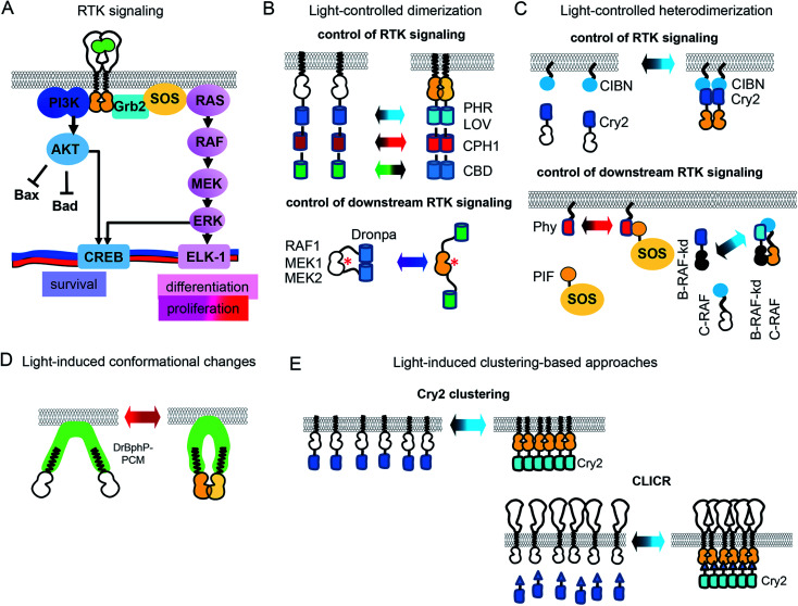
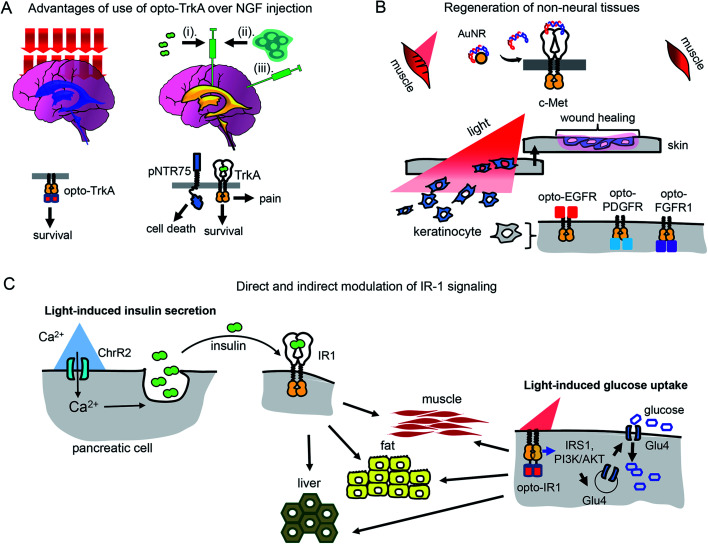
Inhibition of receptor tyrosine kinases (RTKs) by small molecule inhibitors and monoclonal antibodies is used to treat cancer. Conversely, activation of RTKs with their ligands, including growth factors and insulin, is used to treat diabetes and neurodegeneration. However, conventional therapies that rely on injection of RTK inhibitors or activators do not provide spatiotemporal control over RTK signaling, which results in diminished efficiency and side effects. Recently, a number of optogenetic and optochemical approaches have been developed that allow RTK inhibition or activation in cells and in vivo with light. Light irradiation can control RTK signaling non-invasively, in a dosed manner, with high spatio-temporal precision, and without the side effects of conventional treatments. Here we provide an update on the current state of the art of optogenetic and optochemical RTK technologies and the prospects of their use in translational studies and therapy.


Figures

Fig. 1
Fig. 1. Design of opto-RTKs and ways to light-control RTK downstream signalling. (A) Activation of major RTK signalling pathways by growth factors. GF binding leads to the dimerization of the RTK and activation of the downstream signaling. (B) Light-controlled dimerization. Top: RTK intracellular domains are fused to photoreceptors, which dimerize upon action of light. This leads to dimerization and activation of RTKs. Bottom: Dimerization is used for the photocaging of the MEK catalytic center. (C) Light-controlled heterodimerization. Top: Heterodimerization for control of RTK signalling. Intracellular RTK domains are fused to cryptochrome 2 (Cry2). Illumination with blue light leads to the simultaneous translocation of Cry2-RTK to the PM and its activation. Bottom: Control of downstream RTK signalling. Light-controlled translocation of the SOS to the PM leads to activation of downstream ERK cascade starting from RAS. Heterodimerization of Cry2-B-RAF and CIBN–C-RAF-kd leads to the activation of the ERK cascade starting from MEK. (D) Light-induced conformational changes. RTK intracellular domains are attached to the photosensory core (PCM) of bacterial phytochrome of D. radiodurans (DrBphP). Upon action of near-infrared light DrBphP-PCM undergoes conformational changes, leading to RTK activation. (E) Light-induced clustering and CLICR. Top: RTK intracellular domains are fused to Cry2 photoreceptor. Light-induced clustering of Cry2 leads to the activation of opto-RTKs. Bottom: Endogenous RTK activation using CLICR. PLCγ-SH2-motif is fused to Cry2. Upon action of light SH2-Cry2 fusions cluster and interact with endogenous RTKs. Inactive RTK domains are shown in white while activated RTK domains are shown in orange.
Fig. 2
Fig. 2. Optochemical means of controlling RTK activation. (A) Photocaging of amino acid residues. Top: Upon UV illumination ONB is photocleaved. Photodeprotection of Tyr in the CDR of nanobody enables binding of the extracellular EGFR domain and inhibition of its signalling. Bottom: Upon near-infrared illumination coumarine derivative is photocleaved. Photodeprotection of Lys in the active center of MEK1 results in kinase activation. (B) Development of semi-genetically encoded opto-RTKs. Left: Venus-flytrap (VFTD) based optochemical RTK activation. Ligand-binding domains of insulin receptor 1 (IR1) and c-Met are changed to VFTD of the GPCR mGluR2 with snap-tag. Labeling with BGAG8 makes VFTD-RTK chimeras photoactivatable. Upon illumination with UV light uncaged glutamate from BGAG8 binds VFTD which results in the VFTD-RTK chimera activation. Right: Photocaged rapamycin is able to induce dimerization of RTK domains only upon UV illumination. (C) DNA aptamer uncaging. Left: c-Met agonist (DNA aptamer) is linked to blocker aptamer with photocleavable linker. UV illumination leads to linker cleavage, agonist release and c-Met activation. Right: c-Met agonist (DNA aptamer) is conjugated to golden nanorods (AuNRs). Near-infrared illumination leads to heating of AuNRs, c-Met agonist release, endogenous c-Met activation. (D) CALI. VEGFR-2 binding peptoid is conjugated to Ru(ii) (tris-bipyridil)2+. Illumination leads to production of singlet oxygen, which inactivates VEGFR-2. (E) Opto-PROTAC approach. Ceretinib, specific to ALK (“warhead”) is connected to opto-POMA, UV illumination causes cleavage of NVOC group, interaction of POMA with E3 ligase and destruction of RTK intracellular domain.
Fig. 3
Fig. 3. Cancer therapy with photoactivatable antibodies and all-optical screening of RTK inhibitors. (A) Photobodies in cancer therapy. Comparison of the regular anti-RTK mAbs therapy and therapy with photobodies. Top: Therapy with regular anti-RTK mAbs: injected antibodies interact with RTKs expressed both on the surface of normal (left) and oncogenic (right) cells. This results in the reduction of the concentration of mAbs, reaching the tumor and adverse effects in normal tissues, due to partial inhibition of RTK signaling in normal cells. Bottom: Therapy with photobodies. Injected antibodies interact only with RTKs expressed on the surface of tumour cells after illumination of tumour with UV light (right). There is no mAbs loss in normal tissues and there is no inhibition of normal RTK signaling. There are no adverse effects either.. (B) Screening of RTK inhibitors using opto-RTKs. Cells expressing opto-RTKs (opto-FGFR1, opto-EGFR or opto-ROS1) and a MAPK/ERK pathway-responsive GFP reporter (SRE-GFP) are activated with light, and pathway activation is detected using GFP reporter. Cells are treated with prospective small molecule RTK inhibitors. If the substance inhibits RTK signalling, then GFP signal reporting MAPK/ERK signal activation is absent. The approach requires not contact to the cells, solution exchange, reagent addition with exception of addition of prospective RTK inhibitors.
Fig. 4
Fig. 4. Perspective of optical control of RTK activity in humans and model animals. (A) Advantages of usage of opto-TrkA over NGF injection. Non-invasive activation of opto-TrkA in cholinergic neurons in patients with Alzheimer's disease. Left: Activation of opto-TrkA in the forebrain can be performed non-invasively. Light activates only opto-TrkA, promoting survival of cholinergic neurons. Right: (i) NGF delivered through injection in the choroid plexus diffuses to peripheral nervous system promoting adverse effects. (ii) NGF produced by genetically modified autologous patient fibroblasts injected in the nucleus basalis of Meynert activates not only TrkA, but “death receptor” p75NTR. (B) Regeneration of non-neural tissues. Top: Example of repair of muscle damage in rodents by activation of endogenous c-Met with light. The similar approach involving delivery of photo-caged c-Met activator can be applied for treatment of muscle damage in humans. Bottom: Wound repair in skin or cornea by light-activation of opto-EGFR, opto-PDGFR and opto-FGFR1 can be performed by delivery of opto-RTKs into keratinocytes and their migration towards the wound. (C) Treatment of diabetes mellitus with help of optical manipulation of insulin secretion and opto-RTKs. Ex vivo: autologous pancreatic cells are transformed with ChR2 and injected back to the patient. Illumination with light causes insulin release, which activates IR1 in key insulin-sensitive tissues. In vivo: optically controlled IR1 is delivered to key insulin-sensitive tissues. Light activation of opto-IR1 induces activation of PI3K/Akt signalling cascade, translocation of the Glu4 glucose transporter to the cell membrane and glucose uptake.
Fig. 5
Fig. 5. Challenges of using of opto-RTKs in translational research and possible solutions. (A) Delivery of opto-RTKs to target tissues. In vivo: viral and non-viral vectors are directly injected into the patient. Ex vivo: autologous patient cells are directly transfected with opto-RTKs or differentiated into pluripotent stem cells (iPSCs), differentiated into specialized cells and injected back into the patient. (B) Overcoming host immune response. Autologous host cells are engineered to be invisible to host immune system by inactivation of major histocompatibility complex (MHC) and over-expression of CD47. Cells subsequently are transduced with opto-RTKs and are injected back into the patient. (C) Delivery of light towards RTK action sites. Use of up-conversion light-absorbing nanoparticles allows to activate opto-RTKs in deep organs with near-infrared light and focused ultrasound.
None
Anna V. Leopold
None
Vladislav V. Verkhusha

Similar articles

Cited by

References

    1. Maruyama I. N. Cells. 2014;3:304–330. - PMC - PubMed
    1. Al-Tabakha M. M., Arida A. I. Indian J. Pharm. Sci. 2008;70:278–286. - PMC - PubMed
    1. Capsoni S., Cattaneo A. Cell. Mol. Neurobiol. 2006;26:619–633. - PMC - PubMed
    1. Kim H. S., Yoo H. S. Acta Biomater. 2013;9:7371–7380. - PubMed
    1. Fauvel B., Yasri A. mAbs. 2014;6:838–851. - PMC - PubMed