. 2020 Sep 9;7(Pt 6):985-994.
doi: 10.1107/S2052252520011008. eCollection 2020 Nov 1.

Structural insights into the effect of active-site mutation on the catalytic mechanism of carbonic anhydrase

Affiliations

Structural insights into the effect of active-site mutation on the catalytic mechanism of carbonic anhydrase

Jin Kyun Kim et al. IUCrJ. .

Abstract

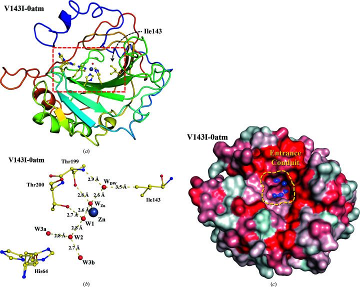
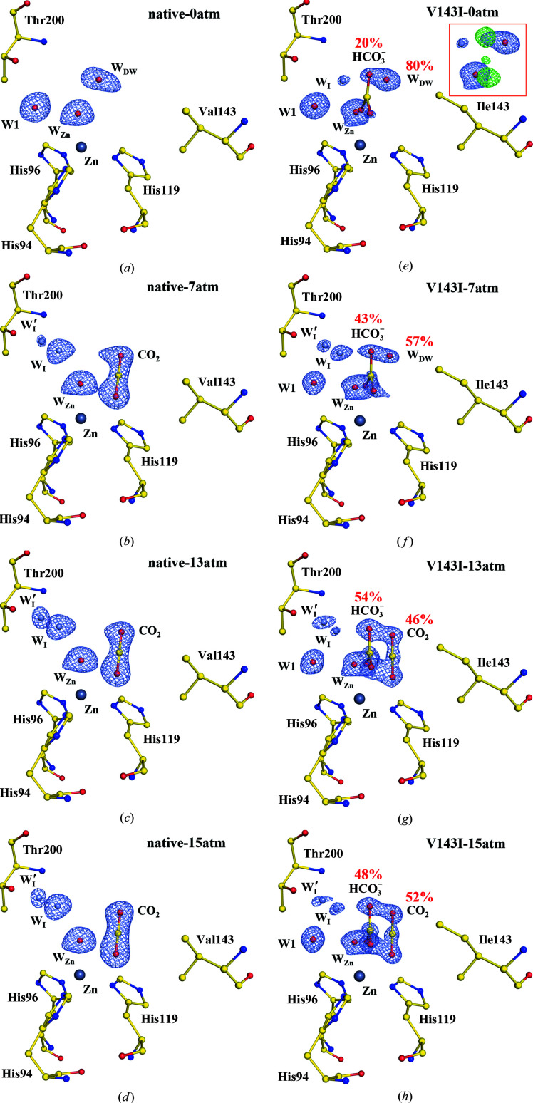
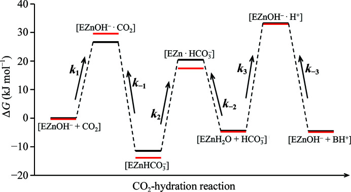
Enzymes are catalysts of biological processes. Significant insight into their catalytic mechanisms has been obtained by relating site-directed mutagenesis studies to kinetic activity assays. However, revealing the detailed relationship between structural modifications and functional changes remains challenging owing to the lack of information on reaction intermediates and of a systematic way of connecting them to the measured kinetic parameters. Here, a systematic approach to investigate the effect of an active-site-residue mutation on a model enzyme, human carbonic anhydrase II (CA II), is described. Firstly, structural analysis is performed on the crystallographic intermediate states of native CA II and its V143I variant. The structural comparison shows that the binding affinities and configurations of the substrate (CO2) and product (HCO3 -) are altered in the V143I variant and the water network in the water-replenishment pathway is restructured, while the proton-transfer pathway remains mostly unaffected. This structural information is then used to estimate the modifications of the reaction rate constants and the corresponding free-energy profiles of CA II catalysis. Finally, the obtained results are used to reveal the effect of the V143I mutation on the measured kinetic parameters (k cat and k cat/K m) at the atomic level. It is believed that the systematic approach outlined in this study may be used as a template to unravel the structure-function relationships of many other biologically important enzymes.

Keywords: X-ray crystallography; active-site mutation; active-site water dynamics; carbonic anhydrase II; enzyme mechanism; metalloenzymes; structural biology; zinc ion.


Figures

Figure 1
Figure 1
Structure of V143I CA II. (a) Overall structure of V143I-0atm: V143I CA II with no CO2 pressurization. The active site (red box) is located at a depth of 15 Å from the surface. Note that Ile143 is located at the hydrophobic pocket in the active site. (b) Ordered water network in the hydrophilic region serving as a proton-transfer pathway. (c) Surface rendition of V143I-0atm. The entrance conduit (diameter of 7–10 Å, guided with a yellow dotted line) connects the active site to the bulk solvent outside, forming the replenishment pathway. The electron density of the entrance-conduit waters is contoured at 1.5σ. Hydrophobic amino acids are shaded in red, while hydrophilic amino acids are coloured white.
Figure 2
Figure 2
CO2/HCO3 -binding site of native and V143I CA II. The intermediate waters (WI and WI′) are coloured steel blue for clarity. The electron density (2F o − F c) is contoured at 1.5σ where not indicated otherwise. (a)–(d) Native CA II structures. (e)–(h) V143I CA II structures. WI′ at 7, 13 and 15 atm is contoured at 1.25σ and WI at 0 atm is contoured at 1.0σ. Partial occupancies of HCO3 and WDW were determined in V143I-0atm and V143I-7atm, and partial occupancies of HCO3 and CO2 were determined at the higher CO2 pressures (see the supporting information). The inset (red box) in V143I-0atm shows the difference map (F oF c contoured at 3.0σ; green) when the HCO3 molecule is not included in the structure refinement.
Figure 3
Figure 3
Proton-transfer pathway including the water network and His64. The entrance-conduit waters (WEC) are coloured cyan and the intermediate waters (WI and WI′) are coloured steel blue for clarity. The electron density (2F oF c) is contoured at 1.5σ where not indicated otherwise. The major hydrogen bonds between water molecules are represented by dashed lines, while alternative hydrogen bonds that are mutually exclusive are represented by dotted lines. (a)–(d) Native CA II structures. (e)–(h) V143I CA II structures. WI′ at 7, 13 and 15 atm and W′EC1 at 15 and 13 atm are contoured at 1.25σ, and WI at 0 atm is contoured at 1.0σ. The inset (red box) in V143I-0atm shows the difference map (F oF c contoured at 3.0σ; green) when the HCO3 molecule is not included in the structure refinement.
Figure 4
Figure 4
The water-replenishment pathway including entrance-conduit waters (cyan) and intermediate waters (steel blue). The electron density (2F oF c) is contoured at 1.5σ where not indicated otherwise. (a)–(d) Native CA II structures. WEC3 at 7 and 13 atm and WEC4 at 15 atm are contoured at 1.25σ, and WEC3 at 15 atm is contoured at 1.0σ. (e)–(h) V143I CA II structures. WI′ at 7, 13 and 15 atm and W′EC1 at 15 and 13 atm are contoured at 1.25σ and WI at 0 atm is contoured at 1.0σ. Compared with native CA II, V143I CA II shows significantly perturbed positions for WEC2.
Figure 5
Figure 5
Estimated free-energy profiles for the CO2-hydration reaction catalyzed by CA II. The energy states of native CA II (black) are from a previous study (Behravan et al., 1990 ▸). The energy states of V143I CA II (red) are qualitatively estimated with respect to the native form by considering the structural information and the variations in the reaction rate constants. Note that the energy level of [EZnH2O + HCO3 ] in the V143I variant is assumed to be the same as that in native CA II. The depicted energy gaps are not to scale.

References

    1. Aggarwal, M., Kondeti, B., Tu, C., Maupin, C. M., Silverman, D. N. & McKenna, R. (2014). IUCrJ, 1, 129–135. - PMC - PubMed
    1. Alexander, R. S., Kiefer, L. L., Fierke, C. A. & Christianson, D. W. (1993). Biochemistry, 32, 1510–1518. - PubMed
    1. Alexander, R. S., Nair, S. K. & Christianson, D. W. (1991). Biochemistry, 30, 11064–11072. - PubMed
    1. Behravan, G., Jonsson, B.-H. & Lindskog, S. (1990). Eur. J. Biochem. 190, 351–357. - PubMed
    1. Chegwidden, W. R., Carter, N. D. & Edwards, Y. H. (2013). The Carbonic Anhydrases: New Horizons. Basel: Birkhäuser.