. 2020 Oct;8(20):1291.
doi: 10.21037/atm-20-5182.

Artesunate alleviates myocardial ischemia/reperfusion-induced myocardial necrosis in rats and hypoxia/reoxygenation-induced apoptosis in H9C2 cells via regulating the FAK/PI3K/Akt pathway

Affiliations

Artesunate alleviates myocardial ischemia/reperfusion-induced myocardial necrosis in rats and hypoxia/reoxygenation-induced apoptosis in H9C2 cells via regulating the FAK/PI3K/Akt pathway

Shunyang Fan et al. Ann Transl Med. 2020 Oct.

Abstract

Background: The various anti-inflammatory, anti-apoptotic, and antioxidant effects of Artesunate (Art) have been explored in numerous studies. This study aimed to evaluate the function of Art on myocardial necrosis in apoptotic cardiomyocytes in vivo and in vitro.

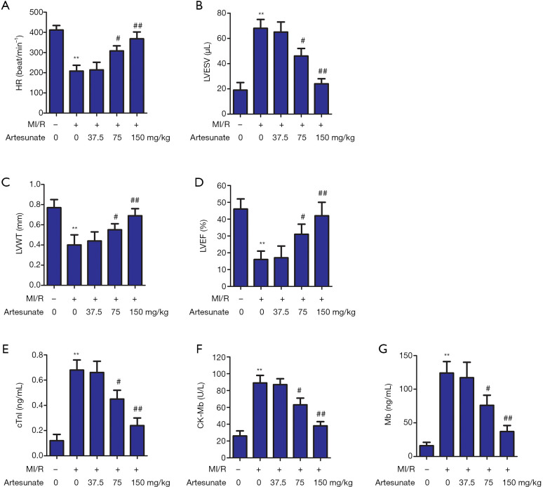
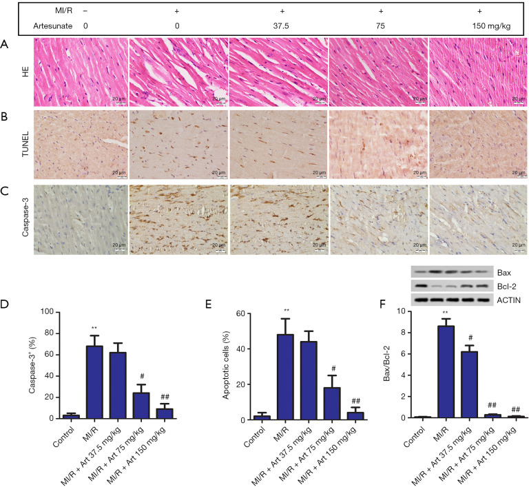
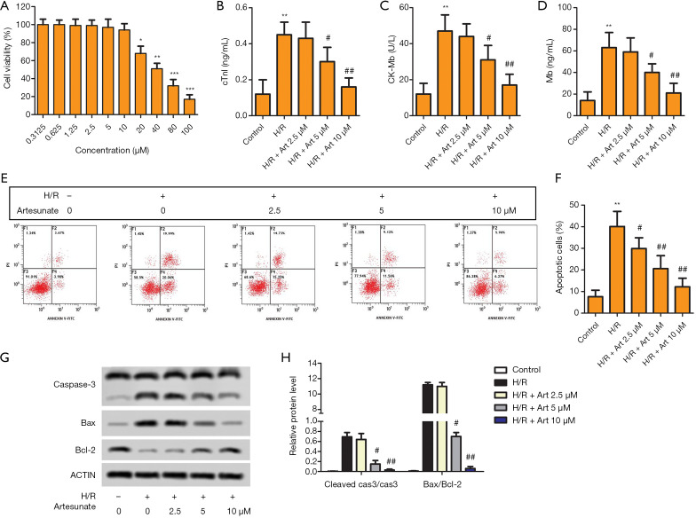
Methods: Sprague Dawley (SD) rats were randomly divided into groups: a control group, a myocardial ischemia reperfusion (MI/R) group, and MI/R+ Art groups. To establish a MI/R model, rats were subjected to left anterior descending artery ischemia for 45 minutes, and then reperfusion for 2 hours. Hypoxia was induced in H9C2 cells by subjecting them to hypoxic conditions at 37 °C for 4 hours, before placing them in a normoxic chamber for 2 hours. The test methods were used in this test, such as echocardiography, enzyme-linked immunosorbent assay (ELISA), HE staining, TUNEL staining, immunohistochemistry, flow cytometry, western blot, and CCK-8 assay.

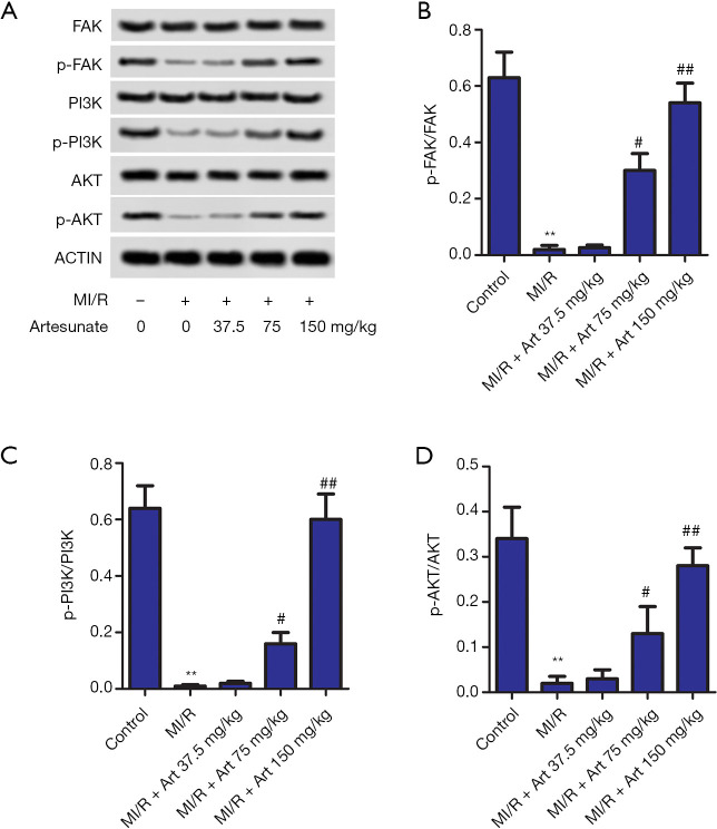
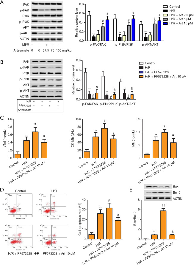
Results: Art improved myocardial systolic function caused by MI/R injury in vivo. Simultaneously, Art reduced the levels of cardiac troponin I (cTnl), creatine kinase-MB (CK-MB) and myohemoglobin (Mb) in vivo and in vitro. Moreover, Art inhibited cardiomyocyte apoptosis in vivo and in vitro. The focal adhesion kinase (FAK)/phosphatidylinositide-3 kinases (PI3K)/AKT signaling pathway was also activated by Art in vivo and in vitro. Furthermore, after inhibitor PF573228 was added, Art inhibited apoptosis in H9C2 cells via activation of the FAK/PI3K/AKT signaling pathway in vitro.

Conclusions: This study confirms that Art alleviated MI/R injury and inhibited cardiomyocyte apoptosis in vivo and in vitro. Art exerted an inhibitory effect on cardiomyocyte apoptosis by activating the FAK/PI3K/AKT signaling pathway. Therefore, Art may serve as an alternative treatment for MI/R injury.

Keywords: Artesunate; apoptosis; focal adhesion kinase/phosphatidylinositide-3 kinases/Akt pathway (FAK/PI3K/Akt pathway); hypoxia/reoxygenation; myocardial ischemia/reperfusion.


Conflict of interest statement

Conflicts of Interest: All authors have completed the ICMJE uniform disclosure form (available at http://dx.doi.org/10.21037/atm-20-5182). The authors have no conflicts of interest to declare.

Figures

Figure 1
Figure 1
Effect of Art on cardiac injury in the MI/R model. Rats were randomly divided into 5 groups: the control group; the MI/R group; the 37.5 mg/kg Art group; the 70 mg/kg Art group; and the 150 mg/kg Art group. (A) HR (beat/min), (B) LVESV (µL), (C) LVWT (mm), and (D) LVEF (%) were detected by echocardiography. An enzyme-linked immunosorbent assay (ELISA) was carried out to measure the levels of (E) cTnl, (F) CK-MB, and (G) Mb. **, P<0.05 vs. the control group; #, P<0.05 vs. the MI/R group; ##, P<0.01 vs. the MI/R group. MI/R, myocardial ischemia reperfusion; HR, heart rate; LVESV, left ventricular end systolic volume; LVWT, left ventricular wall thickness; LVEF, left ventricular ejection fraction; cTnl, cardiac troponin I; CK-MB, creatine kinase-MB; Mb, myohemoglobin.
Figure 2
Figure 2
Effect of Art on myocardial apoptosis in the MI/R model. (A) H&E staining of heart tissues from the in MI/R rats revealed that the cardiomyocytes were disordered, the cells were swollen, and some cells were dissolved. Magnification 200×; (B) the apoptotic cells were measured by terminal deoxynucleotidyl transferase-mediated dUTP-biotin nick end labeling (TUNEL) staining. Magnification 200×; (C) the expression levels of caspase-3 were detected by immunohistochemical staining. Magnification 200×; (D) the relative level of caspase-3 was analyzed by image 6.0; (E) the relative level of cell apoptosis was analyzed by image 6.0; (F) the protein expression levels of Bax and Bcl-2 were detected by western blot. **, P<0.05 vs. the control group; #, P<0.05 vs. the MI/R group; ##, P<0.01 vs. the MI/R group. MI/R, myocardial ischemia reperfusion.
Figure 3
Figure 3
Effect of Art on the FAK/PI3K/Akt pathway in the MI/R model. (A) The protein expression levels of p-FAK, p-P13K, and p-AKT were detected by western blot. Semi-quantitative analysis of the relative levels of p-FAK (B), p-P13K (C), and p-AKT (D) in each group of rats. **, P<0.05 vs. the control group; #, P<0.05 vs. the MI/R group; ##, P<0.01 vs. the MI/R group. MI/R, myocardial ischemia reperfusion.
Figure 4
Figure 4
Effect of Art on H9C2 cell apoptosis in the H/R model. (A) Cell viability was detected by CCK-8 assay. H9C2 cells were divided into 5 groups: the control group; the H/R group; the low-dose group (2.5 µM Art); the medium-dose group (5 µM Art); and the high-dose group (10 µM Art). (B) cTnl, (C) CK-MB and (D) Mb were measured by ELISA. (E,F) Cell apoptosis was detected by flow cytometry. The representative column diagrams showing results of number of F2 + F4 as apoptotic cells. (G) The protein expression levels of cleaved caspase-3, Bax, and Bcl-2 were detected by western blot. (H) Semi-quantitative analysis of the relative levels. *, P<0.05 vs. the control group; **, P<0.01 vs. the control group; ***, P<0.001 vs. the control group; #, P<0.05 vs. the H/R group; ##, P<0.01 vs. the H/R group.
Figure 5
Figure 5
Effect of Art on the FAK/PI3K/Akt pathway in the H/R model. (A) The protein expression levels of p-FAK, p-P13K, and p-AKT were detected by western blot. H9C2 cells were divided into 4 groups: the control group; the H/R group; H/R + PF573228 group; and the H/R + PF573228 + Art group. (B) The protein expression levels of p-FAK, p-P13K, and p-AKT were detected by western blot. (C) ELISA was carried out to measure cTnl, CK-MB, and Mb. (D) Cell apoptosis was detected by flow cytometry. The representative column diagrams showing results of number of F2 + F4 as apoptotic cells. (E) The protein expression levels of Bax and Bcl-2 were detected by western blot. **, P<0.05 vs. the control group; #, P<0.05 vs. the H/R group; &, P<0.05 vs. the H/R + PF573228 group.

Similar articles

Cited by

References

    1. Hausenloy DJ, Yellon DM. Myocardial ischemia-reperfusion injury: a neglected therapeutic target. J Clin Invest 2013;123:92-100. 10.1172/JCI62874 - DOI - PMC - PubMed
    1. Hou X, Fu M, Cheng B, et al. Galanthamine improves myocardial ischemia-reperfusion-induced cardiac dysfunction, endoplasmic reticulum stress-related apoptosis, and myocardial fibrosis by suppressing AMPK/Nrf2 pathway in rats. Ann Transl Med 2019;7:634. 10.21037/atm.2019.10.108 - DOI - PMC - PubMed
    1. Vincent A, Covinhes A, Barrère C, et al. Acute and long-term cardioprotective effects of the Traditional Chinese Medicine MLC901 against myocardial ischemia-reperfusion injury in mice. Sci Rep 2017;7:14701. 10.1038/s41598-017-14822-x - DOI - PMC - PubMed
    1. Braunersreuther V, Jaquet V. Reactive oxygen species in myocardial reperfusion injury: from physiopathology to therapeutic approaches. Curr Pharm Biotechnol 2012;13:97-114. 10.2174/138920112798868782 - DOI - PubMed
    1. Guo CX, Jiang X, Zeng XJ, et al. Soluble receptor for advanced glycation end-products protects against ischemia/reperfusion-induced myocardial apoptosis via regulating the ubiquitin proteasome system. Free Radic Biol Med 2016;94:17-26. 10.1016/j.freeradbiomed.2016.02.011 - DOI - PubMed