. 2020 Oct;8(20):1313.
doi: 10.21037/atm-20-6331.

Long non-coding RNA TRPM2-AS sponges microRNA-138-5p to activate epidermal growth factor receptor and PI3K/AKT signaling in non-small cell lung cancer

Affiliations

Long non-coding RNA TRPM2-AS sponges microRNA-138-5p to activate epidermal growth factor receptor and PI3K/AKT signaling in non-small cell lung cancer

Dong Cui et al. Ann Transl Med. 2020 Oct.

Abstract

Background: Long non-coding RNAs (lncRNAs) can play pivotal roles in tumor progression by acting as microRNA (miRNA) sponges. This study aimed to investigate the association of a novel lncRNA, TRPM2-AS, with the miR-138-5p/EGFR axis in the development of non-small cell lung cancer (NSCLC).

Methods: Sixty NSCLC tissues and paired adjacent non-tumor tissues were analyzed. The relative expression levels of TRPM2-AS, miR138-5p, and epidermal growth factor receptor (EGFR) and the interactions between them were analyzed. The NSCLC cell lines NCI-H1299 and A549 were transfected with TRPM2-AS shRNA/pcDNA, and miR-138-5p mimics. Cell proliferation, migration, invasion, and apoptosis were examined in response to different transfection conditions. Dual-luciferase reporter assay was performed to identify the target interactions between TRPM2-AS, miR-138-5p, and EGFR. A549 cells stably transfected with shRNA were injected into BALB/c null nude mice to establish a tumor xenograft model.

Results: TRPM2-AS was up-regulated in NSCLC tumors and cell lines. Cell proliferation, migration, and invasion were inhibited in NSCLC cells treated with sh-TRPM2-AS, while apoptosis was induced. The targeting of TRPM2-AS by miR138-5p and miR138-5p by EGFR were validated with dual-luciferase reporter assay. TRPM2-AS was found to be negatively correlated with miR138-5p but positively correlated with EGFR. PI3K/AKT/mTOR was activated by pcDNA-EGFR but inactivated by miR-138-5p mimics. In the tumor xenograft mouse model, sh-TRPM2-AS suppressed tumor formation, reduced the expression of EGFR and Ki67, and promoted tumor cell apoptosis.

Conclusions: Our results suggested that TRPM2-AS can increase the levels of EGFR via sponging miR-138-3p; this promoted NSCLC cell proliferation, migration, and invasion in vitro, and exacerbated tumors in vivo. These findings highlight TRPM2-AS/miR-138-5p as a potential target for reducing drug resistance in patients with NSCLC.

Keywords: Epidermal growth factor receptor (EGFR); TRPM2-AS; miR-138; non-small cell lung cancer (NSCLC); phosphatidylinositol 3-kinase (PI3K).


Conflict of interest statement

Conflicts of Interest: All authors have completed the ICMJE uniform disclosure form (available at http://dx.doi.org/10.21037/atm-20-6331). The authors have no conflicts of interest to declare.

Figures
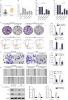
Figure 1
Figure 1
Expression profile and effects of TRPM2-AS in NSCLC. (A) TRPM2-AS expression in NSCLC and adjacent tissues from 60 patients was analyzed by paired t-test. (B) TRPM2-AS expression in normal HBE cells and several NSCLC cell lines. TRPM2-AS expression in NCI-H1299 (C) and A549 (D) cells transfected with the indicated shRNAs were tested with qRT-PCR. Colony formation (stained with crystal violet and directly photographed with a digital camera) (E), apoptosis (F), invasion (stained with crystal violet and observed under 400× scope) (G), and migration (H) in NCI-H1299 and A549 cells transfected with sh-TRPM2-AS were analyzed with colony formation assay, flow cytometry, Transwell assay, and wound-healing assay, respectively. (I) Protein expression in NCI-H1299 and A549 cells transfected with sh-TRPM2-As. Data are shown as mean ± SD. **, P<0.01 versus control and ***, P<0.001versus control with t-test. NSCLC, non-small cell lung cancer; HBE, human bronchial epithelioid; qRT-PCR, quantitative real-time polymerase chain reaction. sh1-TRPM2-As was also marked as sh-TRPM2-As.
Figure 2
Figure 2
TRPM2-AS is targeted by miR-138-5p. (A) miR-138-5p expression in NSCLC and adjacent tissues from 60 patients was analyzed by paired t-test. (B) miR-138-5p expression in HBE cells and several NSCLC cell lines. Targeting of TRPM2-AS by miR-138-5p was verified with luciferase assay in NCI-H1299 (C) and A549 (D) cells. (E) miR-138-5p expression in TRPM2-AS-silenced NCI-H1299 and A549 cells. Expression of TRPM2-AS (F) and miR-138-5p (G) in TRPM2-AS-silenced NCI-H1299 and A549 cells. (H) Pearson’s correlation analysis of miR-138-5p and TRPM2-AS in NSCLC tissues from 60 patients. RNA levels were all detected by qRT-PCR. Data are shown as mean ± SD. **, P<0.01 and ***, P<0.001 versus control by t-test. NSCLC, non-small cell lung cancer; HBE, human bronchial epithelioid; qRT-PCR, quantitative real-time polymerase chain reaction.
Figure 3
Figure 3
EGFR is targeted by miR-138-5p. (A) Relative protein expression of EGFR in NSCLC and adjacent tissues from 60 patients was analyzed with paired t-test. (B) EGFR expression in HBE cells and several NSCLC cell lines. (C) EGFR expression in NCI-H1299 and A549 cells treated with miR138-5p mimic. Targeting of EGFR by miR-138-5p was verified with luciferase assay in NCI-H1299 (D) and A549 (E) cells. (F) RNA expression of EGFR in NCI-H1299 and A549 cells. (G) EGFR protein expression in NCI-H1299 and A549 cells transfected with miR-138-5p mimic and/or pcDNA-EGFR, and (H) the protein level was semi-quantified with Image J. (I) Pearson’s correlation analysis of EGFR and TRPM2-AS in NSCLC tissues from 60 patients. Colony formation (stained with crystal violet and directly photographed with a digital camera) (J) and Transwell invasion (stained with crystal violet and observed under 400× scope) (K) assays in NCI-H1299 and A549 cells transfected with miR-138-5p mimic and/or pcDNA-EGFR. RNA and protein levels were detected by qRT-PCR and western blot respectively. Data are shown as mean ± SD. **, P<0.01 and ***, P<0.001 versus control and ##, P<0.01 versus miR-138-5p by t-test. NSCLC, non-small cell lung cancer; HBE, human bronchial epithelioid; qRT-PCR, quantitative real-time polymerase chain reaction; EGFR, epidermal growth factor receptor.
Figure 4
Figure 4
PI3K/AKT/mTOR was involved in miR-138-5p/EGFR interaction. (A) Protein expression levels of PI3K, AKT, mTOR, and their phosphorylated forms in NCI-H1299 cells transfected with miR-138-5p mimic and/or pcDNA-EGFR. (B) Protein expression levels of PI3K, AKT, mTOR, and their phosphorylated forms in A549 cells transfected with miR-138-5p mimic and/or pcDNA-EGFR. Protein expression was detected by western blot and analyzed with Image J. Data are shown as mean ± SD. *, P<0.01 NC mimic and #, P<0.01 versus miR-138-5p mimic by t-test. EGFR, epidermal growth factor receptor.
Figure 5
Figure 5
Silencing of TRPM2-AS inhibited NSCLC progression in vivo. After injection with transfected A549 cells, the mice were observed every 5 days and sacrificed on day 30. (A) The tumors were harvested on day 30. (B) The tumor volume was measured and analyzed with one-way analysis of variance. (C) Relative TRPM2-AS and miR-138-5p expression in xenograft tumors was tested by qRT-PCR. (D) Expression levels of EGFR, Ki67, and TUNEL in the xenograft tumors were detected with immunohistochemistry under 200× scope (stained with diaminobenzidine). (E) Relative EGFR expression in the xenograft tumors. (F) Relative miR-138-5p expression in the xenograft tumors was tested by qRT-PCR. (G) Cell apoptosis in the xenograft tumors was tested with TUNEL assay. The staining was analyzed with Image Pro Plus. Data are shown as mean ± SD. *, P<0.05, **, P<0.01 versus control t-test or one-way ANOVA. NSCLC, non-small cell lung cancer; qRT-PCR, quantitative real-time polymerase chain reaction; EGFR, epidermal growth factor receptor; ANOVA, analysis of variance.
Figure S1
Figure S1
Targeting of epidermal growth factor receptor (EGFR) by miR-139-5p predicted with TargetScan 7.2.
Figure S2
Figure S2
Representative targeting of mutant epidermal growth factor receptor (EGFR) by miR-138-5p.
Figure S3
Figure S3
Luciferase report vectors of the TRPM2-AS and miR-138-5p mimics used.
Figure S4
Figure S4
Luciferase report vectors of the epidermal growth factor receptor (EGFR) and miR-138-5p mimics used.

References

    1. Global Burden of Disease Cancer Collaboration , Fitzmaurice C, Abate D, et al. Global, Regional, and National Cancer Incidence, Mortality, Years of Life Lost, Years Lived With Disability, and Disability-Adjusted Life-Years for 29 Cancer Groups, 1990 to 2017: A Systematic Analysis for the Global Burden of Disease Study. JAMA Oncol 2019;5:1749-68. 10.1001/jamaoncol.2019.2996 - DOI - PMC - PubMed
    1. Rampinelli C, Minotti M. Improving CT screening for lung cancer with a highly predictive risk model. Ann Transl Med 2018;6:128. 10.21037/atm.2018.01.34 - DOI - PMC - PubMed
    1. Tanzawa S, Ishihara M, Haruyama T, et al. Which is better, EGFR-TKI mono or combination for non-small cell lung cancer with mutated EGFR? Transl Cancer Res 2019;8:2223-9. 10.21037/tcr.2019.08.18 - DOI - PMC - PubMed
    1. Meador CB, Hata AN. Acquired resistance to targeted therapies in NSCLC: Updates and evolving insights. Pharmacol Ther 2020:107522. 10.1016/j.pharmthera.2020.107522 - DOI - PMC - PubMed
    1. Ma LY, Xie XW, Ma L, et al. Downregulated long non-coding RNA TRPM2-AS inhibits cisplatin resistance of non-small cell lung cancer cells via activation of p53- p66shc pathway. Eur Rev Med Pharmacol Sci 2017;21:2626-34. - PubMed