

Investigating the Chronology of Meniscus Root Tears: Do Medial Meniscus Posterior Root Tears Cause Extrusion or the Other Way Around?
- PMID: 33209944
- PMCID: PMC7645763
- DOI: 10.1177/2325967120961368
Investigating the Chronology of Meniscus Root Tears: Do Medial Meniscus Posterior Root Tears Cause Extrusion or the Other Way Around?
Abstract
Background: Meniscus root tears are increasingly being recognized. Meniscal extrusion has previously been associated with medial root tears; however, the relationship between secondary meniscal restraints, such as the meniscotibial (MT) ligament, extrusion, and root tears has yet to be formally evaluated.
Purpose: To better understand the association between MT ligament competence, medial meniscal extrusion, and medial meniscus posterior root tears (MMPRTs) as well as to determine the progression of meniscal extrusion over time.
Study design: Case series; Level of evidence, 4.
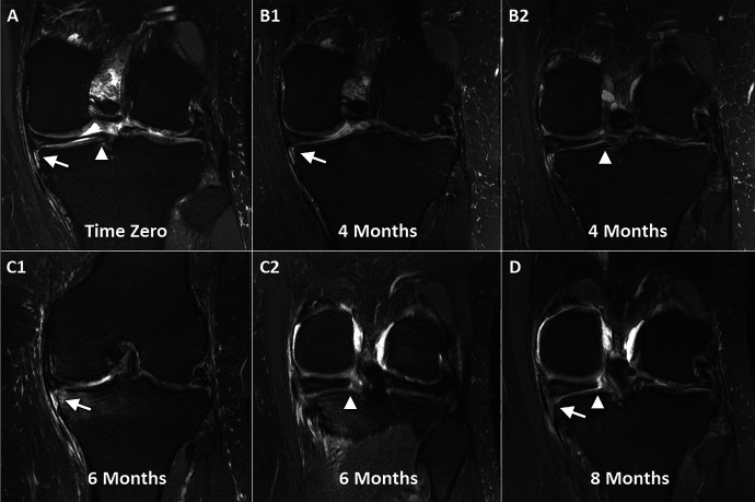
Methods: Serial magnetic resonance imaging (MRI) scans were reviewed for patients who showed evidence of medial meniscal extrusion and MMPRTs on at least 1 of ≥2 available MRI scans. All patients were symptomatic at the time of diagnosis. All MRI scans were analyzed independently by 2 board-certified musculoskeletal radiologists. MT ligament disruption, medial meniscal extrusion, and MMPRTs were recorded for each MRI scan. The time between MRI scans, presence of insufficiency fractures, and Outerbridge classification for the medial femur and tibia were also evaluated.
Results: Overall, 27 knees in 26 patients were included in this study, with a total of 63 MRI scans analyzed (21 knees with 2 MRI scans, 3 with 3 MRI scans, and 3 with 4 MRI scans). All patients demonstrated clear medial meniscal extrusion and MT ligament disruption before the subsequent development of MMPRTs (P < .001). Mean extrusion at the time of initial MRI was 3.3 ± 1.1 mm and increased significantly to 5.5 ± 1.8 mm at the time of first imaging with an identified MMPRT (P < .001). The mean time between initial MRI and the first identification of an MMPRT on later MRI was 1.7 ± 1.6 years.
Conclusion: In a sample of 27 symptomatic knees with serial MRI scans both before and after an MMPRT diagnosis, all patients demonstrated MT ligament disruption and associated meniscal extrusion before the development of subsequent medial meniscus root tears. These findings suggest that MT ligament disruption and medial meniscal extrusion represent early and predisposing events contributing to MMPRTs. Therefore, this provides a possible explanation of why meniscal extrusion is not corrected with medial meniscus root repair.
Keywords: MMPRT; extrusion; medial meniscal extrusion; medial meniscus posterior root tear; meniscotibial ligament.
© The Author(s) 2020.
Conflict of interest statement
One or more of the authors has declared the following potential conflict of interest or source of funding: This study was partially funded by a grant from the National Institute of Arthritis and Musculoskeletal and Skin Diseases for the Musculoskeletal Research Training Program (T32AR56950). A.J.K. has received research support from Aesculap/B. Braun, Ceterix Orthopaedics, Exactech, Gemini Medical, and Histogenics; consulting fees from Arthrex, DePuy, JRF Ortho, and Vericel; speaking fees from Arthrex; and royalties from Arthrex; is a board or committee member for the Musculoskeletal Transplant Foundation; and has stock/stock options in Responsive Arthroscopy. M.D.L. has a family member who has received research support from Smith & Nephew; consulting fees from Arthrex, Linvatec, Ossur, and Smith & Nephew; and royalties from Arthrex, Ossur, Smith & Nephew, and Thieme. M.H. has received hospitality payments from DePuy Synthes. C.L.C. has received educational support from Arthrex and hospitality payments from Arthrex and Zimmer Biomet. M.J.S. has received research support from Arthrex and Stryker, consulting fees from Arthrex, and royalties from Arthrex. AOSSM checks author disclosures against the Open Payments Database (OPD). AOSSM has not conducted an independent investigation on the OPD and disclaims any liability or responsibility relating thereto.
Figures
References
-
- Allaire R, Muriuki M, Gilbertson L, Harner CD. Biomechanical consequences of a tear of the posterior root of the medial meniscus: similar to total meniscectomy. J Bone Joint Surg Am. 2008;90(9):1922–1931. - PubMed
-
- Choi JY, Chang EY, Cunha GM, et al. Posterior medial meniscus root ligament lesions: MRI classification and associated findings. AJR Am J Roentgenol. 2014;203(6):1286–1292. - PubMed
-
- Chung KS, Ha JK, Ra HJ, Nam GW, Kim JG. Pullout fixation of posterior medial meniscus root tears: correlation between meniscus extrusion and midterm clinical results. Am J Sports Med. 2017;45(1):42–49. - PubMed
-
- Costa CR, Morrison WB, Carrino JA. Medial meniscus extrusion on knee MRI: is extent associated with severity of degeneration or type of tear? AJR Am J Roentgenol. 2004;183:17–23. - PubMed
-
- Daney BT, Aman ZS, Krob JJ, et al. Utilization of transtibial centralization suture best minimizes extrusion and restores tibiofemoral contact mechanics for anatomic medial meniscal root repairs in a cadaveric model. Am J Sports Med. 2019;47(7):1591–1600. - PubMed
Grants and funding
LinkOut - more resources
Full Text Sources
